
5 The Discovery of DNA
Learning Objectives
- Explain how DNA was determined to be the molecule of inheritance.
- Identify the contributions of Chargaff, Franklin, Watson, and Crick to the discovery of the structure of DNA
Today, DNA is a household term. We see it referenced in paternity tests, ancestry kits, and crime shows. This molecule is central to our understanding of life itself. Yet the discovery of DNA is relatively modern, really only being solidified by the middle of the 20th century. The story of how it was discovered can help us understand the nature of DNA as well as experimental biology and how human nature can influence the scientific process.
By the early 1900s, biochemists had isolated hundreds of chemicals from living cells. Scientists also knew that certain characteristics were passed from parents to offspring through discrete units (later called genes), but they didn’t know what these units were made of physically. Which of these chemicals was the hereditary material – the physical substance responsible for passing traits from one generation to the next? Proteins seemed like the most promising candidates since they were abundant, diverse, and complex molecules. With 20 different amino acids available for building proteins, they appeared to have the complexity necessary to carry genetic information. In contrast, DNA was known to contain only four different nucleotides (A, T, G, C), making it seem too simple to encode the vast diversity of life. However, a few key experiments demonstrated that DNA, rather than protein, is the genetic material.
Lecture Video: The Discovery of DNA and Subsequent Biology Revolution
Griffith’s Experiment and the basis of inheritance (1929)
Frederick Griffith was a microbiologist interested in what made some bacteria pathogenic and others not. At the time, microbiologists had identified two strains of the bacterium Streptococcus pneumoniae. The R-strain produced rough colonies on a bacterial plate, while the other S-strain was smooth. More importantly, the S-strain caused fatal infections when injected into mice, while the R-strain did not (Figure 1). Neither did “heat-treated” S-strain cells. Griffith in 1929 noticed that upon mixing “heat-treated” S-strain cells together with some R-type bacteria (neither should kill the mice), the mice died and there were S-strain, pathogenic cells recoverable. Thus, some non-living component from the S-type strains contained genetic information that could be transferred to and transform the living R-type strain cells into S-type cells (Figure 1).

Avery, MacLeod and McCarty’s Experiment demonstrate the DNA is the molecule of inheritance (1944)
What kind of molecule from within the S-type cells was responsible for the transformation? To answer this, researchers named Avery, MacLeod and McCarty separated the S-type cells into various components, such as proteins, polysaccharides, lipids, and nucleic acids. Only the nucleic acids from S-type cells were able to make the R-strains smooth and fatal. Furthermore, when cellular extracts of S-type cells were treated with DNase (an enzyme that digests DNA), the transformation ability was lost. The researchers therefore concluded that DNA was the genetic material, which in this case controlled the appearance (smooth or rough) and pathogenicity of the bacteria (Figure 2).

Chargaff’s Rule and base-pairing (1950)
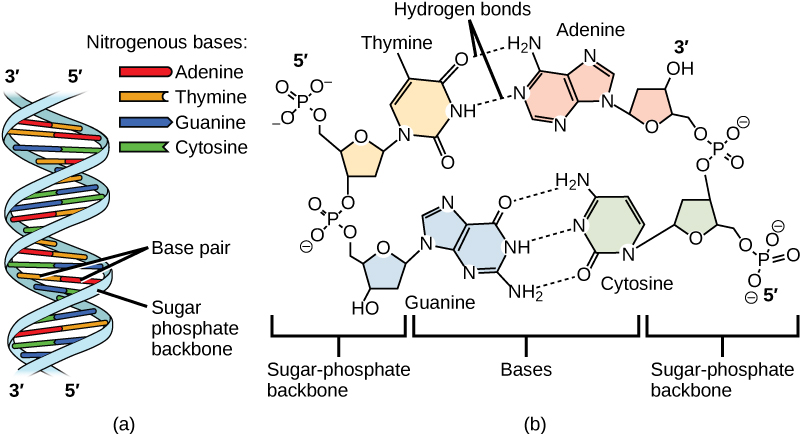
Scientists now were confident that DNA was the molecule of inheritance, but they were not certain what it looked like or how exactly it worked. A critical breakthrough came from Erwin Chargaff, who uncovered the base pairing rules that became essential to understanding DNA’s structure. Chargaff’s experiments showed that in any DNA molecule, the amount of adenine (A) consistently equals the amount of thymine (T), and the amount of cytosine (C) equals the amount of guanine (G) (Table 1).
Chargaff’s findings demonstrated that DNA was not just a random sequence of nucleotides, but rather a built around specific base pairing rules.
The percentages represent the proportion of each base relative to the total DNA content.
Chargaff’s rules are reflected in the nearly equal proportions of adenine to thymine and cytosine to guanine in each organism.
While the exact percentages vary between species, the 1:1 ratio between adenine and thymine, and between cytosine and guanine, remains consistent across different organisms.
This table highlights the universality of Chargaff’s findings across a range of species, which was critical to understanding the structure and function of DNA.
| Table 1. Data from Chargaff’s experiments. Chargaff’s rules are reflected in the nearly equal proportions of adenine to thymine and cytosine to guanine in each organism. While the exact percentages vary between species, the 1:1 ratio between adenine and thymine, and between cytosine and guanine, remains consistent across different organisms. |
||||
| Organism | Adenine (A) | Thymine (T) | Cytosine (C) | Guanine (G) |
| Human | 30.30% | 30.30% | 19.90% | 19.50% |
| Escherichia coli | 24.70% | 23.60% | 26.00% | 25.70% |
| Yeast | 31.30% | 32.90% | 17.10% | 18.70% |
| Calf Thymus | 29.00% | 29.10% | 22.00% | 20.90% |
| Wheat | 27.30% | 27.10% | 22.70% | 22.80% |
| Rat | 28.60% | 28.40% | 21.40% | 21.50% |
A model of DNA
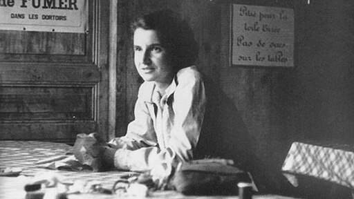
In the 1950s, multiple teams were trying to model the structure of DNA. Linus Pauling discovered the secondary structure of proteins using X-ray crystallography. X-ray crystallography is a method for investigating molecular structure by observing the patterns formed by X-rays shot through a crystal of the substance. The patterns give important information about the structure of the molecule of interest. Maurice Wilkins inspired by Pauling’s work, set about studying the structure of DNA with X-ray crystallography. Rosalind Franklin, who recently joined Wilkins Lab, led the effort to use X-ray crystallography to understand the structure of DNA. Her work produced the famous “Photo 51,” which provided crucial evidence that DNA has a helical structure (Figure 3).
|
Figure 3
|
|
 |
 |
James Watson and Francis Crick used data collected by Franklin, Chargaff, and others to piece together the puzzle of the DNA molecule. In 1953, Watson and Crick proposed a double-helix model for the structure of DNA, which is the model we still use today (Figure 4).
In 1962, James Watson, Francis Crick, and Maurice Wilkins were awarded the Nobel Prize in Medicine for their work in determining the structure of DNA.
Rosalind Franklin passed away from cancer in 1958 and was not recognized by the Nobel Committee, despite her research providing principal evidence toward our understanding of DNA.
(you can learn more about the fascinating life of Rosalind Franklin here and here).
The Life of Rosalind Franklin
The story of Dr. Rosalind Franklin is one marked by triumph and tragedy and illustrates the human nature of science, both good and bad. Franklin was an extraordinary chemist and X-ray crystallographer whose meticulous work produced critical data that contributed to the discovery of the structure of DNA. Despite her brilliance, she often faced barriers as a woman in mid-20th-century science and did not receive full recognition for her contributions during her lifetime. Her story highlights both the collaborative and competitive sides of scientific discovery, showing how advances in knowledge are shaped not only by data and experiments but also by the relationships, biases, and social contexts in which science takes place.
Here are some additional resources to learn more about her as a person and as a scientist.
Defending Franklin’s Legacy (PBS NOVA)
Cobb, M., & Comfort, N. (2023). What Rosalind Franklin truly contributed to the discovery of DNA’s structure. Nature, 616(7958), 657-660.
The DNA Double Helix Discovery (HHMI BioInteractive)
This video provides a great overview of the history discussed in this chapter.
Glossary
Base pairing rules (Chargaff’s rules): The principle that adenine (A) pairs with thymine (T), and cytosine (C) pairs with guanine (G), discovered by Erwin Chargaff.
DNA (deoxyribonucleic acid): The molecule that carries hereditary information in all known living organisms, structured as a double helix.
Double helix: The twisted-ladder shape of DNA, with paired bases forming the rungs.
Genetic material: The substance within a cell that carries information passed from one generation to the next.
Nucleotides: The building blocks of DNA and RNA, composed of a sugar, a phosphate group, and a nitrogenous base.
Photo 51: The famous X-ray crystallography image taken by Rosalind Franklin and Raymond Gosling that provided key evidence for DNA’s helical structure.
X-ray crystallography: A method that uses X-ray diffraction patterns to determine the structure of molecules.
References and Resources
- Cobb, M., & Comfort, N. (2023). What Rosalind Franklin truly contributed to the discovery of DNA’s structure. Nature, 616(7958), 657–660. https://doi.org/10.1038/d41586-023-01313-5
- Judson, H. F. (1996). The eighth day of creation: Makers of the revolution in biology. Cold Spring Harbor Laboratory Press.
- Maddox, B. (2002). Rosalind Franklin: The dark lady of DNA. HarperCollins.
- PBS NOVA. (n.d.). Photo 51: The story of Rosalind Franklin. Retrieved from https://www.pbs.org/wgbh/nova/photo51/
- Watson, J. D. (1968). The double helix: A personal account of the discovery of the structure of DNA. Atheneum.
- Wilkins, M. (2003). The third man of the double helix: The autobiography of Maurice Wilkins. Oxford University Press.
