2 Reproduction
Reproduction
Luciano Caixeta, DVM PhD
College of Veterinary Medicine, University of Minnesota
Rafael Bisinotto, DVM MS PhD
College of Veterinary Medicine, University of Florida
Summary
- Describe the importance of reproductive performance.
- Accurately describe the bovine estrus cycle and how to control the estrus cycle by using exogenous hormones in fertility programs.
- Determine the most appropriate reproductive programs for different dairy herds.
- Develop a management plan for starting and stopping to inseminate cows depending on the herd’s goals and performance.
- Explain how animal health and performance can influence reproductive performance.
Introduction
A good understanding of reproductive physiology and management principles is important to a dairy veterinarian because a successful reproductive program is imperative for the success of dairy enterprises. Dairy farms efficiency and profitability are mostly affected by milk production and the initiation of milk production depends on a cow giving birth to a calf. In addition to milk production, a successful reproductive program increases the number of replacement heifers and contributes to the improvement of herd’s genetics. In summary, improved reproductive performance will improve herd success and profitability.
Bovine Estrous Cycle Review
Review this video of the 2018 presentation by Dr. Caixeta
Reproductive Tract
The repro tract starts with the vagina and vulva, moves up through the cervix, and then to the uterine body. Then, it bifurcates into two horns, the left, and the right, and at the ends of each horn, there are the left and right ovaries. A full palpation of the repro tract would include feeling all of these structures. If you do not know whether the cow has two horns or one, your pregnancy diagnosis is incomplete. Every cow should have two uterine horns but it is possible that you will palpate a cow with only one uterine horn or a heifer without any horns.
There are three layers of the uterus. The serosal layer, the muscle layer, and the inner portion of the uterus which is composed of the mucosa and submucosa and is referred to as the endometrium. The caruncles, which are small, non-glandular protuberances on the surface of the endometrium are where the placenta attaches. The endometrium produces prostaglandin F2alpha if there is no maternal recognition of pregnancy, which causes the Corpus Luteum (also called a CL) to lyse, or die, which stops it from producing progesterone, which is the hormone responsible for sustaining a pregnancy.
The CL is located on the ovary. It produces progesterone and is identifiable by transrectal palpation or ultrasound. 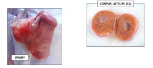 Transrectal palpation has poor accuracy and only poor agreement between clinicians whereas transrectal ultrasound has high agreement and accuracy between clinicians. However, the accuracy of ovarian structures (i.e. CLs and follicles) detection by transrectal palpation is increased with experienced practitioners, especially if they know previous information regarding the animal’s estrus cycle – which is expected for animals enrolled in any fertility program.
Video one is a slowed down video showing a transrectal ultrasound. Near the beginning of the video we see the uterus as the round circles. Next, we observe one of the ovaries and we cover the whole length of the ovary. We briefly see a small, cavitated CL. And finally we see the main CL – about 20-22 millimeters in diameter – which is likely actively producing progesterone.
Video two is the same one we watched previously but at regular speed.
Video 3 is of a different cow at full speed and shows two CLs on the first ovary which are the white cavitary structures seen near the beginning of the video and again at the end. In between, we see the opposite ovary with a follicle which appears as the black, circular structure in the middle of the screen. It is important to notice that the follicle has a thin wall whereas the CLs have a much thicker wall. Recognizing these structures on ultrasound might be difficult at first, but becomes easier with time and practice.
Another important structure in the bovine repro tract and the estrus cycle is the follicle. It harbors the oocyte/egg until ovulation and produces inhibin and estradiol in different stages of the estrus cycle. Inhibin, as its name implies, is produced by a dominant follicle and inhibits other non-dominant follicles from growing. Close to the end of the estrus cycle and associated with other reproductive hormones, a rise in estradiol produced by the follicle causes a cow to begin showing signs of estrus. A follicle is identified through transrectal palpation or through a transrectal ultrasound. They will feel a little like a blister and you might be able to push your finger into it. A CL will be harder and you will be unable to push your finger into it.
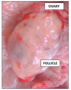
This is a video of an ultrasound of a cow in estrus. At the very beginning of the video you will see the uterus, and the anechoic fluid within the uterus in the upper left corner is mucus. At 6 seconds in the video you see a large dark image in the upper left and this is the follicle; you can see a thin layer around it, the capsule. Compared to the first and second ultrasounds of the CLs, the follicle now looks much different
Hormones involved in the estrus cycle
Two major structures on the ovary are the CL and the follicle. The CL is a solid yellow structure that produces progesterone, which is important to maintain pregnancy up until the endometrium in the uterus placenta begins maintaining it. And the follicle is a blister-like structure that contains the oocyte and thus produces estrogen and also inhibin; one follicle will be the “dominant” follicle.
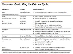
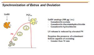
There are seven hormones that are important in controlling the estrus cycle and some of them are used in fertility programs to increase reproductive success in herds. Gonadotropin-releasing hormone, or GnRH, from the hypothalamus, will induce follicle-stimulating hormone, usually called FSH, and luteinizing hormone, called LH. GnRH stimulates FSH and LH at the anterior pituitary. Exogenous GnRH, via the actions of LH, can induce ovulation depending on the stage of the follicle and for this reason, it is used in fertility programs. Because the administration of GnRH can induce ovulation, if GNRH is given when the dominant follicle is not producing enough estradiol, cows will ovulate without showing any signs of estrus.
FSH is released from the anterior pituitary and its primary role is the recruitment of follicular waves & Follicular growth up to deviation. The anterior pituitary also releases LH. LH is important to the growth of the ovarian follicle after deviation. It’s also important for steroidogenesis by the dominant follicle, which in this case is estradiol. The pre-ovulatory LH surge induces ovulation and causes the luteinization of granulosa and theca cells to form the CL.
The next hormone, which we already mentioned, is estradiol. Estradiol is produced by the ovarian follicle. It allows the cow to be sexually receptive, also called estrus behavior, and prepares the repro tract for mating and gestation. In addition, estradiol triggers the pre-ovulatory LH surge and this positive feedback loop leads to ovulation. The dominant ovarian follicle also produces Inhibin which inhibits FSH secretion by the anterior pituitary and stalls the growth of the non-dominant follicles.
Progesterone is produced by the CL and stimulates histotroph secretion by the endometrium. Progesterone maintains pregnancy, prevents estrus behavior, and modulates LH secretion.
The last hormone is PGF2alpha which is produced by the endometrium and induces luteolysis. This regression of the CL decreases the concentration of the circulating progesterone. As a consequence, LH and FSH are released which makes it possible for the dominant follicle of the new follicular wave to fully develop. Thus, the coordinated action of all these ovarian and uterine structures and hormones control the estrous cycle of the cow.
Estrus Cycle
The estrous cycle is divided into four different stages: estrus, metestrus, diestrus, and proestrus. There are minor seasonal effects in dairy cows. The estrous cycle of a cow on average will be 21 days but it can vary from 18 to 24 days. The main reason for this variation is the number of follicular waves that each cow has. Dairy cows can have two, three or four waves within the estrus cycle.
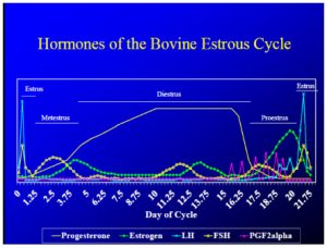
The four different stages of the estrous cycle are divided into two phases: the follicular phase and the luteal phase. The follicular phase is broken into Proestrus, which is the period of luteolysis to the onset of the estrus, and Estrus which is the period of onset of estrus behavior until ovulation. The luteal phase is broken into Metestrus, which is the period from ovulation to the presence of a functional CL (which is about 4 to 5 days), and Diestrus which is the period of having a functional CL to luteolysis – this is the longest stage of the estrus cycle.
Anestrus
Anestrus is not a part of the estrous cycle. Any prepubertal female is in anestrus and there are many cases of anestrus postpartum (including delayed ovulation). If the cow has a luteal cyst, the high concentration of circulating progesterone will likely halt the normal function of the ovaries and cows will not cycle properly and will not likely show estrus. Lastly, there’s nutritional anestrus: When the cow is not getting the proper nutrition they will likely diverge their nutrient reserves to the maintenance of essential biological systems and will likely stop maintaining regular estrus cycle because if they cannot maintain themselves to stay alive then they are unlikely to be able to successfully reproduce and maintain their offspring.
Anestrus is a period of reproductive quiescence. Meaning that when the cow or a heifer is in anestrous, she does not exhibit estrous cycles or estrus behavior. The ovaries are inactive and there are not any ovulatory follicles nor functional CLs present. There is insufficient GnRH release from the hypothalamus which results in too little or no stimulation at the pituitary for LH or FSH secretion. You need to be able to distinguish true anestrus (insufficient hormonal stimuli) from apparent anestrus (failure to detect estrus or failure to detect pregnancy). Anestrus can be caused by pregnancy, lactation, presence of offspring, season, stress (for example, nutritional stress where the cow is not meeting its nutritional needs and therefore cannot reproduce), and pathology (such as ovarian cysts).
Anestrus is simply a nondescript uterus and no ovarian structures, although sometimes there will be cysts palpated on the ovaries. On palpation, the uterus feels very flaccid because it bears no tone, which sometimes makes it difficult to find and identify structures. The ovaries will feel very smooth, without any distinct structures.
Estrus
Estrus is the stage when the female accepts mating. Estradiol is the dominant hormone in this stage and causes physical and behavioral changes. An LH spike results in ovulation and lasts between 12 to 18 hours. When palpating dairy cows it is difficult to assess estrus behavior since they are locked in a chute and cannot express standing estrus. So assessing estrus through palpation relies on assessing uterine tone. A cow in estrus will have a turgid, or hard-toned uterus, a regressing or absent CL, and a large follicle. The follicle is especially prominent right before ovulation and can be accidentally burst during palpation or ultrasound.
Metestrus
Metestrus immediately follows estrus and ovulation and it is the stage of the developing corpus luteum preparing for pregnancy. Progesterone starts to be produced and there needs to be enough progesterone to maintain a pregnancy for the first 21 days. The follicular cavity fills with blood and is termed a corpus hemorrhagicum (also called a CH) that develops into CL. If you remember from the ultrasounds shown at the beginning of this presentation, they showed a CL but there was a cavity. The CL starts filling from the outside in, that’s why you see cavitated CLs. And sometimes when on ultrasound you see cobweb-like images inside them. The corpus hemorrhagicum has many blood vessels and blood inside which makes it look like a cobweb on ultrasound. The CL is actively producing progesterone so there is a steady rise of progesterone levels and then there is an FSH surge which causes a new follicular wave. This takes about 3 to 4 days and on palpation, the uterus will still be turgid, but not as toned as when in estrus.
Diestrus
The next stage is diestrus. The CL is dominant so there are high levels of progesterone – as high as you get in that cow. The uterus is prepared for receiving the pregnancy and there are between 2 and 4 follicular waves depending on the cow. It takes about 10 to 14 days and at palpation the uterus would feel normal and flaccid in comparison to estrus. There is one or multiple CLs present on one or both ovaries and a small follicle on one or both ovaries.
Proestrus
Lastly is proestrus. At this stage, there are PGF pulses which will remove the CL causing a drop in progesterone concentrations. Then you can have an FSH spike and LH surges and you have the beginning of ovulation. On palpation, the uterus will feel normal to turgid as the cow approaches estrus. The regressing CL will feel hard on palpation and a large follicle will be present.
Progression through the stages
Starting at estrus, at day zero, the cow ovulates. Then there is an FSH surge which rises for about two days and is then followed by follicular development and emergence of new follicular waves. Follicular deviation occurs next. The follicles start growing and the CLs start to produce enough progesterone to be considered functional. One follicle – sometimes multiple – will become dominant and produce inhibin and estradiol. The inhibin will be inhibiting the non-dominant follicles from growing and the estradiol will induce the other hormone, LH which will take over the growth of the dominant follicle.
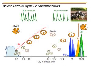
LH pulses will cause the follicle to grow which in turn releases more estradiol to aid in the maturation of the oocyte up to a point where it can be ovulated. Once it ovulates, the follicle will become a CL which produces progesterone in order to maintain the pregnancy. Progesterone prevents the hormones that are controlling follicles from reaching the levels needed for ovulation.
The CL capacity to produce progesterone increases during the next 4 to 5 days and for the rest of the diestrus period, or part of the pregnancy, it will prevent another ovulation. The CL will not be responsive to PGF2a until full maturity in about five days after estrus.
Because there is high progesterone and atresia of the dominant follicle, another surge of FSH occurs which starts a new follicular wave. A dominant follicle starts producing enough inhibin and estradiol to prevent the other follicles from growing. LH takes over the growth of the dominant follicle and if there is no maternal recognition of pregnancy – the cow doesn’t become pregnant – the endometrium produces PGF2alpha that lyses the CL. When progesterone levels decrease, the LH pulses can go even higher, inducing estradiol to go even higher leading to estrus. Cows show estrus about 8-12 hours after they ovulate.
Ovulation happens 27 hours after the onset of estrus behavior. So if estrus started around 8 AM, it would last until, or a little after 8 PM. For this reason, many people will breed their cow 12 hours after they observe estrus behavior. By doing so, they improve their chances of having a capacitated sperm ready to fertilize the egg that was just ovulated. This timeline is based on the fact that the sperm needs about eight hours for capacitation and it’s in the tract and (can) move all the way to the tip of the horn to find the oocyte. This is the main strategy used for breeding cows using estrus detection. In this case, it is important to time breeding to the timing of ovulation, considering how long the sperm cells and the oocytes are viable in the repro tract.
Estrus Cycle Manipulation
So why do we manipulate the estrus cycle? On the dairies, one of the main drivers is because it is very difficult to detect estrus in high producing dairy cows. On average, only 46% of estrus on dairy farms are identified. Manipulation of estrus cycle and fertility programs gives us more control over breeding and allows synchronized timing of when to inseminate the cow. Fertility programs reduce the interval to estrus and insemination and ensure that cows are inseminated at an optimal time – and sometimes inseminated at all. Depending on the protocol and management chosen, we can either synchronize estrus or synchronize ovulation. In the first case, we will inseminate cows based on their expression of estrus and in the second we can AI cows without detection of estrus. It is very important to reinforce that fertility programs do not replace proper management.
The three hormones used in the US for synchronization of estrus or ovulation are GnRH, PGF2alpha, and progesterone. The difference between protocols that synchronize estrus and ovulation is the fact that when we synchronize estrus we still need to check cows for estrus signs before breeding. While when using ovulation synchronization protocols we have greater control of the estrus cycle and we have a good idea of when the cow will ovulate and for this reason, we can schedule the time for AI.
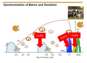
Ovsynch is an ovulation synchronization protocol and the basis for almost all fertility protocols in the US. Briefly, GnRH is given on day zero to induce FSH and LH in order to induce ovulation and a new follicular wave. On day seven, PGF2alpha is given to lyse the CL and consequently decrease progesterone concentration prior to breeding. On day nine, GnRH is given again which will start a new follicular wave but most importantly trigger the ovulation of the dominant follicle. After that, cows should be bred between 16 to 18 hours after the second GnRH administration.
Without any previous estrus manipulation, cows can be at any stage of their estrus cycle making it hard to determine when a cow will express estrus following a PGF2a injection. For example, if the PGF2a shot is given too early after ovulation and the CL is not mature–for example, 3-4 days after ovulation– the CL will not respond. If given on day seven of the cycle, the CL will be lysed and another dominant follicle will deviate which will then be followed by a surge of LH and estradiol and subsequently ovulation. If PGF2a is given on day ten, the cow doesn’t have a follicle that is mature enough to ovulate, but the CL will still be inactivated. Then a follicle will grow under low progesterone levels and the oocyte will have a lower quality when compared to oocytes that grow under high progesterone levels. So if you do give a shot at day ten, you might be hurting the chances of the cow getting pregnant just because this follicle is not as good as it could have been. For this reason, pre-synchronization protocols were developed to be administered prior to a fertility program.
Fertility programs that use pre-synchronization are used to pre-synchronize the estrus cycle and have the repro tract at the ideal stages to start the ovulation synchronization protocol. Pre-synchronization protocols intend to allow the follicle to grow under high progesterone concentrations and then have low progesterone at the time of ovulation and breeding in order to increase the chances of the cows to get pregnant.
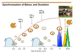
High progesterone during the development of the mature follicle is important for the quality of the oocyte. Nonetheless, low progesterone levels are needed for successful ovulation. If we are only using PGF2a for our fertility protocols, we are only synchronizing estrus. For the synchronization of ovulation, we will need to use GnRH based protocols. Giving GnRH releases LH, and then a new follicular wave is induced. Once you give GnRH you know you’re inducing a follicular wave. So by knowing that you can actually predict ovulation because you know when you started a follicle. Especially if you give PGF2alpha and inactivate the CL and allow the follicle to ovulate.
The other hormone we use is progesterone. If a follicle grows under low progesterone the oocyte is not as fertile as the one growing under higher progesterone levels. If we get rid of the CL, or if the CL is absent, in order to grow the follicle under progesterone, we can increase circulating progesterone levels by using exogenous progesterone vaginal implants. The commercial inserts with progesterone are usually left inside the cows for seven days. The use of exogenous progesterone implants allows the oocyte to grow under progesterone influence and also prevents the expression of estrus. Recently, research results have shown better results when using two inserts (off-label use of the implants). The reason for that is because by using multiple inserts we increased the progesterone level to levels exceeding the 1ng/mL traditionally used to define a CL as an active CL by previous research. Although this strategy increases the reproductive success of dairy cows, it is off-label and it doubles the costs of the protocol. So a lot of farmers will only use one per animal despite the better results of using two inserts.
There are many different synchronization protocols and the job of the veterinarian is to work with their clients to choose a protocol that best fits their budget and goals.
As an example, a synchronization protocol would be: At day zero GNRH is given to induce an LH surge to ovulate the dominant follicle and start a new follicular wave. Seven days later PGF2a is given to induce Luteolysis. Two days later on day nine, the second dose of GnRH is given to cause ovulation, and then 16 to 18 hours later the cows are bred by artificial insemination. The figure below shows the alignment of a fertilization program and the estrous cycle. The first GnRH dose induces FSH and induces a new follicular wave. Then PGF2alpha gets rid of the CL. And the second dose of GnRH will induce ovulation.
