
The Mechanics of Hearing
51 Inner and Outer Hair cells
Learning Objectives
Be able to explain how sound transduction happens from inner hair cells to the brainstem.
Be able to describe the motile response (electromotility) of the outer hair cells.
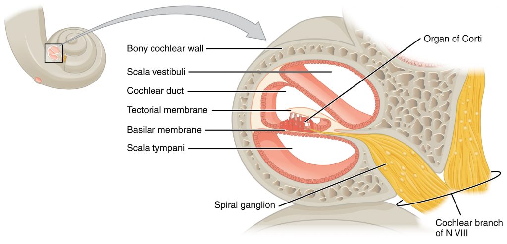
The inner hair cells are the sensory neurons. We have about 3500 of them, running the whole length of the basilar membrane. Each hair cell connects to about 10 auditory nerve endings! These auditory neurons live in the Organ of Corti, which rides on the basilar membrane and houses one row of inner hair cells and three rows of outer hair cells. The top of the Organ of Corti is the tectorial membrane. When the basilar membrane waves, the tectorial membrane shifts sideways relative to the basilar membrane, and stimulates the hair cells. This causes the hair cells to shift and release neurotransmitters. The actual motion of the hair cells are quite tiny. If a hair cell were as tall as the Eiffel tour, then the displacement of the cilia would only be 1 cm.
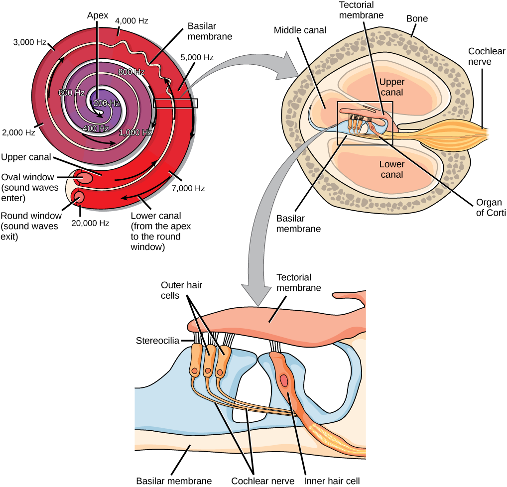
The motile response (electromotility) of the outer hair cells amplifies the vibration of the basilar membrane and sharpens frequency tuning. Prestin is the name of the molecule that makes the cell move. This feedback loop actually makes the ear generate sounds as well! “Otoacoustic emissions” can be measured: these are sounds coming out of your auditory canal. They can be used as hearing screening in newborn babies: play a sound, and measure echo coming back out. If you don’t measure them, maybe it’s just a plugged up middle ear. Noticing hearing loss early is crucial for assisting language development (most common treatment is a cochlear implant, but sign language and hearing aids are also used). After all, outer hair cells are most susceptible to damage and their loss is often the root of hearing damage—you lose amplification for quiet sounds, and you stop being able to “hear out” separate frequencies.
As hair cells become activated, secondary afferent neurons with cell bodies in the spiral ganglion pick up neurotransmitters from inner hair cells and send signals along the auditory nerve to the brain. Auditory information is shuttled to the inferior colliculus in the brainstem, the medial geniculate nucleus of the thalamus, and finally to the auditory cortex in the temporal lobe of the brain for processing. Like the visual system, there is also evidence suggesting that information about auditory recognition and localization is processed in parallel streams (Rauschecker & Tian, 2000; Renier et al., 2009).
Cheryl Olman PSY 3031 Detailed Outline
Provided by: University of Minnesota
Download for free at http://vision.psych.umn.edu/users/caolman/courses/PSY3031/
License of original source: CC Attribution 4.0
Adapted by: Kori SkrypekOpenStax, Psychology Chapter 5.4 Hearing
Provided by: Rice University.
Download for free at http://cnx.org/contents/4abf04bf-93a0-45c3-9cbc-2cefd46e68cc@5.103.
License: CC-BY 4.0
Adapted by: Kori Skrypek
