EEG – electroencephalography
Physical and physiological basis
Learning Objectives
- Be able to describe what signals electroencephalography detect
- Be able to summarize how long the technology has been around


Electroencephalography (EEG) was first used on a human in 1924, credited to Hans Berger. EEG serves the purpose of gaining an understanding of the overall electrical activity of a person’s brain, without needing information on the precise location of the activity. To measure the difference in electrical charge (the voltage) between pairs of points on the head, up to 256 electrodes are placed around the person’s head. The signals received by the electrodes result in a printout of the electrical activity of their brain, or brainwaves, showing both the frequency (number of waves per second) and amplitude (height) of the recorded brainwaves, with millisecond accuracy. Such information is especially helpful to researchers studying sleep patterns among individuals with sleep disorders.
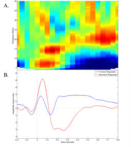
The electrodes are typically fastened to a flexible cap (similar to a swimming cap) that is placed on the participant’s head. The electrodes might also be placed directly on the scalp without a cap. From the scalp, the electrodes measure the electrical activity that is naturally occurring within the brain. They do not introduce any new electrical activity. In contrast to fMRI, EEG measures neural activity directly, rather than correlate to that activity. Also in contrast to fMRI, one major advantage of EEG is its temporal resolution. Data can be recorded thousands of times per second, allowing researchers to document events that happen in less than a millisecond. EEG analyses typically investigate the change in amplitude or frequency components of the recorded EEG on an ongoing basis or averaged over dozens of trials.
EEG is excellent for elucidating the temporal dynamics of neural processes. For example, if someone is reading a sentence that ends with an unexpected word (e.g., Michelle is going outside to water the book), how long after he or she reads the unexpected word does he or she recognize this as unexpected? In addition to these types of questions, EEG allows researchers to investigate the degree to which different parts of the brain “talk” to each other. This allows for a better understanding of brain networks, such as their role in different tasks and how they may function abnormally in psychopathology.
Section 1.“Brain Image” by lumen. Licensed CC BY 4.0“Psychophysiological Methods in Neuroscience” by NOBA. Licensed CC BY-NC-SA 4.0Section 2.

