10 Head
Revised Spring 2024 by T. Clark
Additional Resources for this Chapter:
- Related Supplemental Large Animal Surgery links: LAS Paranasal sinus disease; Ocular Nerve Blocks and Enucleations; LAS Upper respiratory endoscopy; LAS Salivary disorders; LAS Nostrils and nasal conchae; LAS Guttural pouch disorders, LAS Disbudding and dehorning
- Veterinary Gross Anatomy Ungulate Dissection (Head Labs 18-20)
- Associated Video Links:
- Teeth Aging (8:13)
- Equine Skull Bones (10:01)
- Bovine Skull Bones (10:39)
Associated Clinician topics: equine dentition and aging, dental blocks – (The most relevant objectives: A10.1, A10.2, A10.5)
Objective
A10.1 Identify and describe the bone terms associated with the head (skull, mandible, hyoid apparatus).
Skull (lateral surface) (Figs. 1, 14B-1 through 14B-6)
- The main bones of the skull we will focus on include: the frontal (over the forehead), temporal (over the temples), the maxilla (upper jaw), palatine (caudal part of hard palate), mandible (lower jaw), basioccipital (caudal part of ventral braincase), and basisphenoid (rostral part of ventral braincase). (Figs. 1, 14B-6)
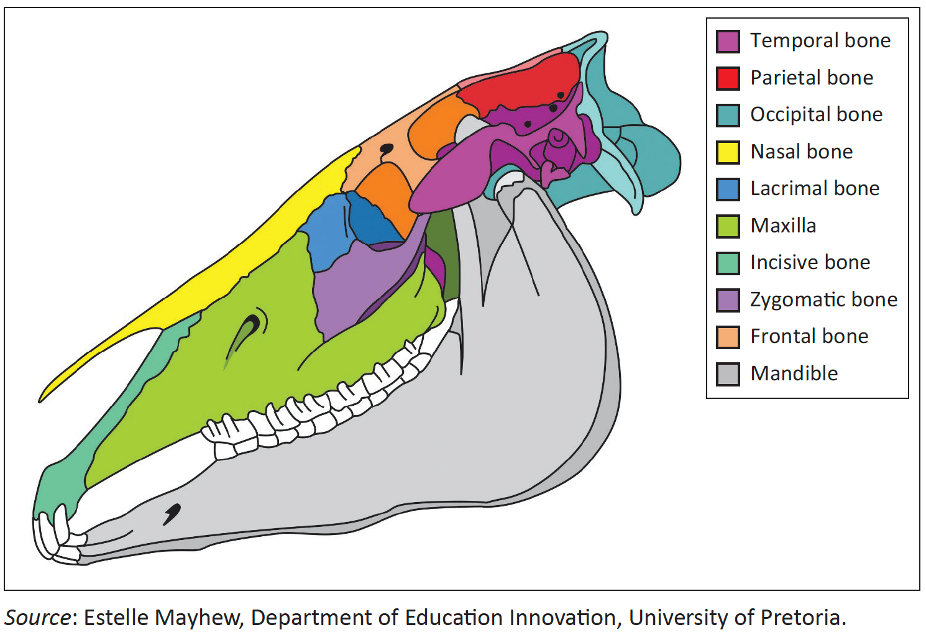
FIGURE 1: Diagrammatic representation of the equine cranium from the lateral perspective illustrating the craniofacial suture lines. https://journals.jsava.aosis.co.za/index.php/jsava/article/view/1764
- The orbit is a complete bony circle in the HORSE, PIG, and RUMINANTS (whereas in the DOG it is completed laterally by the orbital ligament).
- In the HORSE, rostrally the zygomatic arch is continuous with a prominent facial crest (Fig. 14B-1/3), which is not present in the other species we study.
- In BOVINE, the facial crest is absent but there is a well-developed bony prominence called the facial tuberosity (Fig. 14B-3). In the HORSE, the rostral end of the facial crest is the facial tuberosity.
- Rostral and dorsal to the facial tuberosity find the infraorbital foramen where the infraorbital branch of the maxillary nerve (CN V Trigeminal) exits from the infraorbital canal.
- Rostral to the infraorbital foramen find the palpable nasoincisive notch, between the incisive and nasal bones at the rostrum (i.e., nose area).
- The temporal fossa is caudal to the orbit in BOVINE, and is relatively deep. (Fig. 14B-3) The temporal fossa is shallow on the skull of small RUMINANTS and HORSES. (Fig. 14B-2/3)
- Dorsal to the temporal fossa there is a ridge called the temporal line. (Figs. 14B-2, 3)

Figure 14B-1 Horse skull, lateral view. 1, temporalis m.; 2, occipitomandibularis part of digastricus muscle; 3, facial crest; 4, nasoincisive notch; 5, infraorbital foramen; 6, paracondylar process; 7, mental foramen; 8, bar; 9, 9’, supraorbital process and foramen; *, mastoid process of the temporal bone.
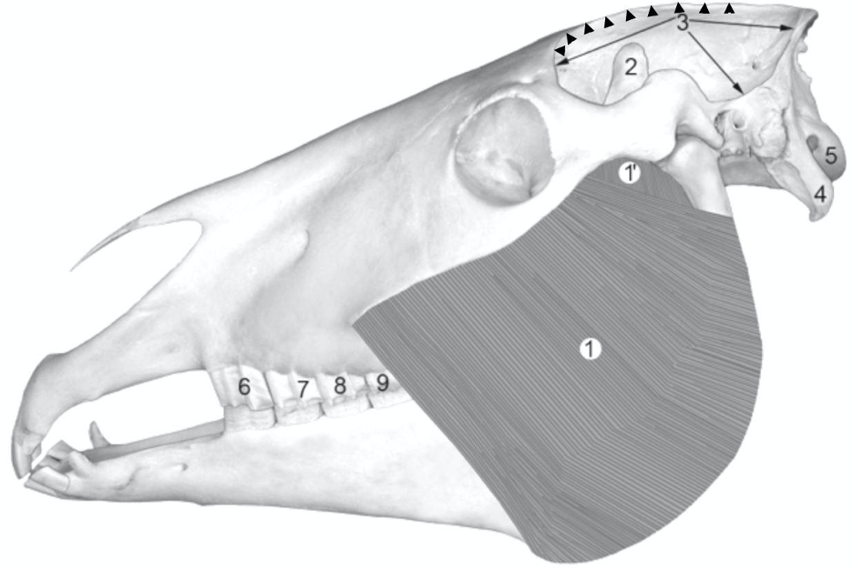
Figure 14B-2 Horse skull, lateral view. 1, masseter muscle; 2, coronoid process; 3, temporal fossa; arrowheads, temporal line; 4, paracondylar process; 5, occipital condyle; 6-9 cheek teeth (Triadan numbers).
- Near the caudal edge of the BOVINE skull are two cornual processes which project from the frontal bone and form the bony parts of the horns. (Figs. 14B-3, 4)
- The cornual processes arise from the frontal bone, but are not present in polled animals (a dominant trait that is often bred for). In horned animals, the cornual process is hollow due to extension of the frontal sinus into this process. The horn sheath covers the cornual process.
- Caudodorsal to the orbit is the supraorbital foramen through which a sensory nerve from the ophthalmic nerve (CN V Trigeminal) courses. (Fig. 14B-1/9′) In horses, this nerve may be blocked (anesthetized) to facilitate ocular examination.
- Ventrally in the orbits of the BOVINE skull, there is a very thin-walled expansion of the maxillary sinus called the lacrimal bulla. (Fig. 14B-3/5)
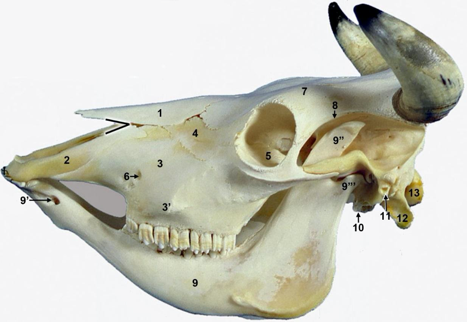
Figure 14B-3 Bovine skull, lateral view. 1, nasal bone (missing in some skulls); 2, incisive bone; black angle, nasoincisive notch; 3, maxilla; 3′, facial tuberosity; 4, lacrimal bone; 5, lacrimal bulla (broken on most skulls); 6, infraorbital foramen; 7, frontal bone; 8, temporal line; 9, mandible; 9′ mental foramen; 9″, coronoid process; 9”’ condylar process; 10, tympanic bulla; 11, external acoustic meatus; 12, paracondylar process; 13, occipital condyle. Note the upper cheek teeth are present, but there are no other upper teeth in ruminants. (TVA 2-38)
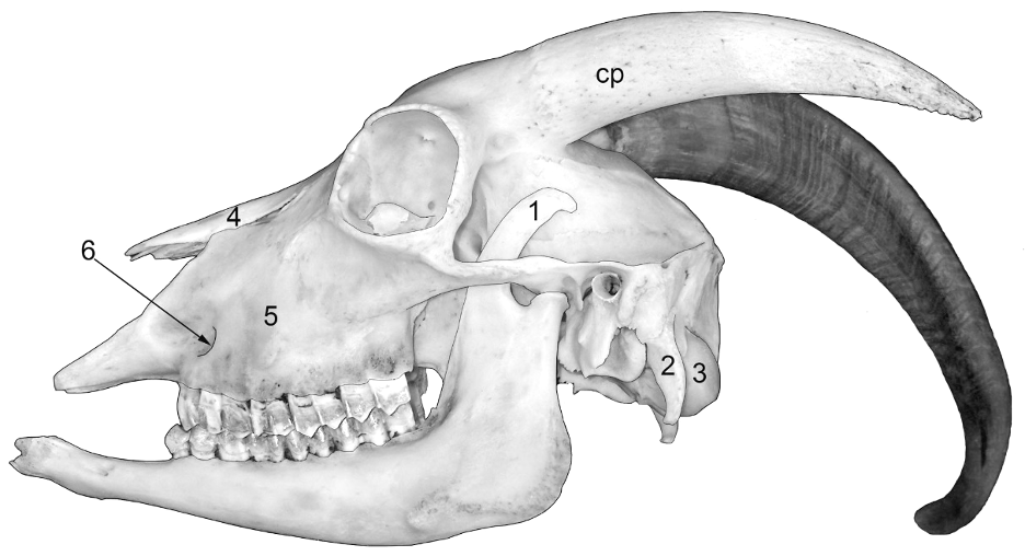
Figure 14B-4 Goat skull, lateral view. (The specimen is rotated so that the horns appear asymmetric.) cp, cornual process arising from the frontal bone; 1, coronoid process of the mandible; 2, paracondylar process; 3, occipital condyle; 4, nasal bone; 5, maxilla; 6, infraorbital foramen. Note the close proximity of the cornual process to the orbit. On the right side the cornual process is covered with a horn.
Skull (ventral surface) (Fig. 14B-6)
- Most of the bony hard palate belongs to the maxilla, but the caudal edge of it is formed by the palatine bone (Fig. 14B-6/13). The palatine bones surround spaces known as the right and left choanae, which are separated by the nasal septum. The septum is formed by the vomer bone and the rest is cartilaginous. The choanae are the transition region between the nasal cavity and the nasopharynx. The soft palate is caudal to the hard palate and attaches to the edges of the palatine bone. There are 6 cheek teeth on either side of the bony palate if the specimen is mature.
- The nasal cavity is dorsal to the hard palate (and the nasopharynx is dorsal to the soft palate). There are large palatine foramina near the suture line between the maxilla and the palatine bones (Fig. 14B-6/14’). Palatine aa. emerge from these foramen and supply blood to the soft tissue of the palate.
- On the caudal ventral edge of the skull are the rounded occipital condyles, which are paired in MAMMALS, but singular in BIRDS (allowing birds to rotate their head more than mammals can).
- Lateral to the occipital condyles are the paracondylar processes (Fig. 14B-6/2) which give rise to muscles that attach to the mandible.
- Rostral to the occipital condyles, there is the basioccipital bone (Fig. 14B-6/7). Rostral to the basioccipital bone is the basisphenoid bone (both create the floor of the brain case), and in young animals the suture line between these two bones is not yet fused (this may lead to the bones pulling apart/fracture if stress is put on the skull or a young animal flips over backwards). This suture line is the spheno-occipital synchondrosis. This is the last cranial suture to fuse.
- Lateral to the basioccipital bone is the petrous portions of the temporal bone (Fig. 14B-6/5), which contains the tympanic bulla, the internal ear, and the vestibular apparatus. The temporal bones have 3 parts: squamous (flat), petrous (contains middle and inner ear), and the mastoid processes (neck muscle insertion).

Figure 14B-6 Caudal part of equine skull, ventral surface. C, choanae; 1, foramen lacerum; 2, paracondylar process; 3, occipital condyles; 4, hypoglossal canal; 5, petrous temporal bone and tympanic bulla; 6, mastoid process of temporal bone; 7, basioccipital bone; 8, basisphenoid bone; 9-11, Triadan teeth 9-11 (M1-M3); 12, 12’, vomer; 13, 13’, horizontal and vertical parts of palatine bone; 14, maxillary bone; 14’, palatine foramen; 15, facial tuberosity; 16, articular facet for mandibular condyle; 16’, retroarticular process. The suture line between 7 & 8 is the spheno-occipital synchondrosis (dotted line).
Mandible (Figs. 14B-1 through 4)
- The mandibular symphysis (on the midline) is the fibrous connection of the right and left mandibles.
- The coronoid process is found dorsally on the ramus of each mandible, and projects into the temporal fossa. Slightly more ventral, note the condylar process which articulates with the skull to form the temporomandibular joint.
- On the medial side of the mandible, near the angle, is the mandibular foramen where the inferior alveolar nerve, a branch of the mandibular n. (CN V Trigeminal), enters the mandible.
- On the rostrolateral aspect of the mandible there is the mental foramen through which the mental n. (a terminal branch of the inferior alveolar nerve) courses. (Figs. 14B-1, 3).
Paranasal Sinuses (Figs. 14B-7 to 14B-10)
- In ungulates, the paranasal sinuses are relatively large. Ungulates need a large head functionally, to allow attachment for the of muscles of mastication, but there is a balance with how dense and solid the bony skull can be. The sinuses allow for the large head to be lighter.
- The nasal turbinates/conchae are bilateral sets of ridges projecting into the nasal cavity and function to humidify incoming air, warm incoming air, and catch particulates before they enter the lower respiratory tract.
- Knowing the extent and species variation of the paranasal sinuses can be important if humane euthanasia requires stunning by mechanical means (e.g., captive bolt). Also, paranasal sinuses can become infected and may need to be surgically drained.
- In BOVINE there is a cornual diverticulum (Fig. 14B-8), which is an extension of the frontal sinus into the cornual process (i.e., if there is trauma to the horn, there can be an open sinus). Numerous septa divide the BOVINE frontal sinus, one of which almost completely and divides the frontal sinus into a rostral and a caudal compartment (Fig. 14B-7).
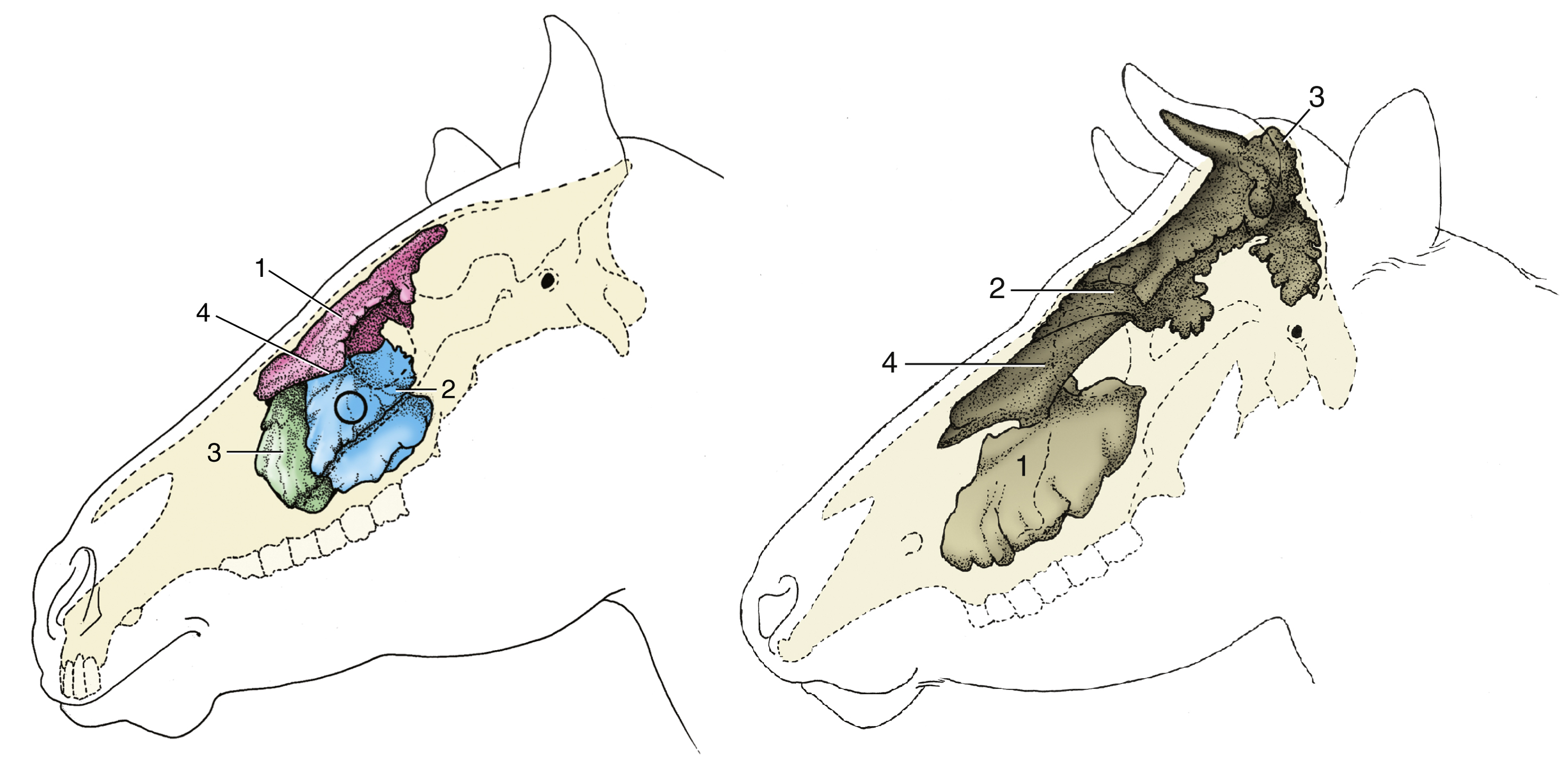
Figure 14B-7 (Left, equine) Topography of the paranasal sinuses. The circle indicates where the caudal maxillary sinus can be trephined. 1, Conchofrontal sinus; 2, caudal maxillary sinus; 3, rostral maxillary sinus; 4, position of frontomaxillary opening between 1 and 2. (Right, bovine) 1, Maxillary sinus; 2, rostral frontal sinuses; 3, caudal frontal sinus; 4, dorsal conchal sinus. (from TVA figs. 18-13, 25-11).
Figure 14B-8 Caudal portion of a bovine skull to show the frontal sinus in the center extending into an opened cornual process as the cornual diverticulum on the right . (On the left the cornual process has not been opened.) Numerous septa divide the frontal sinus into a labyrinth of connected spaces.
- The small frontal sinus of EQUINE is continuous with the dorsal conchal sinus rostrally and is called the conchofrontal sinus. In EQUINE, ventrally the frontal sinus opens into the maxillary sinus via a large frontomaxillary opening. (Fig. 14B-7/4) Basically, all sinuses drain to the maxillary sinus in EQUINE.
- In RUMINANTS, the infraorbital canal separates the maxillary sinus from the more medial palatine sinus (Fig. 14B-9/3, 10/6). The palatine sinus is best seen on off center split heads, as the palatine sinus does not cross the midline. (Figs. 14B-10/7, 25-9)
- The maxillary sinus of the BOVINE extends into the lacrimal bulla. Dorsal to the maxillary sinus is the dorsal conchal sinus (Fig. 14B-7/4).
- The maxillary sinus of the HORSE lies deep to the facial crest. It is divided into separate rostral and caudal compartments (Fig. 14B-7/2, 3) and is partially divided into medial and lateral parts by the infraorbital canal. The maxillary sinus drains into the nasal passageways via an opening in the rostral portion of the maxillary sinus. (Supplemental Clinical LAS Paranasal sinus disease)
- Maxillary sinus gets larger with age in EQUINE. They are smallest in the first year, and then gradually get larger. Once the teeth have fully erupted the maxillary sinuses are the biggest. Since the maxillary sinus is adjacent to the teeth alveoli, the sinus gets larger as the teeth get shorter due to continuous eruption. As the teeth shorten the alveoli around them remodel and the maxillary sinus enlarges “by default.” Unfortunately, since some roots of teeth are in/near the sinus the sinuses can become infected if a tooth root is infected. Younger animals have longer roots in the sinus. (more on teeth below)
- Clinical Notes: sinus cysts are similar to conchal cysts and are congenital anomalies with similarly slow development and clinical signs (e.g., blockage of the nasal passages and deformation of the skull). (Supplemental Clinical LAS Paranasal sinus disease)
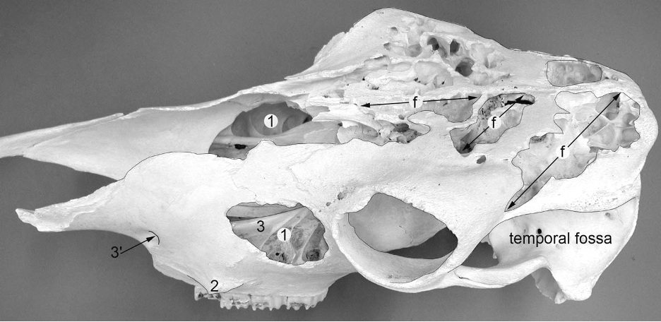
Figure 14B-9 Bovine skull, dorsal lateral view; surface bone removed to expose the frontal sinuses (f) and the maxillary sinus (1). 2, facial tuberosity; 3, 3’, infraorbital canal and foramen. The nasal bones are missing so the maxillary sinus can be seen on both sides. Cornual processes are absent.
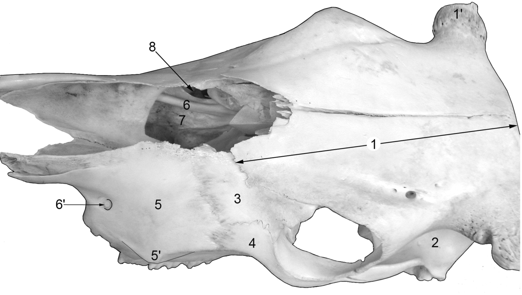
Figure 14B-10 Bovine skull, dorsal view; nasal bones missing. 1, frontal bone; 1’, cornual process; 2, temporal fossa; 3, lacrimal bone; 4, zygomatic bone; 5, maxillary bone; 5’, facial tuberosity; 6, 6’, infraorbital canal and foramen; 7, palatine sinus; 8, maxillary sinus.
Hyoid Apparatus
- The hyoid apparatus helps to support the larynx and several tongue muscles.
- In EQUINE the basihyoid bone, the central bone of the hyoid apparatus, has a prominent lingual process (Fig. 4.8/5) that is embedded in the root of the tongue. The thyrohyoid bones project caudodorsally from the basihyoid bone and articulate with the thyroid cartilage of the larynx. Dorsally, projecting off of the basihyoid are the ceratohyoid bones. There may be a small epihyoid bone at the junction of cerato- and stylohyoid bones (although it often fuses with the stylohyoid bone). The stylohyoid bones are long and flat, create compartments in the guttural pouches, and articulate with the styloid processes at the base of the skull.
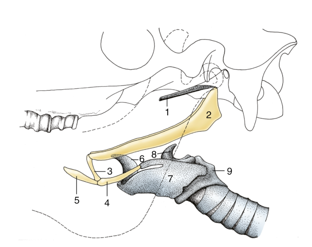
Figure. 4-8 Hyoid apparatus suspending the larynx from the base of the skull (horse). The broken line indicates the mandible. 1, Cartilage of auditory tube; 2, stylohyoid; 3, keratohyoid; 4, thyrohyoid; 5, lingual process of basihyoid; 6, epiglottic cartilage; 7, thyroid cartilage; 8, arytenoid cartilage; 9, cricoid cartilage.
- In BOVINE the basihyoid bone is also the central bone, but it has a short lingual process. The thyrohyoid bones articulate with the thyroid cartilage of the larynx. The ceratohyoid bones articulate with the basihyoid and rod-shaped epihyoid bone, which in turn articulates with the long, flattened stylohyoid bone. The angle of the stylohyoid is drawn out in the form of a hook.
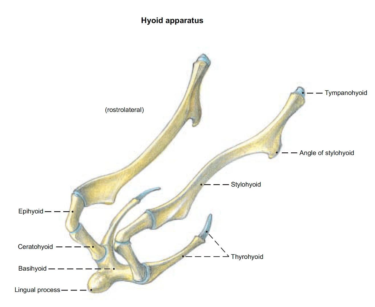
Figure above – Bovine hyoid apparatus. Labeled structures: tympanohyoid, stylohyoid, angle of stylohyoid, epihyoid, ceratohyoid, basihyoid, lingual process, thyrohyoid. (Budras and Habel Bovine Anatomy)
Objective
A10.2 Identify and describe the bony landmarks associated with commonly utilized nerve blocks and venipuncture in bovine and equine; summarize the arteries, veins, and nervous system structures of the head.
Cornual Nerve Block (Fig. 25-6, 25.2 and below)
- In BOVINE the temporal line is an important landmark for locating the cornual n., which is important for blocking the nerves near the base of the horn for disbudding and dehorning. (Supplemental Clinical LAS Disbudding and dehorning)
- The cornual n. is a branch of CN V Trigeminal, ophthalmic division, carrying sensation from the cornual dermis. Additional ophthalmic branches can be anesthetized near the orbit and temporal line.
- GOATS have two nerves receiving sensation from each horn (both are ophthalmic branches), and both need to be blocked appropriately. (see figure below)
- Near the caudal and dorsal edge of the BOVINE skull, the cornual processes project from the skull and form the bony part of the horn. The cornual processes arise from the frontal bone, but they are not present in polled animals (i.e., those without horns). In horned animals, the cornual process is hollow due to an extension of the frontal sinus called the cornual diverticulum (Fig. 14B-8). Due to this connection, if there is trauma to the horn, there can be an open sinus.
- The poll is a protuberance between the ears or just caudal to the ears. Typically, animals are now bred and selected to have no horns.
- In contrast, antlers (in cervids or the deer family) are solid and covered with skin (velvet) rather than horn, but still arise from the frontal bone. Antlers differ from horns in that they are solid and are shed annually after the fall breeding season (the rut).
- Clinical Note: Dehorned animals may have a small remnant of the cornual process. The size of the remnant will depend on the age of the animal at the time of dehorning and the skill of the person who did the procedure. Note the more rostral position of the cornual process of small RUMINANTS, as the frontal bone does not extend as far caudally. (Supplemental Clinical LAS Disbudding and dehorning)
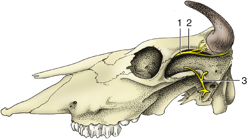
Figure. 25-6 Cornual nerve (1) follows the temporal line (2) on bovine skull. The auriculopalpebral nerve (3) is palpable where it crosses the zygomatic arch.
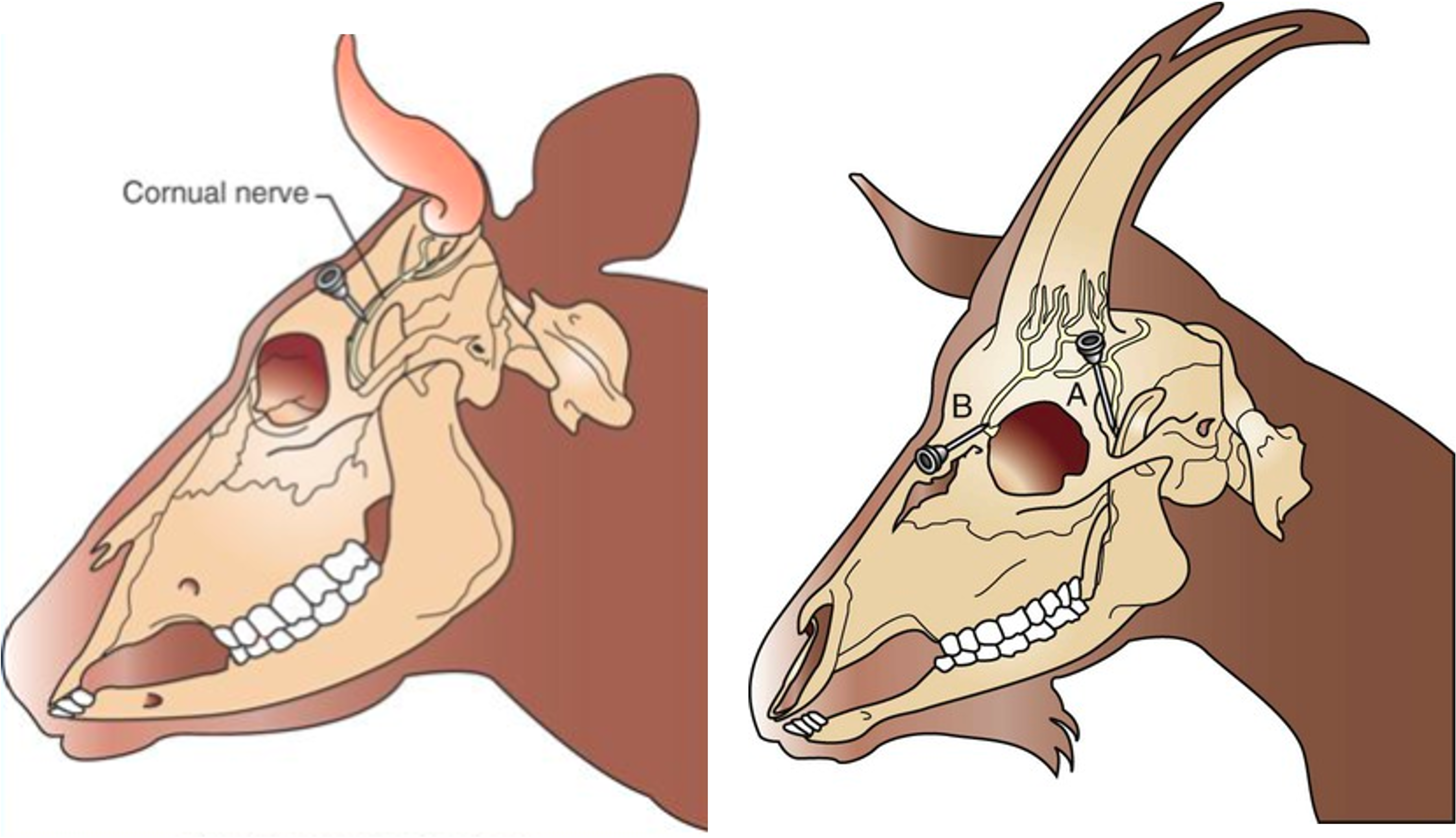
Figures above (left) Bovine: needle placement for desensitizing the cornual n. bovine. The cornual n. follows the temporal ling/ridge to the base of the horn. (right) Caprine (goat): A, Needle placement for desensitizing the cornual branch of the lacrimal n. B, Needle placement for desensitizing the cornual branch of the infratrochlear n. (Modified from Muir WW III, Hubbell JAE, Bednarski RM, Skarda RT: Veterinary anesthesia, 4th ed. St. Louis, Mosby, 2000.)
Dental Nerve Blocks
- Clinical Note: The infraorbital foramen can be palpated in the HORSE and is a useful injection site for dental blocks of the upper teeth.
- To reach the upper arcade of teeth (upper incisors and maxillary premolar teeth), the infraorbital n. (CN V Trigeminal, maxillary division) enters the maxillary foramen (inside the bony orbit), then runs within the infraorbital canal, and finally exits the infraorbital foramen. If the anesthetic is introduced into the foramen and canal, you are able to block sensory signal from the entire upper arcade of teeth.
- The nasoincisive notch and facial tuberosity are useful in locating the infraorbital foramen on a live HORSE since the foramen is approximately midway between the facial tuberosity and the nasoincisive notch.
- Additionally, the levator labii superioris m. (Fig. 18-8) covers the infraorbital foramen and must been pushed aside to locate the infraorbital n.
- The levator labii superioris m. arises over the maxilla and runs dorsorostrally to form a common tendon with its counterpart on the other side. The combined tendon of these bilateral muscles travels between the nostrils, and splays out within the upper lip, allowing for the lip curl (i.e, Flehmen response – part of the vomeronasal organ system to sense pheromones).
- To reach the maxillary nerve (for standing sinus surgeries and full upper arcade of teeth), the zygomatic arch can be used as a landmark. (Supplemental Clinical DVM 360 Maxillary nerve block)
- Clinical Note: the inferior alveolar n. (CN V Trigeminal, mandibular division) enters the mandibular foramen and runs within the body of the mandible. This nerve is the focus for dental blocks of the lower arcade (mandibular) of teeth. The inferior alveolar n. is reached from the axial (i.e., medial) aspect of the mandible.
- The mental n., which exits through the mental foramen, is a terminal branch of the inferior alveolar nerve. Anesthetizing the mental n. would desensitize the lower lip, gingiva and part of the interdental space, but not the teeth. To anesthetize any of the mandibular teeth, a needle would need to be advanced into the mental foramen and local anesthetic injected into the canal that courses through the mandible. This is not an extensive block (mostly incisors and canines). A muscle of the lower lip (the depressor labii inferioris muscle) may need to be displaced dorsally to gain access to the mental foramen. (Figure 18-8/3′) (Supplemental Clinical AAEP Mental nerve block)
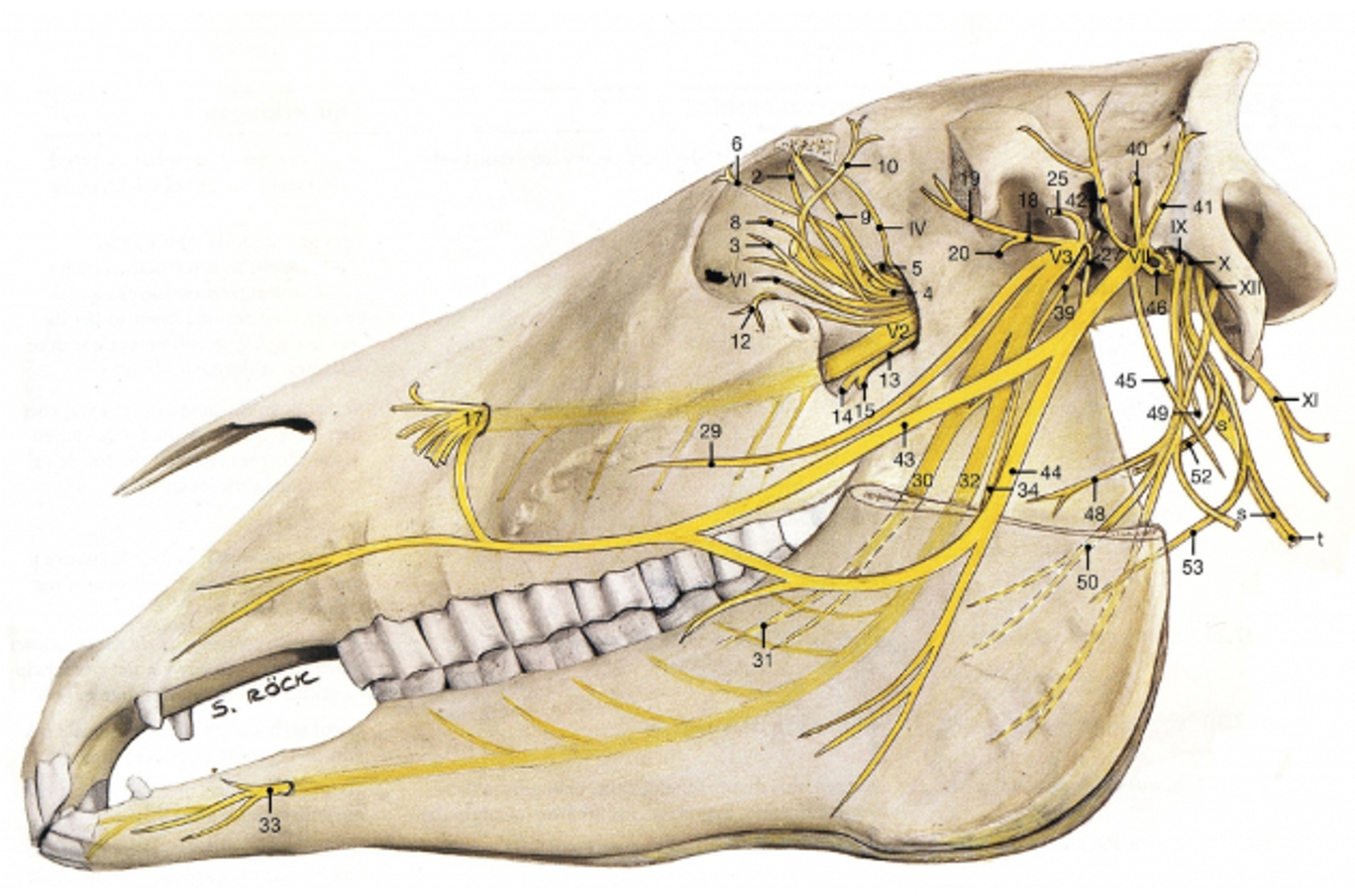
Figure above – Lateral view of horse skull. Trigeminal nerve branches: V1 – ophthalmic (supraorbital, lacrimal), V2 – maxillary (infraorbital), V3 – mandibular (inferior alveolar, mental). IV – trochlear, VII – facial (auriculopalpebral), IX – glossopharyngeal, X – vagus, XI – accessory, XII – hypoglossal nerve branches also shown. (S. Rock)
Figure 18-8 Deep dissection of the equine head. 1, Infraorbital nerve; 1′, levator labii superioris; 2, dorsal buccal branch of facial nerve; 3, mental nerve; 3′, depressor labii inferioris; 4, facial vein; 5, deep facial vein; 6, buccal vein (5 & 6 are part of the venous sinuses); 7, buccinator; 8, masseter; 9, occipitomandibularis; 10, sternocephalicus; 11, external jugular vein; 12, mandibular salivary gland; 13, linguofacial vein; 14, maxillary vein.
Ocular Nerve Blocks (Figs. 18-36, above, and below)
- During standing eye surgeries, the focus is to decrease the movement (i.e., motor innervation) to the eyelids (palpebrae) as well as sensation from and around the eye.
- The supraorbital n. arises from the frontal n., a branch of CN V Trigeminal, ophthalmic division, and carries sensation from the superior palpebra and skin of the forehead. A branch of the maxillary division carries sensation from the inferior palpebra.
- The lacrimal n., a branch of CN V Trigeminal, ophthalmic division, carries sensation from the skin and conjunctiva of the eye (medial aspect), while a separate ophthalmic division branch carries sensation from the lateral skin and conjunctiva. The cornea can detect sensation as well, and it too is carried by the ophthalmic division.
- The auriculopalpebral n., a branch of CN VII Facial, carries motor fibers to the muscles in the superior palpebra and muscles around the eye.
- Clinical Notes: There are different nerve blocks used to assist in enucleation or procedures of the eye. More information can be found in the Supplemental Clinical LAS Ocular Nerve Blocks and Enucleations. These blocks can include: a 4 point block, a Peterson block, and retrobulbar blocks. When doing procedures near or around the eye you need to be aware of the oculocardiac reflex. Traction on the eye can lead to cardiac asystole due to this reflex. Blocking CN V Trigeminal branches (via retrobulbar nerve blocks) that carry sensory information from the structures of the eye can prevent this reflex.
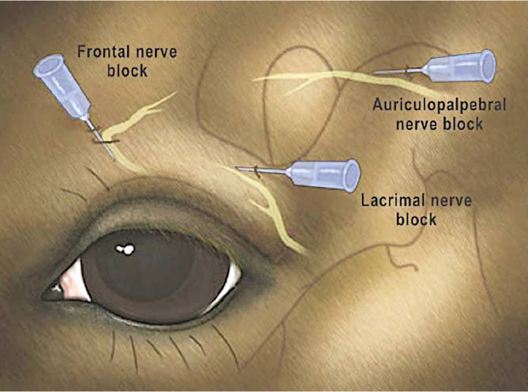
Figure above – Lateral view of the horse eye, with frontal, auriculopalpebral, and lacrimal nerve block locations labeled.
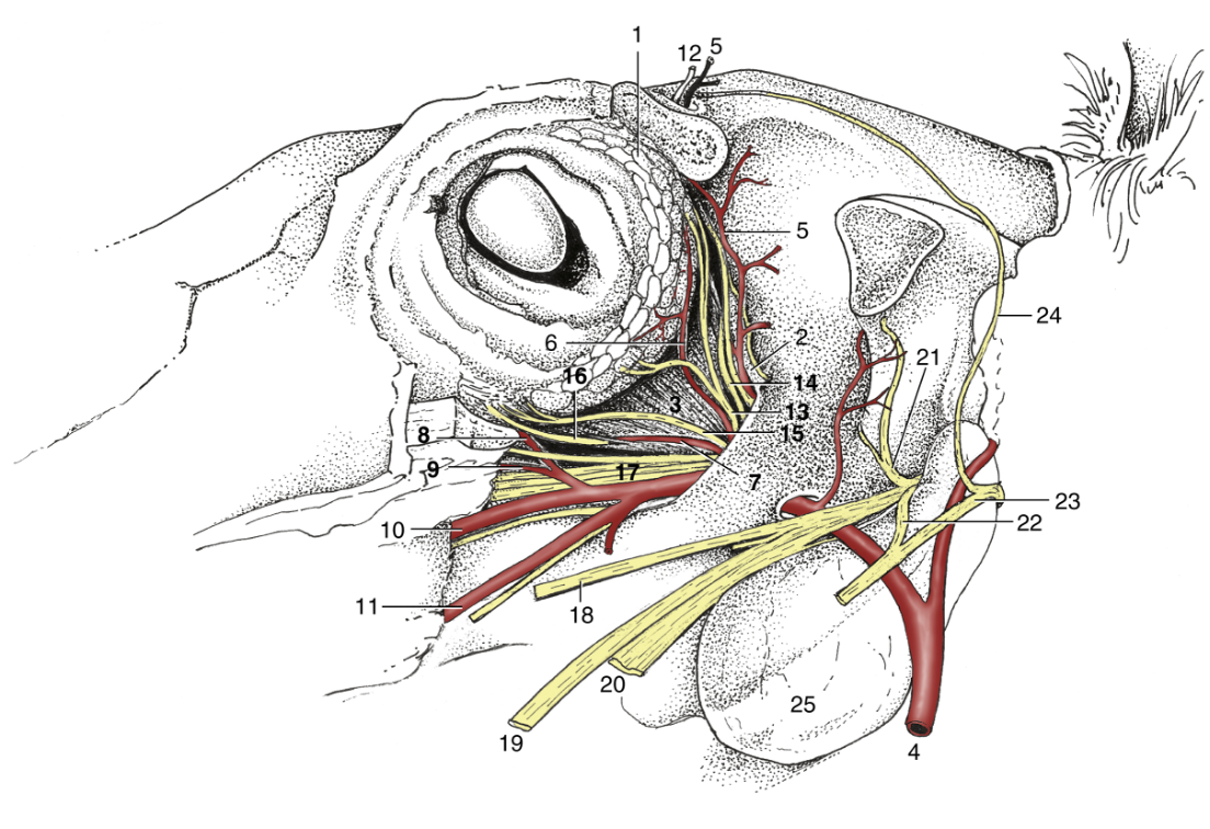
Figure 18-36 Dissection of the orbit; the zygomatic arch and periorbita have been removed. 1, Lacrimal gland; 2, periorbita; 3, lateral rectus; 4, maxillary artery (a.); 5, supraorbital a.; 6, lacrimal a.; 7, muscular branch of external ophthalmic a.; 8, malar a.; 9, infraorbital a.; 10, major palatine a.; 11, buccal a.; 12, supraorbital nerve (frontal n.); 13, lacrimal n.; 14, trochlear n.; 15, zygomatic n.; 16, oculomotor n.; 17, rostral branches of maxillary n.; 18, buccal n.; 19, lingual n.; 20, inferior alveolar n.; 21, masticatory n.; 22, auriculotemporal n.; 23, CN VII facial n.; 24, auriculopalpebral n.; 25, guttural pouch.
Cranial Nerves
- CN V Trigeminal – has several sensory branches from the face and head that need to be blocked for clinical procedures (see nerve blocks above) – branches include: cornual, maxillary (infraorbital), mandibular (inferior alveolar, mental). It also provides motor innervation to the muscles of mastication. (Fig. above, lateral view of horse skull)
- CN VII Facial – provides motor innervation to the muscles of the face via the dorsal and ventral buccal branches (coursing over the masseter m.) and the auriculopalpebral n. The facial n. also provides parasympathetic innervation to some of the glands of the face.
- CN X Vagus – provides parasympathetic innervation to thoracic and abdominal viscera, motor innervation to (and sensory from) the larynx and pharynx. The recurrent laryngeal n. provides motor to many intrinsic laryngeal muscles via the caudal laryngeal n. branch, while the cranial laryngeal n. is a direct branch from the vagus and innervates the cricothyroideus m. (more below in Laryngeal Paralysis)
- CN XII Hypoglossal – provides motor innervation to the tongue muscles.
- The vagosympathetic nerve trunk is the combination of the sympathetic trunk and vagus nerve in the neck (the trunk is wrapped within the carotid sheath). The cervical sympathetic trunk contains a large cranial cervical ganglion near the base of the skull near the site of separation of the sympathetic trunk and the vagus nerve.
Venipuncture and Arterial Access
Veins
- In all species, the facial v. joins the lingual v. to form the linguofacial v. The maxillary v. joins the linguofacial v. to form the external jugular v. (Figs. 18-8, 25.2)
- The facial v. runs along the ventral edge of the mandible alongside the facial a.
- In EQUINE, there are three veins that course across the face deep to the masseter muscle. These veins have dilated portions called sinuses. The transverse facial vein and sinus courses just ventral to the facial crest. The deep facial vein and sinus courses ventral to the transverse facial vein and sinus. The buccal vein and sinus courses most ventrally across the face. (Fig. 14-6). These venous sinuses are important as they are an alternative site where blood samples can be taken.
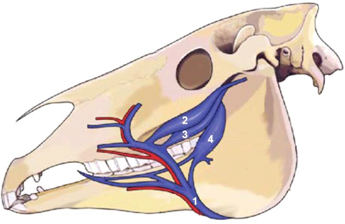
Figure 14-6 Venous sinuses in the horse. 1, Facial v.; 2, transverse facial vein; 3, deep facial vein; 4, buccal vein. Dilations of 2, 3 and 4 form venous sinuses. (Modified from Walesby et. al. The transverse facial venous sinus: an alternative location for blood collection in the horse. Equine Veterinary Education. 19(2). 100-102, 2007.
Arteries
- The external and internal carotid aa. are the main branches of the common carotid a., which runs within the carotid sheath in the neck. In the EQUINE, the internal carotid a. is smaller and runs dorsally up and into the cranial cavity. (Fig. 18-40) In the BOVINE, the extra-cranial part of the internal carotid a. regresses to become a connective tissue strand.
- One of the main branches of the external carotid a. is the linguofacial trunk, which gives rise to two arteries, lingual and facial.
- The lingual a. provides blood to the tongue.
- The facial a. provides blood to the face. As it courses over the ventral edge of the mandible, it is palpable and is a site for taking the pulse. (Fig. 18-40)
- The transverse facial a. is a branch of the superficial temporal a., which is near the transition of the external carotid to the maxillary a. It is important due to its use for arterio-puncture (i.e., arterial blood sample). It is located caudal to the lateral palpebral commissure and ventral to the zygomatic arch. (Fig. 18-40)
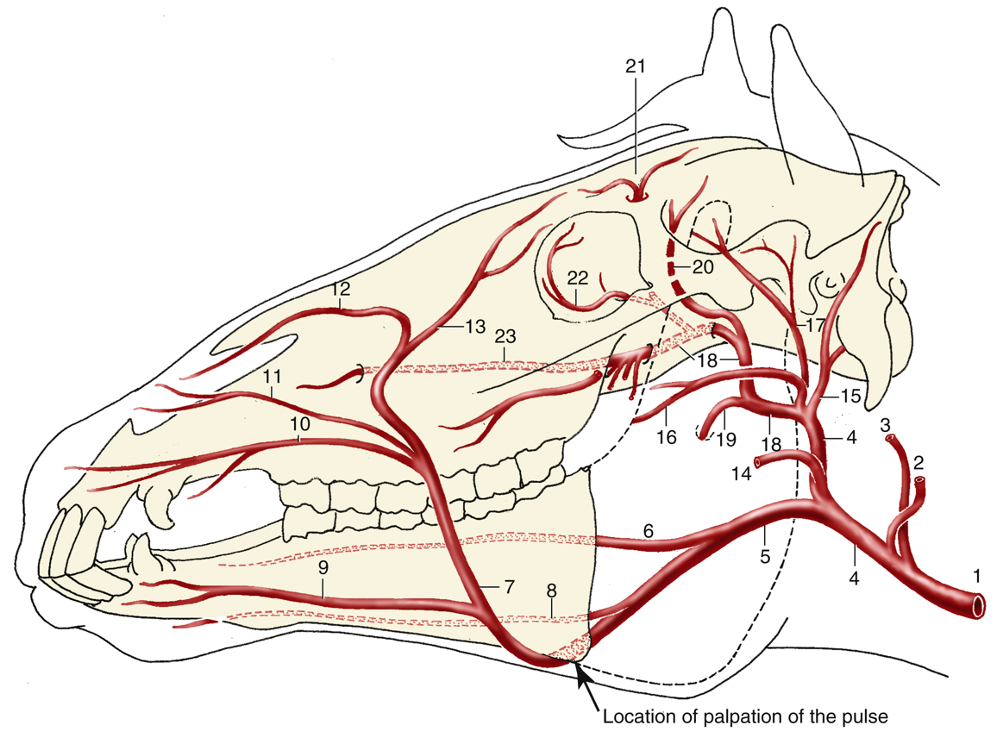
Figure 18-40 Principal arteries of the head (schematic). 1, Common carotid artery (a.); 2, occipital a.; 3, internal carotid a.; 4, external carotid a.; 5, linguofacial trunk/a.; 6, lingual a.; 7, facial a.; 8, sublingual a.; 9, inferior labial a.; 10, superior labial a.; 11, lateral nasal a.; 12, dorsal nasal a.; 13, angularis oculi a.; 14, masseteric a.; 15,caudal auricular a.; 16, transverse facial a., displaced ventrally for clarity; 17, superficial temporal a.; 18, maxillary a.; 19, inferior alveolar a.; 20, caudal deep temporal a.; 21, supraorbital a.; 22, malar a.; 23, infraorbital a.
Nervous System Structures
Eye Structures (Fig. 5-36)
- The superior and inferior palpebrae are the upper and lower eyelids, respectively. The separation between the eyelids is the palpebral fissure. The medial and lateral corners, where the eyelids meet, are called medial and lateral palpebral commissures (or canthi), respectively.
- In the lateral and dorsal aspect of each orbit there is a lacrimal gland that produces the aqueous component of tears. At the medial edge/margin of both palpebrae, there are small openings, called lacrimal puncta, that lead to lacrimal canaliculi and eventually the nasolacrimal duct (which extends through the lacrimal and maxillary bones, as well as the nasal cavity) which empties into the ventral floor of the nasal cavity (all structures are bilateral). The opening into the ventral floor is called the orifice of the nasolacrimal duct. (More on procedures utilizing the nasolacrimal duct below.)
- The inner lining of the palpebrae and outer covering of the anterior portion of the sclera is called conjunctiva. The cavity created by the lining between the sclera and the palpebrae is called the conjunctival sac. The conjunctiva on the inside of the eyelids is called palpebral conjunctiva, while the conjunctiva covering of the anterior portion of the sclera is called bulbar conjunctiva. The junction at which the two layers meet is called the conjunctival fornix.
- Medially, and deep to the inferior palpebra is an additional membrane called the third eyelid (aka nictitating membrane).
- The eye sits within the bony orbit, but is surrounded by a post-orbital fat pad dorsally. Surrounding the extraocular muscles is a membrane called the periorbita.
- The muscles controlling the movements of the eye include the four recti muscles: medial, lateral, dorsal, and ventral rectus. Additionally, there is a dorsal and ventral oblique m., as well as the retractor bulbi mm.
- The eye (aka eyeball, bulbus oculi) is formed by three main layers (aka coats, tunics).
- External fibrous coat: this layer includes the “white of the eye,” the sclera and the transparent rostral portion, the cornea. The junction where the cornea and sclera meet is called the limbus (aka corneoscleral junction).
- Middle vascular coat (aka uvea): this layer includes the iris (containing two configurations of smooth muscle – the dilator and constrictor pupillae mm.), with a hole called the pupil. The pupil lets light into the eye. The choroid is the vascular layer and is usually very dark colored, except for the region called the tapetum lucidum which is a shiny and colorful reflective region that gives animals “eye shine” at night. The ciliary body encircles the lens and has a circular ciliary m. that controls the zonular fibers attaching to the lens (allowing for accommodation – near and far vision).
- Extending from the free border of the iris in ungulates are iridic granules (corpora nigra). (FIG. 9.9) These structures are thought to help shade the eye and may provide nutrients to the anterior chamber of the eye. Iridic granules are better developed on the superior region of the iris.
- Internal coat (aka retina): this is the nervous layer of the eye. It includes the point where all ganglion cells of the retina exit each eye, the optic nerve. Where the optic nerve exits there are no photoreceptors, hence it is called the “blind spot” or optic disc. The rounded inner caudal region of the eye is called the fundus, and is what is seen in a fundoscopic examination of the eye.
- The lens sits posterior to the iris and focuses light onto the retina.
- The different compartments of the inner eye are filled with fluid to maintain the shape of the eye. The anterior (cornea to iris) and posterior (iris to lens) chambers are filled with aqueous humor, while the large posterior compartment (posterior to the lens) is called the vitreous chamber and is filled with vitreous humor (or vitreous body).

Fig. 5-36 Sagittal section of dog eye. 1, tapetum lucidum; 2, nontapetal nigrum; 3, lens; 4, ciliary body; 5, posterior chamber; 6, anterior chamber; 7, iris; 8, cornea; 9, superior eyelid/palpebra; 10, palpebral conjunctiva; 11, bulbar conjunctiva; 12, fornix; 13, sclera; 14, choroid; 15, retina; 16, superior retinal vein; 17, optic disk/disc; 18, optic nerve; 19, dura and arachnoid mater; 20, inferior medial retinal vein; 21, third eyelid. (from Guide to Dissection of the Dog)
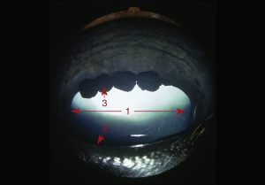
FIG. 9.9 Anterior surface of the equine iris with characteristic iridic granules. 1, Pupil; 2, pupillary margin; 3, iridic granule. TVA
Brain Structures (Figs. 3-2 and below)
- The brain is formed by right and left cerebral hemispheres, which create the cerebrum. The surface of the cerebrum is thrown into ridges called gyri, and depressions called sulci.
- There are several (bilateral) lobes of the cerebrum, each with different primary functions. These include the frontal lobe (motor planning and execution, the rostral portions of the brain), occipital lobes (integration of vision, the caudal portions of the brain), and piriform lobes (olfaction, ventral portions of the brain) along with the olfactory bulbs and tracts of CN I Olfactory.
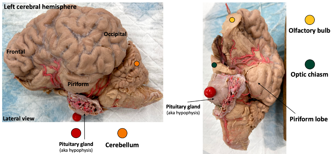
Figure above – Equine brain, lateral and ventral views. (left) Left cerebral hemisphere with frontal, piriform, and occipital lobes, and cerebellum and pituitary gland (aka hypophysis) labeled. (right) Ventral view with olfactory bulb, optic chiasm, piriform lobe, and pituitary gland labeled. (RLarsen)
- The brainstem houses many important pathways and relay structures to and from the brain. The four main regions of the brainstem are:
- The diencephalon, which includes the thalamus (a motor and sensory relay) as well as the hypothalamus (an important regulator of the autonomic system, homeostasis, and the hypophysis/pituitary gland). The optic chiasm (the decussation/crossing of the CNs II Optic) sits just rostral and ventral to the hypothalamus.
- The midbrain, a relay for visual and auditory information and reflexes.
- The pons, a relay between the cerebellum and the brain, brainstem, and spinal cord.
- The medulla, a location where respiratory, cardiovascular, and feeding responses are regulated.
- The cerebellum is a regulator and assists with balance and motor coordination.
- The spinal cord is protected by the vertebrae and sits within the vertebral canal. The dorsal roots enter (with sensory information) and ventral roots exit (with motor information) and form the spinal nerves. The pia mater is directly on the surface of the spinal cord, the arachnoid mater is in the middle, and the tough dura mater forms the outer meningeal layer. Cerebrospinal fluid is found in the subarachnoid space, between the arachnoid and pia mater.
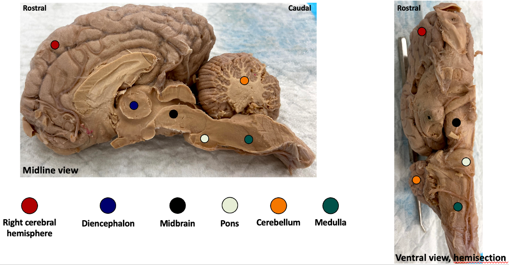
Figure above – Equine brain, midline and ventral views. (left) Right cerebral hemisphere with diencephalon, midbrain, pons, cerebellum, and medulla labeled. (right) Ventral view with the same structures labeled. (RLarsen)
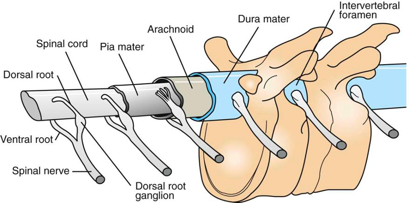
Figure 3-2 Spinal cord and vertebrae diagram. Spinal cord running within the vertebral foramen and spinal nerves exiting the intervertebral foramen. Parts forming a spinal nerve: dorsal and ventral roots, dorsal root ganglion. Meningeal layers: dura mater = tough mother; arachnoid mater = spider-like mother; subarachnoid space = cerebrospinal fluid (CSF); pia mater = tender mother. (from Gardner E: Fundamental of Neurology & Cunningham’s Textbook of Veterinary Physiology)
Objective
A10.3 Identify and describe the muscles (and associated structures) of the larynx, tongue, and of mastication; describe the clinical disorders related to these regions.
Larynx (Figs. 8-20 and below)
- The laryngeal cartilages from rostral/cranial to caudal are: epiglottic (flap that protects the larynx and airway, sits cranial and dorsal to the thyroid cartilage), thyroid (largest, shield-like, open dorsally), arytenoids (paired, butterfly-shaped, sit dorsally between the thyroid and cricoid cartilages), and the cricoid (the only cartilage to encircle the larynx and airway).
- The space between the thyroid and cricoid cartilages is the thyroid notch, and it is covered by the cricothyroid ligament.
- The formation of sound by the larynx is associated with the vocal folds (includes the vocalis m. and ligament within), which surround the glottic cleft and help form the glottis. These folds are attached to the vocal processes of the arytenoid cartilages.
- The HORSE, similar to the DOG, has laryngeal ventricles that are bilateral lateral pouches that are located between the vestibular and vocal folds.
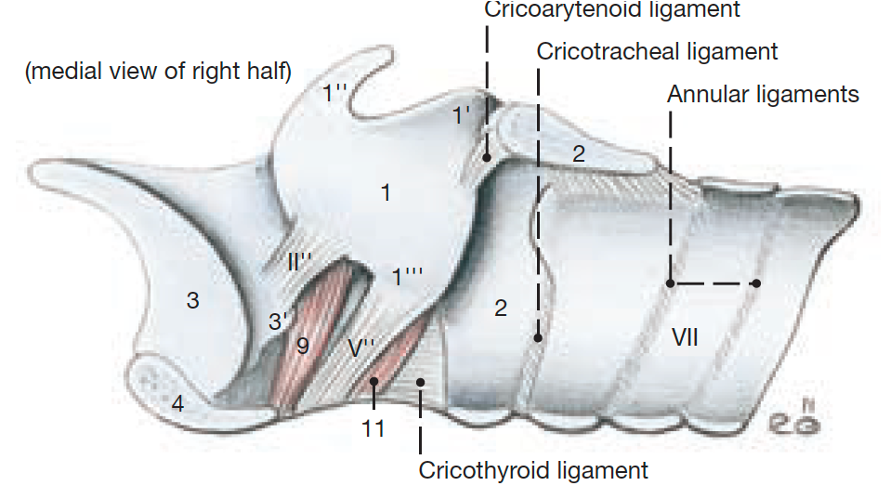
Figure above – Midline view of larynx of horse. 1, arytenoid cartilage; 1’, ; 1”, corniculate process; 1’”, vocal process; 2, cricoid cartilage; 3, epiglottic cartilage; 3’, cuneiform process; 4, thyroid cartilage ; 9, ventricularis m.; 11, vocalis m.; II”, vestibular ligament; V”, vocal ligament (vocal fold covers this); VII, tracheal cartilage; Labeled structures: cricothyroid ligament (covers the thyroid notch), cricoarytenoid ligament, cricotracheal ligament, annular ligaments. (Budras et al., Anatomy of the Horse)
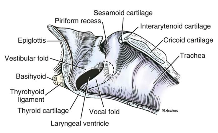
FIGURE 8-20 Median section of the larynx. (The dotted lines show the extent of the laryngeal ventricle.) Labeled structures: epiglottis, thyroid cartilage, cricoid cartilage, trachea; vestibular fold, piriform recess, laryngeal ventricle, vocal fold; basihyoid, thyrohyoid ligament. (Miller’s Anatomy of the Dog)
- The cricoarytenoideus dorsalis m. (CAD) is the only muscle that can abduct (or open/widen) the vocal folds and glottis. It originates on the dorsolateral surface of cricoid cartilage and inserts on the lateral surface of arytenoid cartilage. Almost all of the intrinsic muscles of the larynx (including CAD) are innervated by the caudal laryngeal n., a continuation of the recurrent laryngeal n. The recurrent and caudal laryngeal nn. ascend alongside the trachea to reach the laryngeal mm.
- The cricoarytenoideus lateralis (CAL) muscle sits rostrolateral on the cricoid cartilage and narrows the glottic cleft. It is innervated by the caudal laryngeal n.
- The cricothyroideus muscle sits ventrolateral on the cricoid cartilage (sometimes called the “bow tie” muscle) and can narrow the glottic cleft as well as put tension on the vocal folds. It is innervated by the cranial laryngeal n. which is a direct branch from CN X Vagus.
- The cranial laryngeal n. also receives sensory information from the mucosa rostral to the vocal folds and is associated with the cough reflex.
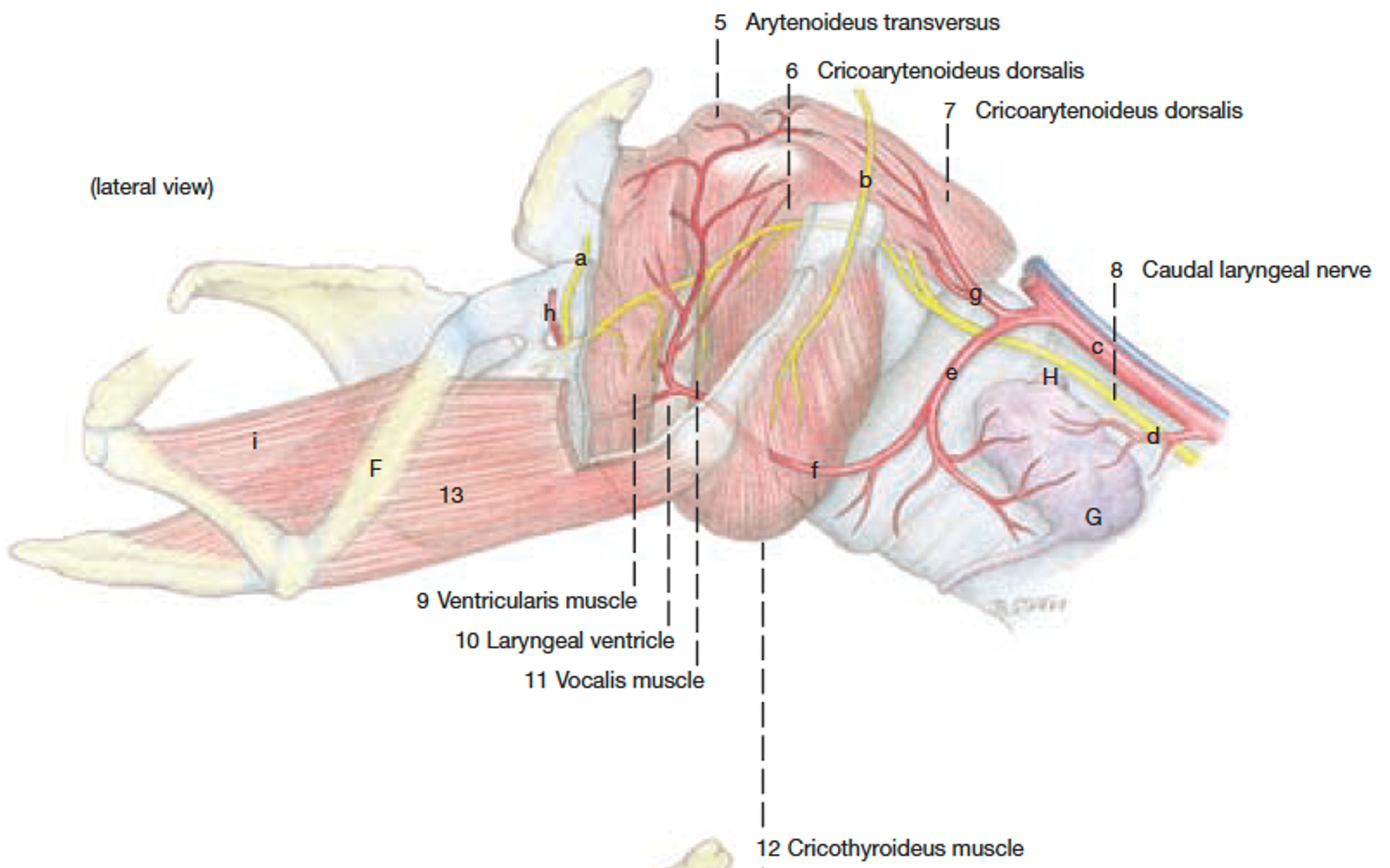
Figure above – Muscles, nerves, and vessels of the equine larynx, lateral view. Labeled structures: F, thyrohyoid bone; 13, thyrohyoid m.; H, parathyroid gland; G, thyroid gland. Labeled structures: 5, arytenoideus transversus; 6, cricoarytenoideus lateralis; 7, cricoarytenoideus dorsalis; 8, caudal laryngeal n.; 9, ventricularis m.; 10, laryngeal ventricle; 11, vocalis m.; 12, cricothyroideus m.; a & b, cranial laryngeal n. branches; c, common carotid a. (Budras et al. Anatomy of the Horse)
- Clinical Notes: HORSES can have a condition called laryngeal hemiplegia or ‘roaring‘ (hemi = unilateral), which is somewhat similar to laryngeal paralysis (aka LarPar) in DOGS. Laryngeal paralysis is due to damage of the recurrent laryngeal n. (the somatic motor branch of CN X Vagus).
- About 90% of cases are on the left side only (i.e., hemiplegia – see images below via laryngoscope/endoscope). Remember, the pathway of the recurrent laryngeal n. differs from left to right sides of the body and can provide clues to the site of a lesion.
- If there is damage to these nerves, the laryngeal muscles may become paralyzed. For the cricoarytenoideus dorsalis m., this results in an inability to abduct the arytenoid cartilage and vocal fold on the affected side. This leads to inspiratory dyspnea (i.e., difficult breathing) and a roaring sound from air passing through the narrowed glottis. This typically is only a problem when the horse is exerted.
- As a clinician, you will likely use endoscopy to view the larynx and becoming familiar with the anatomy from an endoscopic (i.e., rostral) view will be helpful. (Supplemental Clinical LAS Upper respiratory endoscopy)
- From the endoscopic view below identify the: epiglottis, arytenoids, vocal folds, glottis.
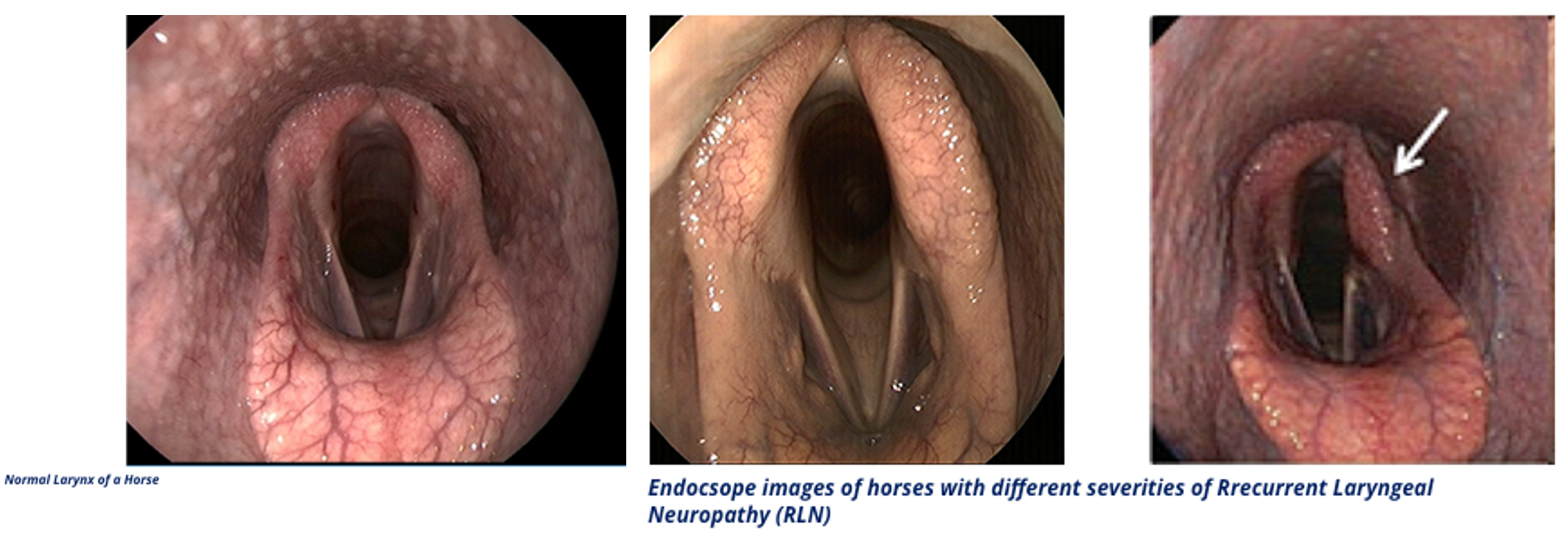
Figure above – (left) normal endoscopic view of the laryngeal inlet of a horse; (middle and right) different severities of recurrent laryngeal n. damage in horses causing roaring. (http://www.southwestequine.com.au/roarers/)
Tongue and Hyoid (Figs. 3.9, 25.2, 25-9, 5-32)
- The lingual a. provides blood to the tongue, while CN XII hypoglossal nerve provides motor innervation to the tongue muscles.
- The tongue has three regions: root, body, and apex (from proximal to distal).
- RUMINANTS have special features of the tongue including:
- The lingual fossa/fissure, a transverse crease in the middle of the tongue.
- The torus linguae, a raised portion of the tongue caudal to the lingual fossa.
- The buccal papillae, conical papillae lining the inside of the cheeks.
- The surface of the tongue is covered in papillae, including the large round vallate papillae (with a moat encircling each one) near the root of the tongue.
- RUMINANTS have special features of the tongue including:

FIG. 3.9 Dorsal view of the tongue and epiglottis of cattle (bo) and horse (eq). 1, Palatine tonsil; 2, median groove; 3, filiform papillae; 4, foliate papillae; 5, epiglottis; 6, tonsillar sinus/crypt; 7, root of tongue; 8, vallate papillae; 9, torus linguae; 10, fossa linguae/lingual fossa/fissure; 11, fungiform papillae. TVA, modified
- Mucosal folds cover the muscles that support the tongue and include the palatoglossal fold (between the soft palate and root of the tongue) and the palatopharyngeal fold (between the soft palate and pharyngeal wall).
- Palatal ridges cover the inner (ventral) surface of the hard palate, and in RUMINANTS the upper incisors (I) and canine (C) teeth are absent and are replaced by a dental pad.
- There are several salivary glands that have ducts that enter into the oral cavity. The parotid salivary gland is on the lateral aspect of each cheek just ventral to the ears (in EQUINE it covers part of Viborg’s triangle). The parotid salivary gland has a prominent parotid duct that courses rostrally along the ventral border of the mandible before turning dorsally on the side of the head to course with the facial a. and v. on their way through the cheek and into the buccal cavity.
- The prominent mandibular salivary gland is curved and is located caudal to the mandible (BOVINE, Fig. 25.2).
- Clinical Notes: the parotid ducts can be lacerated or become blocked. (Supplemental Clinical LAS Salivary disorders)

FIG. 25.2 Superficial dissection of the head, bovine. 1, Masseter; 2, zygomaticus; 3, buccinator; 4, facial vein; 5 and 6, dorsal and ventral buccal branches of facial nerve, respectively; 7, auriculotemporal nerve; 8, cornual nerve; 9, infraorbital nerve; 10, parotid duct and facial artery and vein; 11, parotid gland; 12, mandibular salivary gland; 13, parotid lymph node; 14, lateral retropharyngeal lymph node; 15, spinal accessory nerve; 16, maxillary vein; 17, external jugular vein; 18, linguofacial vein; 19, common carotid artery; 20, (sub) mandibular lymph node; 21, cornual diverticulum of frontal sinus. TVA
- A few of the main muscles of the tongue are: (Fig. 5-32)
- The genioglossus m. is found at the ventral floor of the tongue and originates on the inside of the chin (near the mandibular symphysis) or the genial tubercle, hence the name genio-glossus, from chin to tongue.
- The hyoglossus m. is ventral and caudal to the root of the tongue, spanning from the basihyoid bone to the tongue, hence the name hyo-glossus.
- A few muscles that help support the tongue and are connected to the hyoid apparatus are: (Fig. 5-32)
- The geniohyoideus m. is ventral to the tongue (and genioglossus m.) and extends from the mandibular symphysis (inside of chin) to the basihyoid bone, hence the name genio-hyoid.
- The mylohyoideus m. is ventral to the geniohyoideus m. and extends between the two mandibles from the mandibular symphysis to the hyoid apparatus.
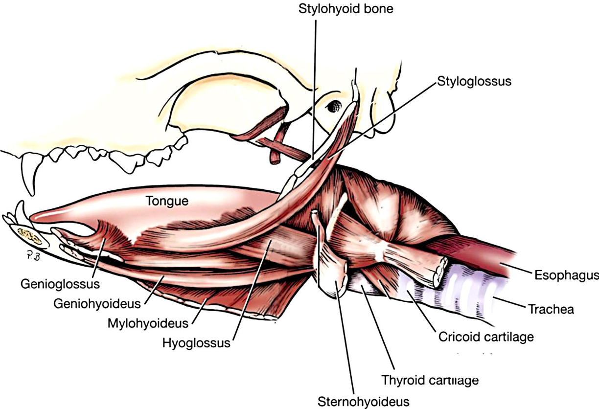
(Fig. 15-1 in Large Animal Dissection eBook) Fig. 5-32 parasagittal view of tongue and hyoid muscles in the dog (similar to ungulates). Labeled structures: tongue, genioglossus, geniohyoideus, mylohyoideus, hyoglossus, sternohyoideus, thyroid cartilage, cricoid cartilage, trachea, esophagus, styloglossus, stylohyoid bone. (from Guide to Dissection of the Dog)
Masticatory Muscles (Figs. 18.23, 25.2, 14B-2, 14B-1, 18-8)
- The muscles of mastication are well-developed in ungulates and are bilateral, and (mostly) innervated by CN V Trigeminal.
- One of the largest and most prominent is the masseter m., which runs from the facial crest (in EQUINE), facial tuberosity, and zygomatic arch to the angle and ramus of the mandible.
- The temporalis m. fills the temporal fossa and inserts on the coronoid process of the mandible. In EQUINE, the temporal line is the most medial border of the temporalis m. The masseter and temporalis mm. act to close the jaw.
- The medial pterygoid m. (medial to the mandible) runs from the pterygoid process to the ramus and condylar process of the mandible and causes side to side movements of the jaw. The masseter (unilaterally) and medial pterygoid mm. act to cause lateral movements of the jaw.
- The digastricus m. runs from the paracondylar process to the medial border of the body of the mandible. It is the primary muscle that opens the mouth.
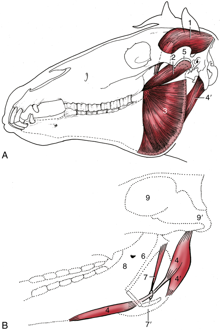
FIG. 18.23 (A) Equine: the deep masticatory muscles of the left side have been exposed by removal of the left mandibular ramus (stippled). (B) Medial view of the right digastricus and some related structures. 1, Temporalis; 2, pterygoideus lateralis; 3, lateral surface of pterygoideus medialis/medial pterygoid; 4, digastricus; 4′, occipitomandibularis; 5, left temporomandibular joint; 6, stylohyoid; 7, stylohyoideus; 7′, insertion of stylohyoideus on thyrohyoid; 8, medial surface of right mandible and mandibular foramen; 9, cranial cavity; 9′, foramen magnum. TVA
Objective
A10.4 Identify and describe the head structures associated with nasolacrimal flushing, the proper placement of a nasogastric tube, and the guttural pouch in equine.
Nasolacrimal Flushing (Figs. 18-3 and below)
- Clinical Notes: A nasolacrimal flush can be used to unblock a clogged nasolacrimal duct, flush debris out of the eye, and/or administer medication into the eye in EQUINE.
- To cannulate the nasolacrimal duct, a tube is passed through the orifice of the nasolacrimal duct found in the rostral and ventral aspect of the nasal cavity (floor of the nostril).
- The nasal diverticulum (aka false nostril) is dorsolateral to the main orifice of the nostril.
- Using the nasolacrimal duct system, fluid can be sent in a retrograde direction toward the eye, since there is access to the eye via the lacrimal sac, lacrimal canaliculi, and the exit at the lacrimal puncta (see images below).
- The nasolacrimal duct may become disrupted by infection of the nearby maxillary sinuses.
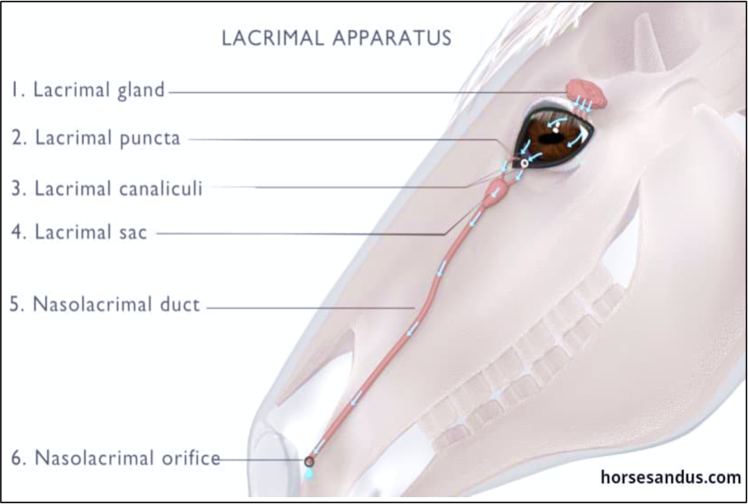
Figure above – View of the nasolacrimal apparatus in equine. Labeled structures: lacrimal gland, lacrimal puncta, lacrimal canaliculi, lacrimal sac, nasolacrimal duct, nasolacrimal orifice/orifice of the nasolacrimal duct.

Figure 18-3 (A) Left nostril opened laterally to expose nasal diverticulum. 1, Alar fold, supported by the lamina (1′) of the alar cartilage; dorsal to the alar fold is the nasal diverticulum (1″); 2, floor of nostril supported by the cornu of the alar cartilage; 3, probe in nasolacrimal duct. (TVA, modified)
Nasal and Pharyngeal Structures and Nasogastric Tube Placement (Figs. 8-6, 25-9, and below)
- In the DOG there were dorsal and ventral nasal conchae with dorsal, middle, and ventral meatuses, as well as a common nasal meatus. Remember, the nasal cavity is split along the midline via a nasal septum (with cartilaginous and bony portions, Fig. 8-6).
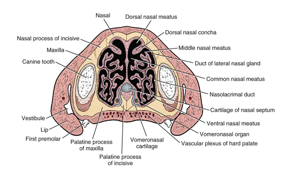
Figure 8-6 Transverse/cross section of the nasal cavities between the canine and first premolar teeth in the dog. Labeled structures: nasal bone, nasal process of incisive bone, maxilla, canine tooth, vestibule, lip, first premolar, palatine process of maxilla, palatine process of incisive, vomeronasal cartilage, vascular plexus of hard palate, vomeronasal organ, ventral nasal meatus, cartilage of nasal septum, nasolacrimal duct, common nasal meatus, duct of lateral nasal gland, middle nasal meatus, dorsal nasal concha, dorsal nasal meatus. (from Guide to Dissection of the Dog)
- The nasal turbinates aka conchae are bilateral sets of thin-walled bony ridges projecting into the nasal cavity. A meatus is the space around the same named concha. In the HORSE there are dorsal and ventral nasal conchae (and ethmoid conchae), with a dorsal, middle, and ventral meatuses.
- Clinical Notes: conchal cysts are congenital anomalies that can slowly develop in the nasal conchae and cause blockage of the nasal passages and deformation of the skull. Hematomas can develop off of the ethmoid conchae and often produce foul smelling discharge, grow into the sinuses, or even be impacted by nasogastric tube placement. (Supplemental Clinical LAS Nostrils and nasal conchae; LAS Paranasal sinus disease)
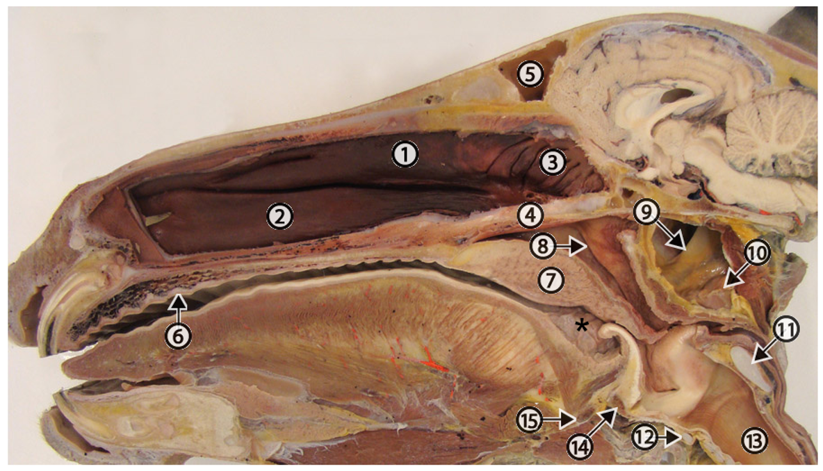
Figure above – Equine split head after removal of the nasal septum to expose the nasal cavity. 1, dorsal concha; 2, ventral concha (ventral nasal meatus ventral to the concha); 3 ethmoidal concha; 4, vomer (bone); 5, conchofrontal sinus; 6, hard palate; 7, soft palate; 8, orifice of the auditory tube on the lateral wall of the nasopharynx. At this place, an endoscope can be passed into the guttural pouch. 9, stylohyoid bone; 10, medial retropharyngeal lymph nodes adjacent to the ventral wall of the guttural pouch; 11, cricoid cartilage; 12, cricoid cartilage (ventral), 13, trachea; 14, ossified rostral edge of the thyroid cartilage; 15, basihyoid bone; asterisk, palatine tonsil. NOTE: In this image the tip of the epiglottis is abnormally positioned ventral to the soft palate. The normal position is dorsal to the soft palate. (http://vanat.cvm.umn.edu/ungDissect/Lab20/Img20-2.html)
- BOVINE have a nasolabial plate at the front of the nose and upper lip that is rigid and hairless (compared to EQUINE, where the upper lip is very mobile). Upon entering the nasal cavity, there are dorsal, middle, and ventral nasal conchae (and ethmoid conchae), with a dorsal, middle, and ventral meatus.
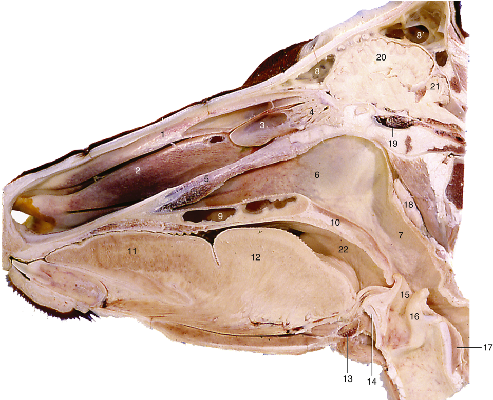
Figure 25-9 Paramedian section of the bovine head. 1, Dorsal nasal concha; 2, ventral nasal concha; 3, middle nasal concha; 4, ethmoidal conchae; 5, vomer; 6, choana; 7, nasopharynx; 8, rostral frontal sinus; 8′, caudal frontal sinus; 9, palatine sinus; 10, soft palate; 11, apex of tongue; 12, torus linguae; 13, basihyoid; 14, thyroid cartilage; 15, epiglottis; 16, arytenoid cartilage; 17, cricoid cartilage; 18, medial retropharyngeal lymph node; 19, venous plexus surrounding hypophysis; 20, cerebrum; 21, cerebellum; 22, entrance to tonsillar sinus. TVA
Pharyngeal Structures
- The pharynx is divided into three main regions: nasopharynx (choanae to the caudal free edge of the soft palate and palatopharyngeal arches), oropharynx (soft palate to the base of tongue and epiglottis), and laryngopharynx (base of epiglottis to esophagus).
- The orifice of the auditory tube is found in the lateral wall of the nasopharynx. (see image of equine split head above)
- The palatine tonsil is found in the lateral walls of the oropharynx. (see image of equine split head above and Fig. 3.9)
- In BOVINE, there is a pharyngeal septum ventral to the bony nasal septum (vomer) and pharyngeal tonsil at the dorsal and caudal wall of the nasopharynx. In BOVINE the ventral meatuses can communicate, while in EQUINE the nasal septum is complete. In EQUINE this allows you to localize the lesion based on unilateral discharge.
Nasogastric Tube Placement
- Clinical Notes: A nasogastric tube is used to relieve gas build up within the stomach (e.g., colic), or to deliver medications or fluids into the stomach (usually in EQUINE). Complications can include bleeding if the tube abrades the conchae/turbinates.
- The nasogastric tube will follow a similar path as was described in small animal anatomy.
- The nasogastric tube first passes through an external naris and enters the nasal cavity, avoiding the nasal diverticulum located dorsolaterally. The tube should be kept in the ventral nasal meatus. The tube has measurements indicated, and the distance from the nostril to the eye helps measure the amount of tube needed to reach to the nasopharynx.
- The tube continues to the nasopharynx, and then into the esophagus (not the larynx and trachea which are more ventral). Remember, the esophagus is dorsal to the trachea and more on the left side in the middle of the neck region. Once the tube reaches the nasopharynx, the swallowing reflex usually initiates and allows the tube to be swallowed. Note, there will be negative pressure in the esophagus.
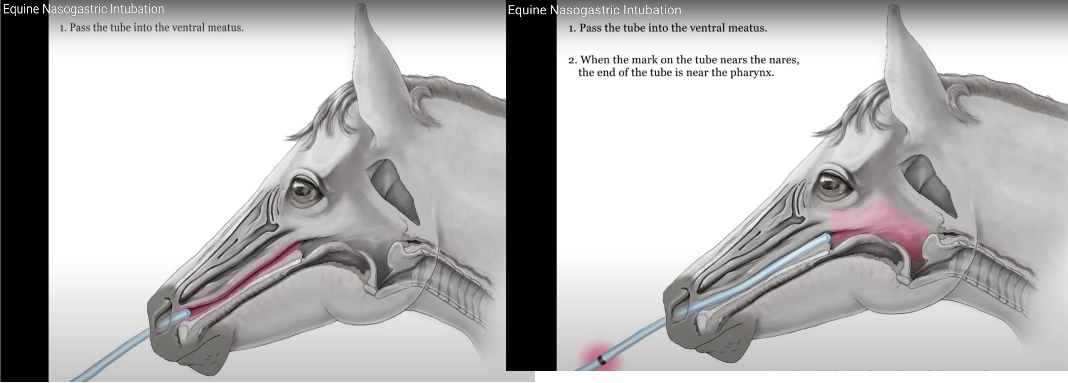
Figures above – Nasogastric tube placement: 1. Pass the tube into the ventral meatus. 2. When the mark on the tube nears the nares, the end of the tube is near the pharynx.
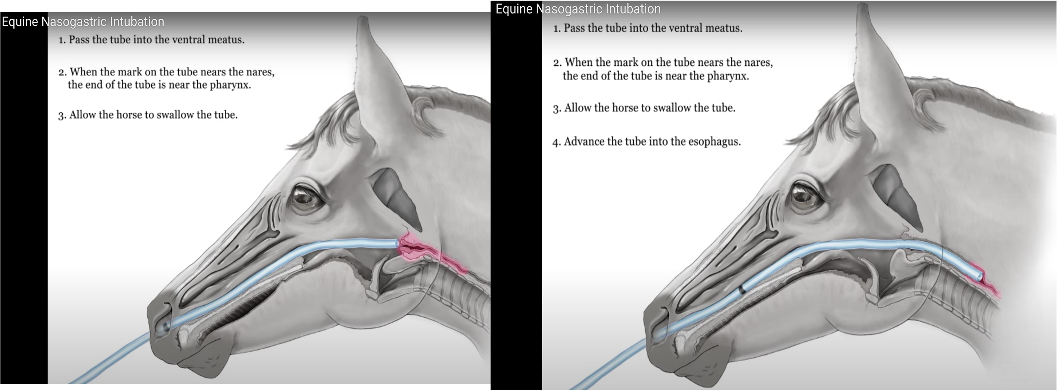
Figures above – Nasogastric tube placement: 3. Allow the horse to swallow the tube. 4. Advance the tube into the esophagus. (https://youtu.be/HR5bHSMUtuo)
Guttural Pouch and Viborg’s Triangle
Guttural Pouch
- In EQUINE, the guttural pouch is an out-pocketing/diverticulum of the auditory (aka Eustachian) tube on each side of the head. The orifice of the auditory tube is located in the nasopharynx, where it appears as a slit-like opening. The cartilage of the auditory tube extends from the middle ear compartment to the nasopharynx. The guttural pouches form from the ventral border of the auditory tube cartilage.
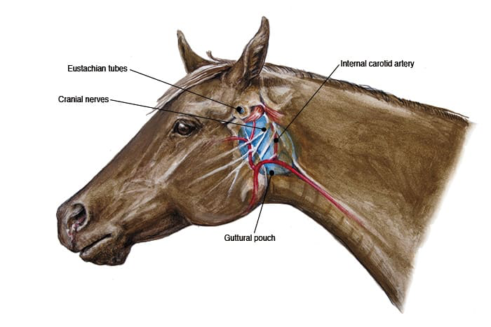
Figure above – View of the left guttural pouch in a horse. Labeled structures: Eustachian tubes, cranial nerves, guttural pouch, internal carotid artery.
- The nasopharynx is rostral to each guttural pouch, while the laryngopharynx is ventral. The longus capitis mm. run along the caudal border of the guttural pouches (see Figure below).
- There is a thin membrane that separates the right guttural pouch from the left. The stylohyoid bone subdivides each guttural pouch into medial and lateral compartments and the pouches are lined by mucosa.
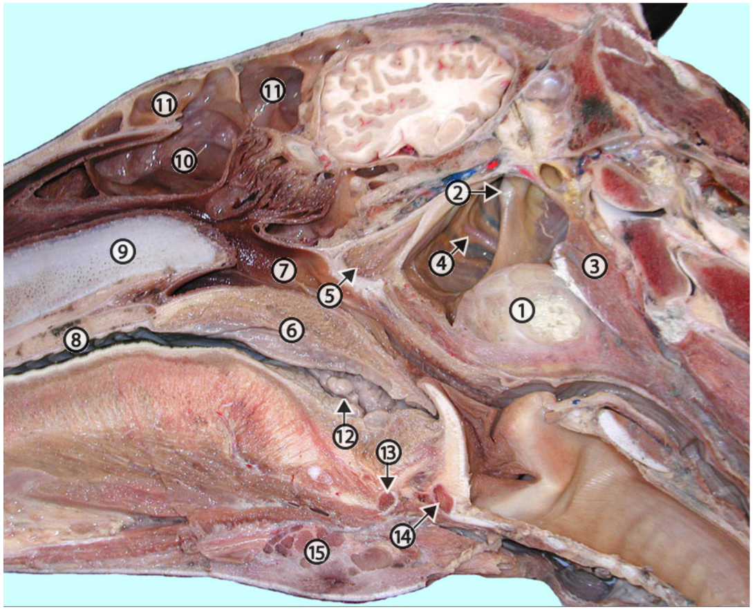
Figure above – Equine split head with a large strangles abscess (1) on the ventral aspect of the guttural pouch in the medial retropharyngeal lymph node. 2, temporohyoid joint; 3, longus capitis m.; 4, maxillary artery; 5, auditory tube; 6, soft palate; 7, nasopharynx; 8, hard palate; 9, cartilage of nasal septum; 10, dorsal conchal sinus; 11, conchofrontal sinus; 12, palatine tonsil; 13, basihyoid bone; 14, ossified rostral edge of the thyroid cartilage; 15, (sub) mandibular lymph nodes. (http://vanat.cvm.umn.edu/ungDissect/Lab20/Img20-5.html)
- There are several important structures running alongside the boundaries of the guttural pouches.
- The common carotid a. splits into the external and internal carotid aa. near the ventral edge of the guttural pouch. The linguofacial trunk (and eventual facial and lingual arteries) is a branch from the external carotid a. and continues to run along the ventral aspect of the guttural pouch.
- The internal carotid a. is on the caudal surface of the pouch briefly and then the medial surface. The internal carotid a. may hemorrhage into the pouch if eroded by infection – see Clinical Notes below.
- The external carotid a. stays on the lateral surface of the pouch.
- The guttural pouches are thought to function as a mechanism to cool the cerebral blood supply (especially during exercise), as the internal carotid a. runs along the thin medial wall of the guttural pouch.
- The common carotid a. splits into the external and internal carotid aa. near the ventral edge of the guttural pouch. The linguofacial trunk (and eventual facial and lingual arteries) is a branch from the external carotid a. and continues to run along the ventral aspect of the guttural pouch.
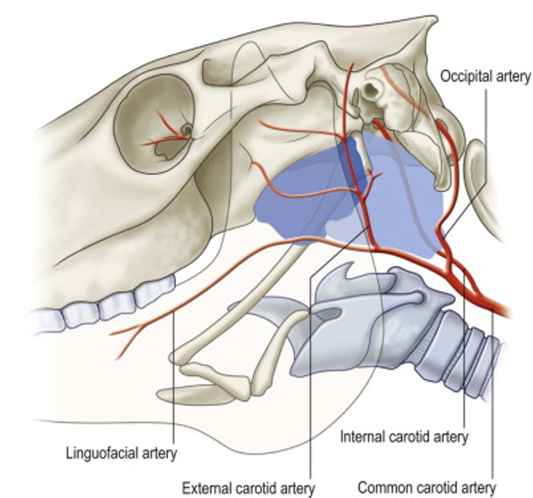
Figure above – Lateral view of equine head, blue indicates left guttural pouch. Labeled structures: common carotid, internal carotid, occipital, external carotid, and linguofacial aa. Note stylohyoid bone running into the ventral boundary of the guttural pouch, forming medial and lateral compartments of the pouch.
-
- There are several cranial nerves that run along the guttural pouch. They include: CNs IX Glossopharyngeal, X Vagus, XI Accessory, and XII Hypoglossal. CN VII Facial and CN V Trigeminal branches are also found near the dorsal and lateral edges of the pouch.
- A medial retropharyngeal lymph node is found on the ventral surface of the pouch (note image above with abscess of the lymph node).
- Clinical Notes: Streptococcus equi can infect the guttural pouch and cause the condition called “strangles,” a highly contagious upper respiratory infection. This infection can cause the medial retropharyngeal lymph nodes to enlarge and abscess. The abscess puts pressure on (i.e., strangles) the airway, including the larynx and trachea which are ventral to the pouches (see Figure above).
- Fungal infections within the pouch (i.e., mycoses, often Aspergillus fumigatus) if left untreated, can erode the walls of the pouches where important cranial nerves and carotid vessels are situated. The vessels can eventually hemorrhage and cause bleeding, which can be life threatening.
- Either type of infection can cause damage to adjacent vessels or nerves, leading to hemorrhaging or dysphagia and/or Horner’s syndrome.
- A region in the neck called Viborg’s triangle may be an access point, where one can avoid major vessels and nerves, to drain fluid from the guttural pouch.
- There are several disorders of the guttural pouch besides strangles. (Supplemental Clinical LAS Guttural pouch disorders).
Boundaries of Viborg’s Triangle (also in A5.2) (Figure below)
- The dorsal/caudal border is the tendon of the sternocephalicus/sternomandibularis m.
- The rostral/cranial border is the angle and ramus of the mandible.
- The ventral border is the linguofacial v. (Note, the linguofacial v. joins with the maxillary v. to form the external jugular v.
- The mandibular lymph nodes (also in BOVINE, Fig. 25.2) are under the chin (and palpable) and run near the linguofacial v. and ramus of the mandible as well.
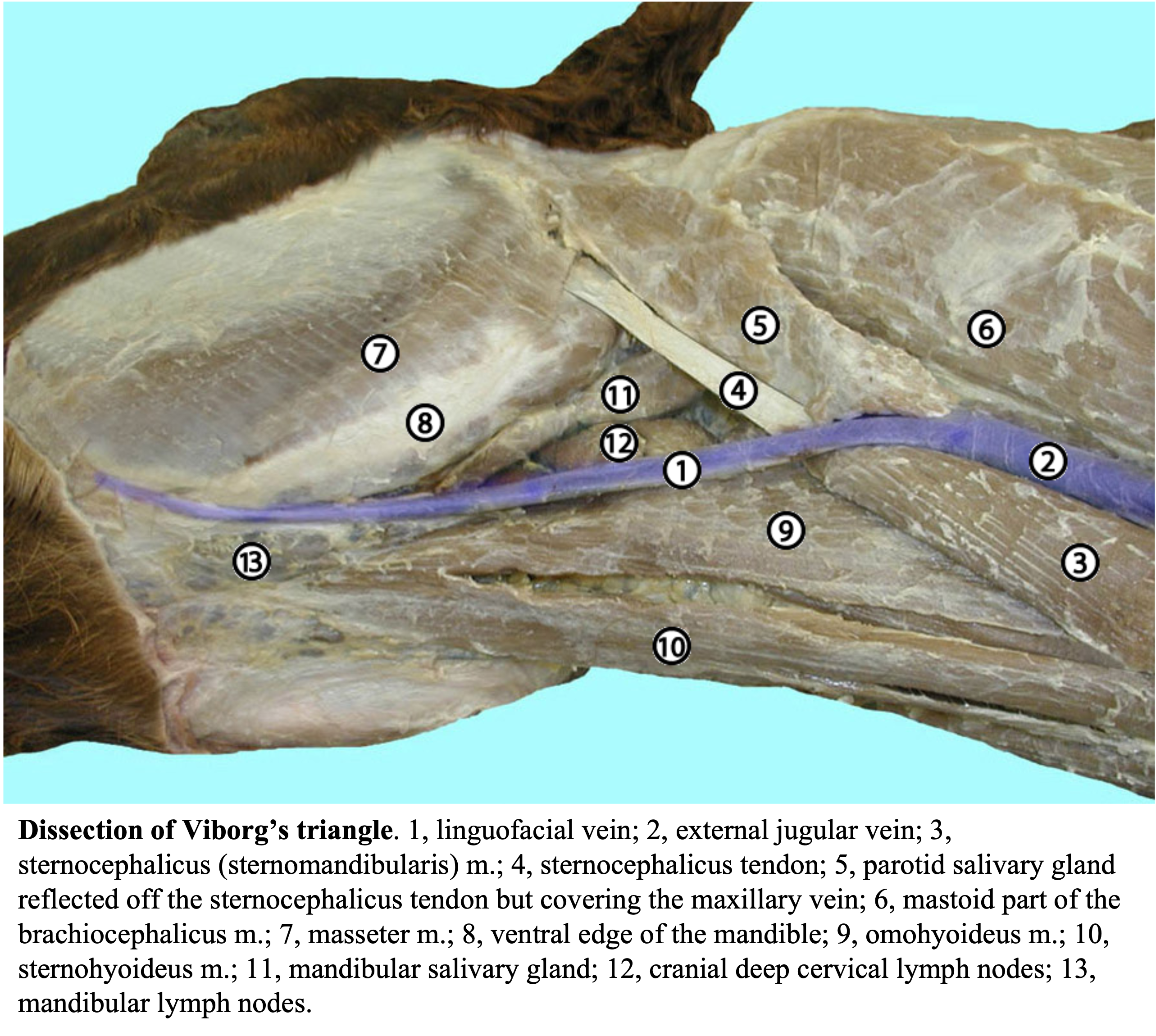
Figure above. Dissection of Viborg’s triangle. 1, linguofacial vein; 2, external jugular vein; 3, sternocephalicus/sternomandibularis m.; 4, sternocephalicus tendon; 5, parotid salivary gland reflected off the sternocephalicus tendon but covering the maxillary vein; 6, mastoid part of the brachiocephalicus m.; 7, masseter m.; 8, ventral edge of the mandible; 9, omohyoideus m.; 10, sternohyoideus m.; 11, mandibular salivary gland; 12, cranial deep cervical lymph nodes; 13, (sub) mandibular lymph nodes. (http://vanat.cvm.umn.edu/ungDissect/Lab08/Img8-13.html)
Objective
A10.5 Identify and describe the listed teeth structures and terms; given a chart, estimate the age of a horse based on the mandibular incisor teeth; be able to assign a Triadan number for any equine tooth.
Teeth (Figs. 14B-11, 14B-12, 18-19, 25-18, 18-20 and below)
- HORSES and CATTLE have brachydont and hypsodont teeth.
- A brachydont tooth has a crown, neck, and root(s). Brachydont teeth in the HORSE include the canines and first premolars. Brachydont teeth in the BOVINE include the mandibular incisors and canines (ruminants do not have upper incisors or canines).
- Unlike brachydont teeth, hypsodont teeth continuously erupt as the animal ages. A hypsodont tooth has an extensive crown, mostly encased in enamel. Hypsodont teeth have small roots and no neck. The cheek teeth in HORSE and RUMINANTS are hypsodont.
- RUMINANTS – The upper incisors (I) and canine (C) teeth are absent and are replaced by a dental pad. (Fig. 25-18)
- The mandibular incisor teeth of RUMINANTS are shovel shaped with a very well-defined neck.
- The mandibular canine teeth of RUMINANTS resemble the incisor teeth, giving the appearance of four incisor teeth on either side. Therefore, the RUMINANT canine tooth is often referred to as an I4 tooth.
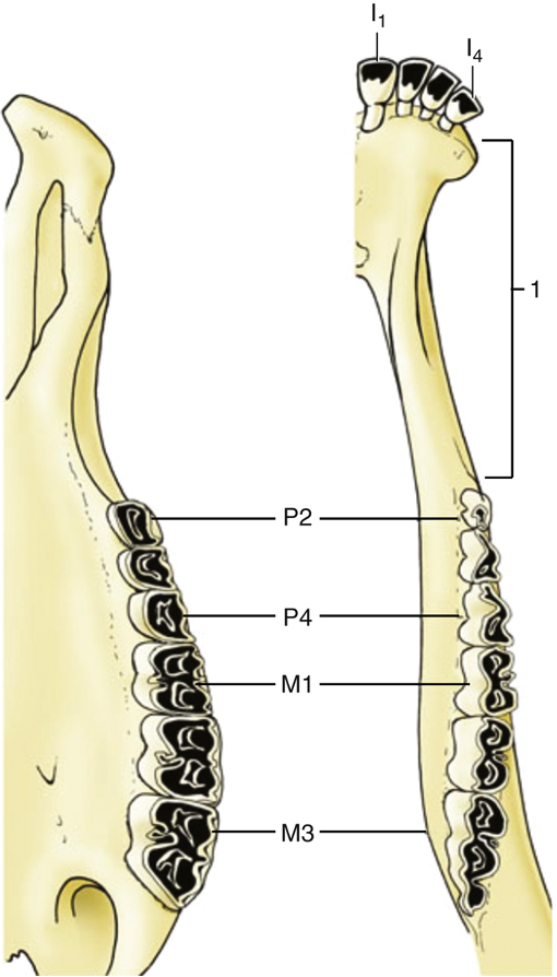
Figure 25-18 Left half of upper and right half of lower jaw of cow. Note the different shapes of the upper and lower cheek teeth and the large diastema (1). I1 and I4, First and fourth incisors; M1 and M3, fourth and sixth upper and lower cheek teeth, respectively; P2 and P4, first and third upper and lower cheek teeth, respectively. TVA
- HORSES – There are upper and lower incisors and canine teeth. (Fig. 18-19)
- The incisor teeth of the HORSE have a rather uniform thickness throughout their length. The incisor teeth of the HORSE are known from medial to lateral as the central (I1), intermediate (I2), and corner (I3) incisors.
- The canine teeth of HORSES are sexually dimorphic, with the canines of mares being vestigial or absent. Mare canines may be seen on skulls but rarely erupt through the gums of the live horse.
- Between the incisors, or canines, and the cheek teeth (premolars (P) and molars (M)) in ungulates is a wide interdental space called the diastema. (Figs. 18-19, 25-18)
- The first premolar tooth (P1) of the HORSE is very small and often absent. This tooth is commonly referred to as the wolf tooth. It is often removed because it is sometimes thought to interfere with the bit. The wolf tooth is NOT the canine tooth. Since the first premolar tooth is small and inconstant, it is often not considered a cheek tooth even though it is adjacent to the cheek teeth. (Fig. 18-19)
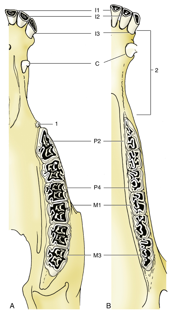
Figure 18-19 The permanent teeth of the (A) upper and (B) lower jaws of a horse. 1, Wolf tooth (P1); 2, diastema (aka bar). I – incisor, C – canine, P – premolar, M – molar.
- In younger HORSES the permanent teeth are erupting, and only incisors and premolars (P2-4) form “caps” that are shed (there is no deciduous precursor of P1 and the molars). Tiny equine deciduous canines are sometimes observed on skulls but never erupt through the gums (i.e., gingiva).
- Caps are the remnant deciduous tooth that is pushed out by the permanent teeth (i.e., they may be seen forming a “cap” on top of the permanent teeth).
- The ventral edge of the mandible of young horses often has prominent temporary tuberosities from the developing premolar teeth P3 and P4 that “don’t fit” into the mandible without causing bulging of the ventral border. These should not be mistaken for fractures.
- The cheek teeth (often include premolars and molars) of HORSES, RUMINANTS, and CAMELIDS, and the incisor teeth of HORSES, are characterized by an in-folding of the enamel layer known as an infundibulum. The infundibulum of the EQUINE incisors is a simple finger-like in-folding, but the infundibula of the cheek teeth are more complex. (Fig. 18-20)
- As a result of wear, the edges of the infundibula become razor sharp, making the occlusal surface of the molar teeth an efficient grinding surface. The greater width of the maxillary cheek teeth and their outer ridges, or cingula (cingulum is singular), are often sharp and need to be filed down or “floated” to prevent injury to the buccal (cheek) mucosa.
- In most species, the molar teeth are larger than the premolar teeth but in HORSES all cheek teeth are of similar size (Fig. 14B-11). This has been referred to as “molarization” of the premolars and reflects the fact that HORSES have the most well-developed cheek teeth of all domestic animals, so some refer to all cheek teeth as molars.
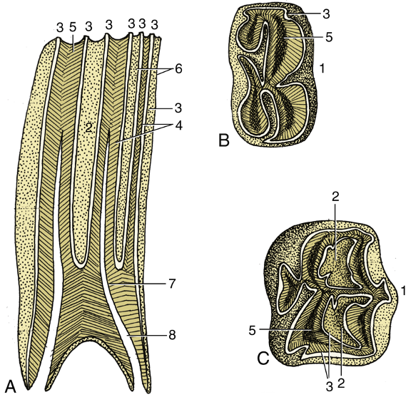
Figure 18-20 Structure of the cheek teeth shown in sagittal section (A) and by views of the occlusal surface of lower (B) and upper (C) molars. 1, Buccal (labial) surface; 2, infundibulum; 3, enamel; 4, dentine; 5, secondary dentine; 6, cement; 7, dental cavity; 8, root canal. TVA
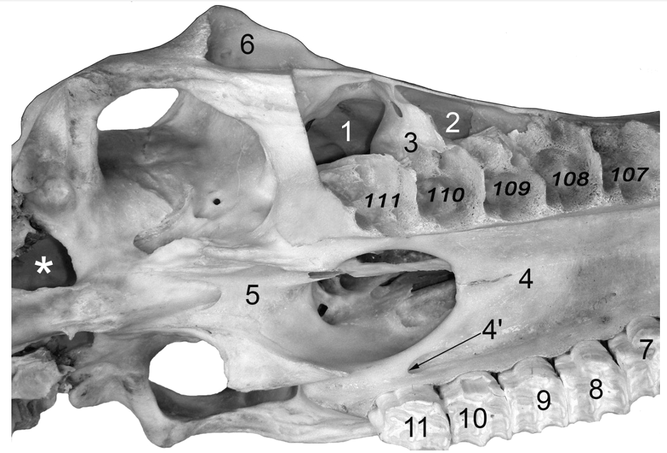
Figure 14B-11 Horse skull, ventral, slightly oblique view. The cheek teeth have been removed on the right side of the skull. The alveoli on the right are labeled by their Triadan numbers. The teeth on the left would then be Triadan numbers 207-211 respectively. 1, caudal maxillary sinus; 2, rostral maxillary sinus; 3, septum between 1 & 2; 4, hard palate; 4’, palatine foramen; 5, vomer; 6, orbit; *, foramen lacerum.
- CAMELIDS (llamas and alpacas) have teeth similar to RUMINANTS with a few significant differences.
- Like RUMINANTS, the mandibular incisors are flattened, shovel shaped teeth that lack infundibula.
- Small CAMELIDS lack upper incisors I1 and I2 but have a canine-shaped third upper incisor. Therefore, the shape of the lower third incisor is significantly different from the upper third incisor.
- In RUMINANTS, the canine teeth resemble the incisor teeth, but in CAMELIDS the canine teeth are sharp and resemble the canine teeth of DOGS. RUMINANTS lack upper canine teeth but CAMELIDS have all four. The canine teeth and the upper third incisor teeth of small CAMELIDS are referred to as the ‘fighting teeth.’
- Like HORSES, the canines of small CAMELIDS are sexually dimorphic but they are less obvious. The fighting teeth of female camelids are smaller than those of the males.
- While RUMINANTS have 3 upper and lower premolar teeth, small CAMELIDS have only 2 upper premolar teeth and one lower premolar tooth (on each side). Old world CAMELIDS have an additional premolar tooth in the space between the canine teeth and the cheek teeth.
Teeth Aging
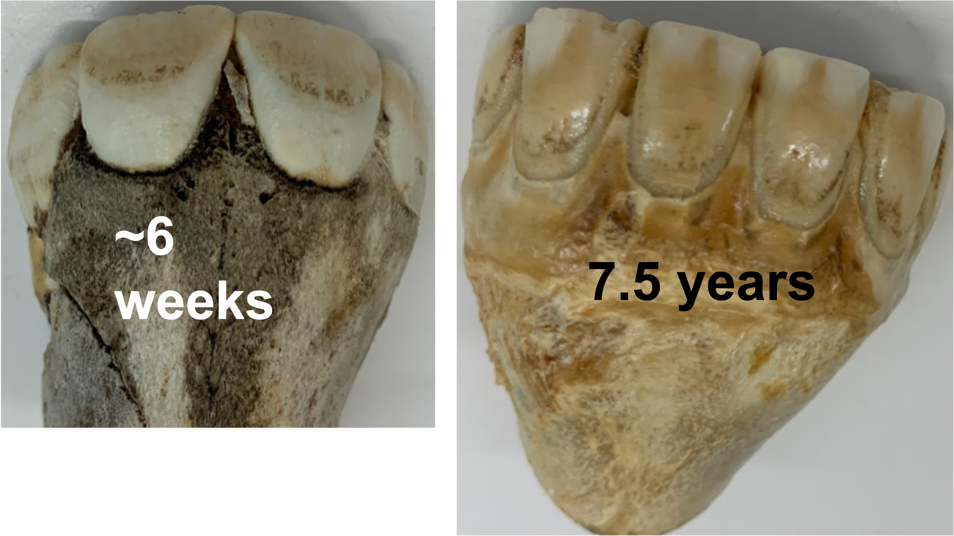
- When estimating EQUINE age by the teeth, the lower incisor teeth are useful. It is important to differentiate between deciduous and permanent incisors. If this is not done, a two-year-old could be confused with a five-year-old. Between 2 and 5 years of age the mistake is easily avoided, because the larger permanent teeth (rectangle-shaped) are easily differentiated from the smaller deciduous teeth (rounded triangle-shaped) when they are present side by side. (Fig. below, left is 6 week old, right is 7.5 year old)
- The infundibula of deciduous teeth are shallow, and the labial surface of the tooth tends to be slightly conical, whereas the same surface of the permanent tooth is more rectangular. Since canines do not erupt until about 5 years, they furnish a quick clue regarding age, but only for males.

Figure 14B-12 Sagittal section through a central incisor tooth (I1) of a young horse showing the internal structure of the tooth. 1, pulp cavity (in life filled with vessels, nerves and connective tissue); 1′, secondary dentine (receding pulp cavity fills in with dark dentine, note that it is located on the labial (lip) side with respect to the infundibulum); 2, dentine; 3, enamel; * cementum in the infundibulum, stippling on portions of the outside of I2 represent cementum as well (often the soft cementum is worn away); A, transection of tooth showing dental star (1′) and cup filled with dark debris; B, transection of tooth showing dental star (1′) and cementum filling the bottom of cup. The central and second incisors are in wear (flat occlusal surface) but I3 is newly erupted and not yet in wear, so the horse is near 5 years old. The dental star is flattened in young horses as in the above figure but more round in older horses after the tooth is worn down past the enamel spot.
- The tooth is said to be ‘in wear’ when enamel is worn off and dentine is exposed on the labial edge of the tooth (e.g., adult incisors are in wear at 3, 4, and 5 years old). When the entire occlusal surface of the tooth is in wear, the tooth is said to be ‘level’.
- (The 7-year “hook” is sometimes referred to and may be visible on the caudal border of the upper corner incisor around 7 years of age, but may be too variable to be useful in aging. The hook represents a lack of wear on the caudal aspect of the upper tooth by the opposing lower incisor. (American Association of Equine Practitioners Guide for Determining the Age of the Horse)
- The lumen of the infundibulum in the EQUINE incisor tooth is known as the cup, or mark. The cup is black due to decaying food and is said to be ‘gone’ when the black mark is no longer present. The cups are lost medial to lateral at 6, 7, 8 years respectively. The bottom of the infundibulum may still, however, be present as a white enamel spot (Fig. 14B-12/4). At first, the enamel spot has a dull white center of cementum, but as wear proceeds, only enamel is seen.
- The infundibulum is surrounded by the more yellow dentine. The pulp cavity fills with darker secondary dentine as the tooth continues to erupt. The dark secondary dentine forms the dental star. On the occlusal surface the dental star will appear as a darker spot just outside of the cup/enamel spot region (Fig. 14B-12/1).
- Loss of tooth substance due to wear is known as dental attrition. As dental attrition occurs, the tooth shortens, but the erupted part of the tooth, known as the clinical crown, does not.
- Galvayne’s groove (Fig. below) is a longitudinal depression on the labial surface of the upper corner incisor. Cementum may remain in part or all of the groove and may or may not have a dark stain. The groove is said to first appear at the gum line at nine to ten years of age and extends the full length of the tooth (i.e., the clinical dental crown) at eighteen to twenty years of age. The presence of Galvayne’s groove is variable and even when present the length in relation to the age of the horse may be inexact.
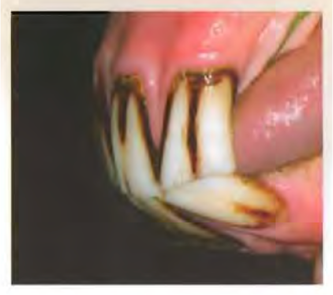
Figure above – Galvayne’s groove. (American Association of Equine Practitioners Guide for Determining the Age of the Horse)
The key below is useful for the determination of EQUINE age based on eruption date (D = deciduous, I = incisor). Given the key on an exam, be able to determine the approximate age of a given EQUINE specimen.
- In general, at 2 years of age all the baby teeth (deciduous) have erupted, and at 5 years of age all permanent incisors have erupted.
- The enamel spot is lost at ages 13-15 years.
- A general way to remember the eruption of deciduous and permanent incisors from I1, I2, I3: 6 days, 6 weeks, 6 months (baby teeth); 2.5, 3.5, 4.5 years (adult); and when the cups are gone from those incisors: 6, 7, and 8 years.
| Di 1 erupted | 1 week (6 days) |
| Di 2 erupted | 1 month (6 weeks) |
| Di 3 erupted | 8 months (6 months) |
| All deciduous lower incisors level | 2 years |
| I1 erupts | 2 1/2 years |
| I2 erupts | 3 1/2 years |
| I3 erupts | 4 1/4 years |
| Canines erupted, I1 and I2 level | 5 years |
| Cup gone from I1 | 6 years |
| All lower incisors level, cup gone I2 | 7 years |
| Cup gone I3 | 8 years |
| Last year all incisors have enamel spot | 13 years |
| All enamel spots gone, occlusal surface triangular | 17 years |
Eruption times of the EQUINE permanent cheek teeth:
| 5-6 months | P1 (wolf tooth) |
| 2 1/2 years | P2 |
| 3 years | P3 |
| 4 years | P4 |
| 1 year | M1 |
| 2 years | M2 |
| 3 1/2 – 4 years | M3 |
- Estimation of BOVINE age is more difficult and less exact. The overall appearance of the animal is useful for differentiating a yearling animal with no permanent teeth from an older animal with permanent teeth well worn. This differentiation, however, is sometimes complicated by the very rapid growth of body size and weight with modern management systems.
- The key below is useful for the estimation of BOVINE age. The eruption pattern of SHEEP is similar but occurs about 1/2 year earlier.
| 3-4 weeks | All deciduous incisors erupted |
| 1 – 1 1/2 years | I1 erupts |
| 2 – 2 1/2 years | I2 erupts |
| 3 years | I3 erupts |
| 3 1/2 – 4 years | C erupts |
| 5 years | All incisors and canine in wear |
| 6 years | I1 level |
| 7 years | I2 level |
| 8 years | I3 level |
| 9 years | C level |
| 12-15 years | “Peg mouth” |
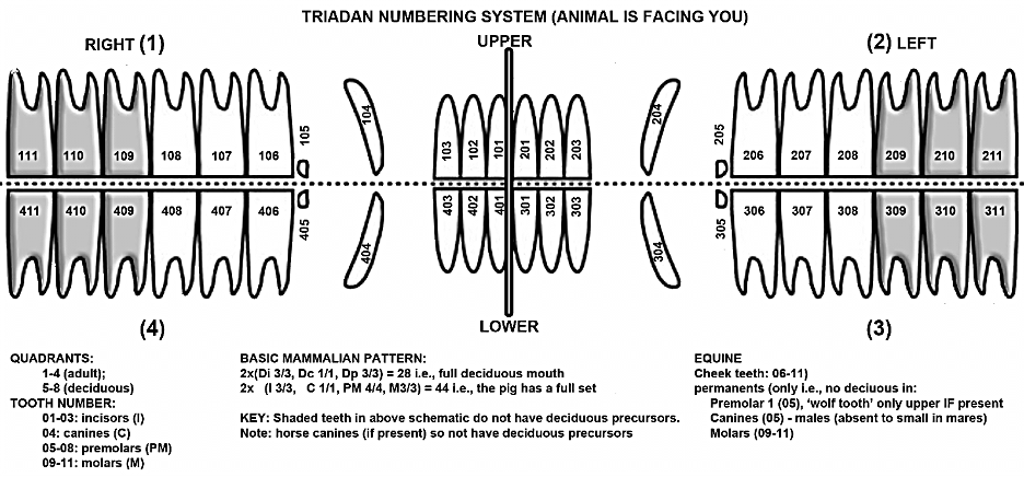
The Triadan System – be able to assign a Triadan number for any EQUINE tooth.
- Similar to small animal anatomy, this system uses 3 digits to describe the location of any tooth. The first digit is for the quadrant, beginning the numbering with the upper right and then moving clockwise (as you are looking at the horse from a rostral view): 1 is upper right, 2 is upper left, 3 is lower left, and 4 is lower right. The other two digits in the Triadan number are for the individual tooth, numbered 1-11, starting from the central incisor through to the last molar in each quadrant. A zero precedes tooth numbers 1 to 9. Quadrants 5-8 are for deciduous teeth.
- Examples: 406 = lower right premolar 2 (first cheek tooth); 806 = deciduous lower right PM2.
- Note the pig has a full set of 44 teeth; dental formula: (I 3/3, C 1/1, PM 4/4, M 3/3) x 2