
3 Pelvic Limb
Revised Spring 2024 by T. Clark
Additional Resources for this Chapter:
- Valuable Supplemental Large Animal Surgery (LAS) course eBook links (starting with Equine Lameness and going through Swine, Small Ruminant, and Poultry Lameness): LAS Osteochondrosis; LAS Gait abnormalities; LAS Nerve and Joint Blocks; LAS Tendon and ligament issues; LAS Hock joint problems; LAS Tendonitis and bowed tendons; LAS Nerve damage; LAS Bovine Foot Anatomy, Lameness, Approaches
- Veterinary Gross Anatomy Ungulate Dissection (Limb Dissection Labs 1-7)
- Associated Video Links:
- Pelvic Limb Bones (7 videos, 28:35)
Objective
A3.1 Describe and differentiate regions and bones of the hind limb by using correct regional/directional terminology.
Note: The terms pelvic limb and hind limb may be used interchangeably.
Directional Terms (repeat from Small Animal Anatomy and Vet Basics)
- Dorsal/ventral, cranial/caudal, medial/lateral, proximal/distal, dorsal/plantar, superficial/deep
- Note: From the tarsus and distally, terminology changes to dorsal and plantar (versus dorsal and palmar from the carpus and distally in the thoracic limb).
Regions of the Pelvic Limb:
- Pelvis (rump): os coxae
- Thigh: femur
- Crus (leg): tibia, fibula
- Pes (hind paw): tarsus (hock), metatarsal bones, digit(s) made up of phalanges and sesamoid bones
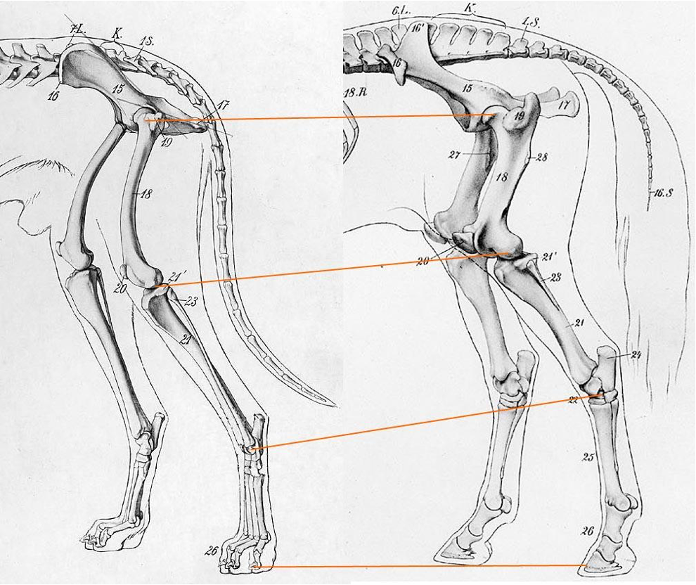
Note: Similar to the thoracic limb, the distal pelvic limb (tarsus) is also lengthened due to unguligrade locomotion. The increased length allows for increased stride length and speed (see image below, right is ungulate forelimb bones scaled to fit canine forelimb bones).
Superficial Structures (There are labeled images in the Appendix/Back Matter with some of these structures as well.)
- Chestnut (eq) – A keratinized structure on the medial side of the pelvic limb, distal to the tarsus. There is also a chestnut on the medial side of the thoracic limb, proximal to the carpus. These are thought to be similar to the tarsal/carpal pads of dogs and cats.
- Ergot (eq) – keratinized growth on the plantar (and palmar) surface of the fetlock.
- Coronet (p) – the skin to hoof (or skin to claw) transition; the ‘crown’ of the hoof. Deep, and distal, to the coronet is a band of dermal tissue (the coronary band). Some may confuse and interchange the terms coronet and coronary band.
- Dewclaws (bov) – underdeveloped digits 2 and 5 (palmar and plantar surfaces).
Bones
Pelvis (Figures 4-3, 4-6, and 4-7)
- The pelvis is composed of two hip bones, which are called the ossa coxae, united ventrally at the pelvic symphysis. Dorsally the two ossa coxae articulate with each side of the sacrum (at the sacroiliac joints), which are the wings of the sacrum that project ventrally.
- Each os coxae is formed by the ilium, ischium, pubis, and a small acetabular bone. The acetabulum creates the socket of the hip and is formed by the fusion of the ilium, ischium, and pubis. There is an acetabular notch ventrally, and the depression where the head of the femur articulates is called the articular surface of the acetabulum. (Fig. 4-6)
- Clinical Note: It will be important to be aware of the dimensions of the bony pelvis when thinking about calving difficulty (i.e., dystocia). Sometimes it is necessary to rotate the calf in the abdomen and pelvis to allow for better positioning and delivery in the COW.
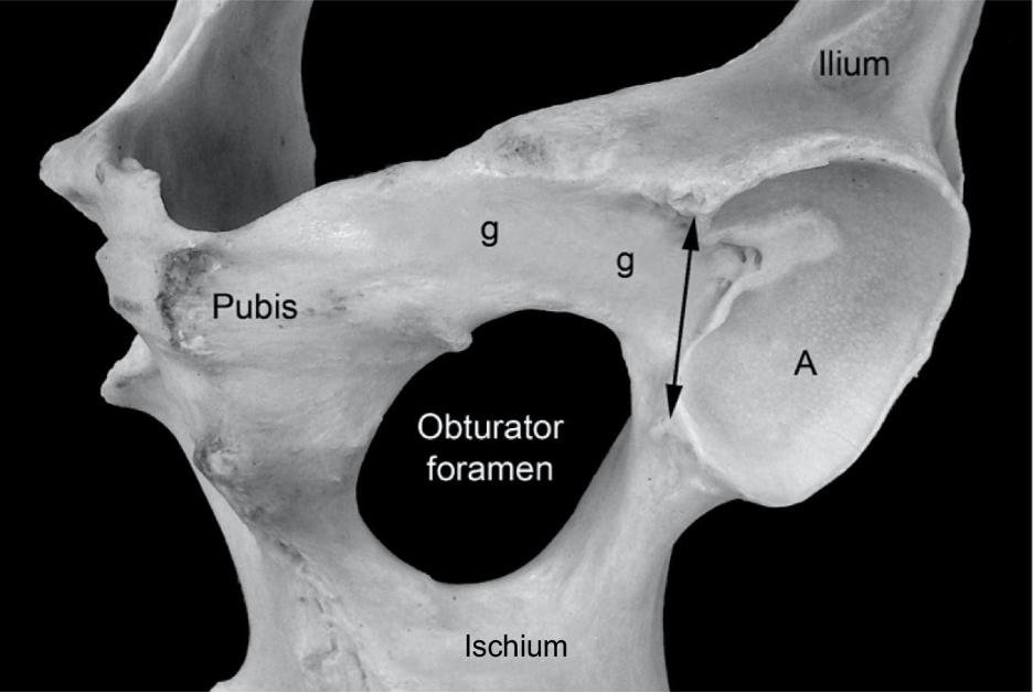
Figure 4-6 Equine left acetabulum, ventral lateral view. A, Articular surface of the acetabulum; g, shallow groove for the accessory ligament of the femoral head; double headed arrow, acetabular notch and the location of the transverse acetabular ligament.
-
- The pubis is the most cranioventral portion of the pelvis. The cranial edge of the pubis forms the pelvic brim, and is the attachment site of the prepubic tendon (i.e., tendinous insertion of the rectus abdominis mm.).
- The obturator foramen (one on each side) is formed by the ischium and pubis, and is a large foramen that is covered by the internal and external obturator mm. The obturator n. runs through the cranial portion of this foramen. (Fig. 4-6)
- The ilium forms the lateral portion of the pelvis and has an iliac body and wing. The medial projection of the wing (towards the sacrum) is the tuber sacrale, and the lateral projection is the tuber coxae (Fig. 4-3). Note that in the CALF, the wing of the ilium is oriented vertically, similar to the DOG (i.e., tuber sacrale dorsal to tuber coxae). However, in the adult RUMINANT and EQUINE the tubera coxae are displaced laterally. The wide spread of the tubera coxae parallels the development of voluminous abdominal organs and an expansive abdominal cavity in herbivores.
- The pubis is the most cranioventral portion of the pelvis. The cranial edge of the pubis forms the pelvic brim, and is the attachment site of the prepubic tendon (i.e., tendinous insertion of the rectus abdominis mm.).
-
-
- Clinical Note: The tubera coxae of CATTLE are called “hook bones” and a clamp-like device called a hook hoist is often attached here to lift recumbent cattle that are unable to stand. The wide spread of the tuber coxae make them high risk areas for fracture, especially in the case of older animals.
- The tubera coxae of the HORSE are difficult to palpate in the live animal because the rump is more muscular than that of RUMINANTS.
- The ischium is the most caudoventral portion of the pelvis and has a projection called the tuber ischii. Between the ilium and the ischium is the ischiatic spine.
- The right and left ischia unite to form an ischial arch along their caudal border. This arch can be especially deep in CATTLE.
- The lateral union of ischium and ilium form a dorsal elevation called the ischiatic spine; greater and lesser sciatic notches (that form foramen) occur cranial and caudal to this spine, respectively (Fig. 4-3).
- Clinical Note: the tubera ischii (known as ‘pin bones’ in CATTLE) form the widest point of the pelvic outlet. Dairy cows are judged on, and selected for, wide spread pin bones. (In the horse, the tubera ischii are covered by hamstring muscles.)
-
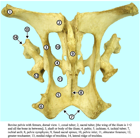
Fig. above – Bovine pelvis with femurs, dorsal view. 1, tuber coxae; 2, tuber sacrale, [the wing of the ilium is 1+2 and all the bone in between]; 3, shaft or body of the ilium; 4, pubis; 5, ischium; 6, tuber ischii; 7, ischial arch; 8, pelvic symphysis; 9, fused sacral spines; 10, pelvic inlet; 11, obturator foramen; 12, greater trochanter; 13, medial ridge of trochlea; 14, lateral ridge of trochlea. (http://vanat.cvm.umn.edu/ungDissect/Lab05/Img5-16.html)
- Sacrosciatic ligament: this broad ligament spanning from the sacrum to the ischium is slightly different in BOVINE and EQUINE. The caudal thickened edge of this ligament is homologous to the sacrotuberous ligament of the DOG. (Fig. 4-3)
- The greater sciatic foramen is formed by the bony greater sciatic notch (of the ilium) and the sacrosciatic ligament. The sciatic n. and the cranial gluteal a. exit the pelvis through this foramen in BOVINE and EQUINE.
- The lesser sciatic foramen is formed by the bony lesser sciatic notch (of the ischium) and the sacrosciatic ligament. The caudal gluteal a. exits the pelvis through this foramen in BOVINE. The tendon of the internal obturator m. exits through this foramen in EQUINE.
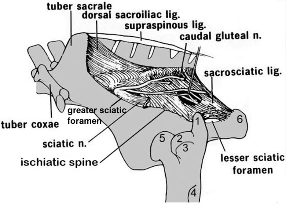
Figure 4-3 Equine pelvis and proximal femur, lateral view. 1, Caudal (middle gluteal) cusp of the greater trochanter; 2, cranial (deep gluteal) cusp of the greater trochanter; 3, facet for insertion of accessory gluteal; 4, third trochanter (superficial gluteal); 5, head of femur (in the acetabulum); 6, tuber ischii. Labeled structures: tuber sacrale, tuber coxae, ischiatic spine, greater and lesser sciatic foramen, sacrosciatic ligament, sciatic n.
- The pelvic canal, or birth canal, is where the fetus passes to exit the female reproductive tract. The pelvic inlet and outlet form the beginning and end of the birth canal, respectively. (Fig. 22.3)
- The pelvic inlet is the ‘opening’ at the cranial aspect of the pelvis, formed by pelvic bones and the sacrum. This includes the wings of the sacrum, the wings of the ilium, and the pelvic brim of the pubis. The pelvic inlet is the size-limited feature of the birth canal as it is bounded by bones and is not expandable. Females of different species (e.g., mare, cow) tend to have larger diameter pelvic inlets than males.
- The pelvic outlet is the ‘opening’ at the caudal aspect of the pelvis formed by the sacrum and caudal vertebrae, sacrosciatic ligament (sacrotuberous part) and the ischial arch (the arch between the tubera ischii). Note that the ischial arch is especially deep in cattle. This means that the pelvic outlet has a ligamentous border and has some ability to stretch.
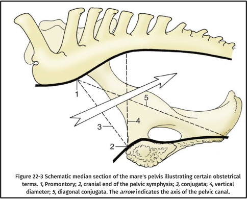
FIG. 22.3 Schematic median section of the mare’s pelvis illustrating certain obstetric terms. 1, Promontory; 2, cranial end of the pelvic symphysis; 3, conjugata; 4, vertical diameter; 5, diagonal conjugata. The arrow indicates the axis of the pelvic canal. (TVA)
Femur (and Patella) (Figs 4-3, 5B-1 through 5B-5)
- Proximally, and on the lateral side of the femur (opposite the head of the femur), there is a greater trochanter with cranial and caudal parts. Slightly distal and medial, there is a lesser trochanter. (Figs. 5B-1 and 5B-5) Several of the gluteal mm. attach to the trochanters of the femur.
- The trochanteric bursa is situated deep to the tendon of the accessory gluteal m. as the tendon crosses over the cranial part of the greater trochanter. (Fig. 3-3)
- Clinical Note: The sciatic nerve exits near the greater sciatic foramen (formed by the sacrosciatic ligament), then passes caudally around the hip joint (medial to the greater trochanter) and courses distally in the pelvic limb. The nerve can be compressed against the caudal surface of the upper femur in downed animals (especially cows).
- The third trochanter (Figs. 4-3, and 5B-2) is well developed on the lateral aspect of the femur in the HORSE, but absent in the OX.
- The head of the femur has a medially located triangular fossa, called the fovea capitis, for attachment of the ligament(s) of the femoral head (Figs. 5B-1 and 5B-2).
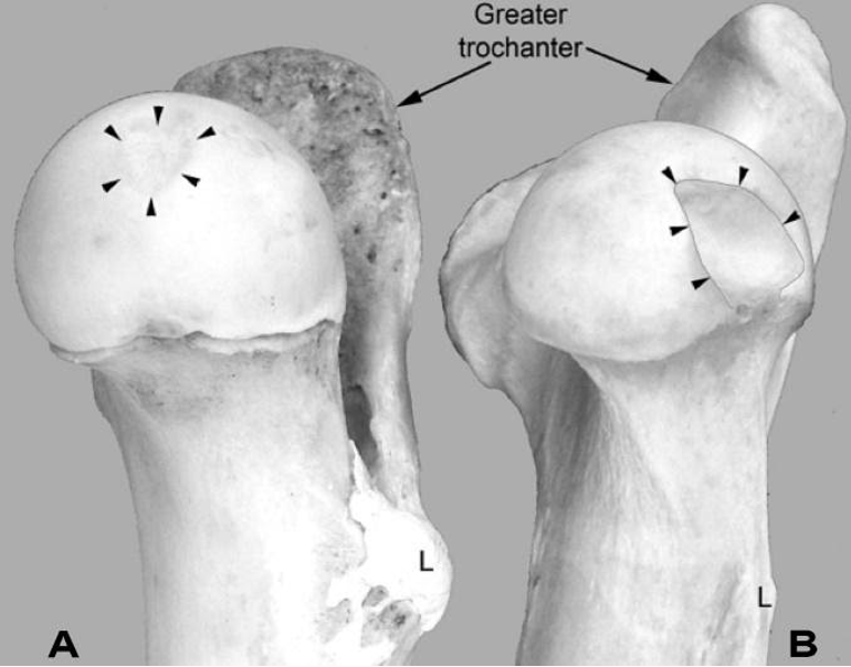
Figure 5B-1 Comparative right femoral head, medial views. A, ox. B, horse. The arrowheads show the location of the fovea capitis where the ligament(s) of the femoral head are attached. The fovea capitis is small and shallow in the ox but large and deep in the horse to accommodate the two ligaments associated with the femoral head in the horse. Two arrows, greater trochanters; L, lesser trochanter, a bony prominence on the medial side of the femur where the iliopsoas muscle inserts. (This bovine femur is immature and hence the physis of the femoral head is present.)
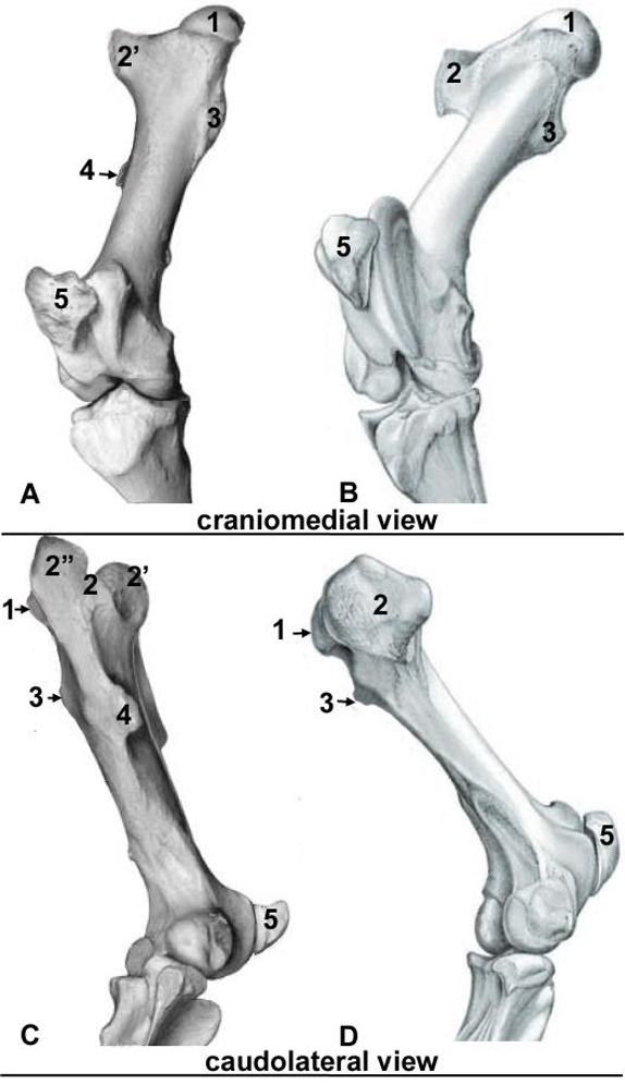
Figure 5B-2 Right Femur and proximal tibia of the (A, C) Equine; (B, D) bovine. 1, head of femur; 2, greater trochanter; 2′ cranial and 2″ caudal parts of the equine greater trochanter; 3, lesser trochanter; 4, third trochanter (equine only); 5, patella. (Modified from: Budras)
- On the distal end of the femur, note the femoral trochlea, which has a large medial ridge and a smaller lateral ridge (Fig. 5B-3/1,2). The femoral trochlea is a common site of osteochondrosis (i.e., joint lesions when cartilage becomes separated from the underlying bone, Supplemental Clinical LAS Osteochondrosis).
-
- The patella is a sesamoid bone which articulates with the femoral trochlea.
- Patellar ligaments loop around the proximal ‘lip’ of the large medial ridge and help lock and stabilize the stifle joint which is key to the EQUINE hind limb stay apparatus. The medial, middle (aka intermediate), and lateral patellar ligaments help keep the patella on track and attach to the tibial tuberosity.
- Clinical Note: Upward fixation of the patella or patellar fixation is a clinical condition of HORSES, and some breeds of CATTLE, in which the stifle cannot be unlocked. As a result, a characteristic stiff-legged gait is seen. (Supplemental Clinical LAS Gait abnormalities, Upward fixation of the patella)
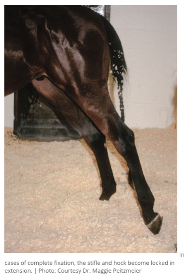
Fig above – Case of complete fixation of the patella. The stifle and hock become locked in extension. Photo: Dr. Maggie Peitzmeier (The Horse: Upward fixation of the patella in horses)
- The distal femur has medial and lateral condyles that articulate with the meniscal cartilages.
- Between the femoral condyles is the intercondylar fossa where the cruciate ligaments (cranial and caudal) attach.
- On the distal part of the lateral condyle of the femur, note the extensor fossa where the long digital extensor and fibularis (peroneus) tertius mm. arise. (Fig. 5B-4)
- The supracondylar fossa is on the caudolateral aspect where the superficial digital flexor m. arises. This fossa is just proximal to the lateral femoral condyle. (Fig. 5B-4)
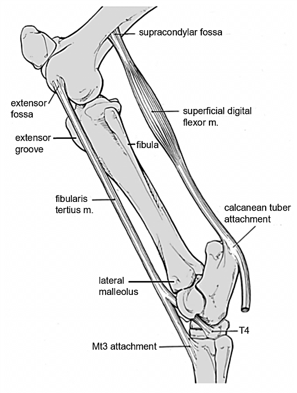
Figure 5B-4 Equine left hind limb, lateral view. Labeled structures: supracondylar fossa, extensor fossa, extensor groove, fibula, lateral malleolus, calcanean tuber(osity); superficial digital flexor m., fibularis tertius m.
Tibia and Fibula (Figures 5B-3 through 5B-5)
- The tibial tuberosity, on the proximal and cranial aspect of the tibia, is where the patellar ligaments attach; caudal to this is the intercondylar eminence on the proximal surface of the tibia between the two relatively flat condyles. (Fig. 5B-3,5)
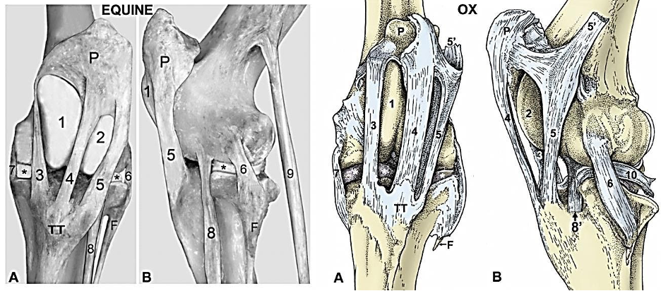
Figure 5B-3 stifle equine (ligamentous preparation- similar image to Figure 5-1) and ox (drawing modified from TVA figure 31-4) A, cranial views; B, lateral views. 1, Medial ridge of femoral trochlea; 2, lateral ridge of femoral trochlea; 3, medial patellar ligament; 4, middle patellar ligament; 5, lateral patellar ligament; 5’, attachment of biceps femoris m. (shown in ox); 6, lateral collateral ligament; 7, medial collateral ligament; 8, fibularis (peroneus) tertius tendon; 8’ combined tendon of long digital extensor and fibularis (peroneus) tertius tendon (ox only); 9, superficial digital flexor tendon (shown in equine, is ~ all tendinous); 10, tendon of popliteus m. (shown in ox); F, fibula; P, patella; TT, tibial tuberosity; *, menisci.
- Lateral to the tibial tuberosity, there is the extensor groove through which the tendons of origin of the long digital extensor and fibularis (peroneus) tertius mm. course. (Fig. 5B-4)
- In the HORSE, on the distal end of the tibia, note the medial and lateral malleolus.
- The medial malleolus is rounded and forms a prominent distal tuberosity that is a significant landmark on the live HORSE and in tarsal radiographs. (Fig. 5B-5)
- The lateral malleolus represents fusion of the distal part of the fibula with the tibia.
- The proximal and/or distal portions of the fibula are reduced on the lateral aspect of the tibia.
- In the OX, the lateral malleolus (distal part of the fibula) is a separate bone. (Fig. 5B-6)
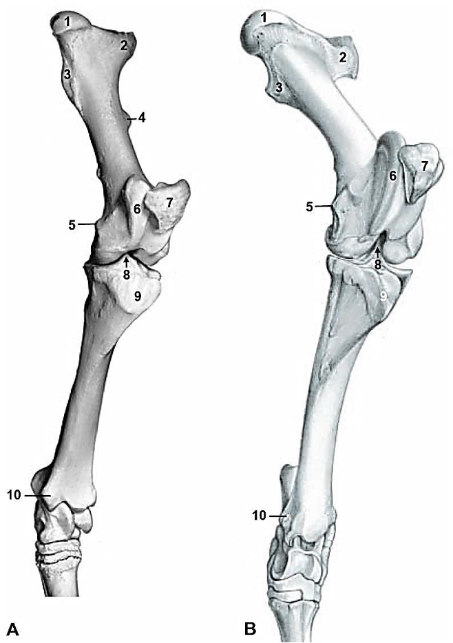
Figure 5B-5 Upper left hind limb of the (A) horse and (B) ox, craniomedial view. 1, femoral head; 2, greater trochanter; 3, lesser trochanter; 4, third trochanter; 5, medial epicondyle; 6, medial trochlear ridge; 7, patella; 8, intercondylar eminence of tibia; 9, tibial tuberosity; 10, medial malleolus.
Tarsus (Figs. 2.60, 5B-6, 5B-7, 5B-8)
- Remember that from (and including) the tarsus distally, the naming changes to plantar surface instead of caudal.
Tarsal bones:
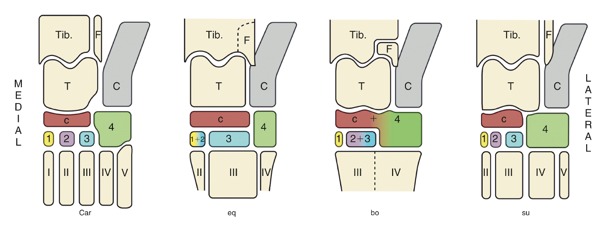
FIG. 2.60 The bones of the tarsal skeleton in the carnivores (Car), horse (eq), cattle (bo), and pig (su) schematic. Roman numerals identify the metatarsal bones, arabic numerals the distal tarsal bones. C, calcaneus; c, central tarsal bone; F, fibula; T, talus; Tib., tibia. (TVA)
HORSE
- Calcaneus and talus: (Figs. 2.60, 5B-6, 5B-7, 5B-8)
- The talus directly articulates with the tibia.
- On the talus, there is a large trochlea with medial and lateral ridges.
- On the calcaneus, note the large calcanean tuberosity (lateral projection), which serves as the place of insertion for the gastrocnemius m., and to a lesser extent, the superficial digital flexor m. The tendon of the superficial digital flexor m. both slides over and inserts on this tuberosity as a part of the common calcanean tendon. A subtendinous calcaneal bursa sits between the calcanean tendon and the calcanean tuberosity.
- The wide distal and medial part of the calcaneus is called the sustentaculum tali. (Fig. 5B-8)
- Tarsal bones: (Figs. 2.60, 5B-6, 5B-7, 5B-8)
-
- The large central (Tc) and third tarsal bones (T3) are the main weight bearing distal tarsal bones.
- On the medial aspect, the first (T1) and second tarsal (T2) bones are most often fused to form one bone, but occasionally there are two separate bones.
- The fourth tarsal (T4) bone sits between the calcaneus and Mt4.
- There is a small vascular canal between the central, third, and fourth tarsal bones. The dorsal pedal a., which passes over the dorsal aspect of the tarsus, gives off the perforating tarsal a. that passes through this vascular canal. (Fig. 5B-7) The vascular canal can be seen on radiographs.
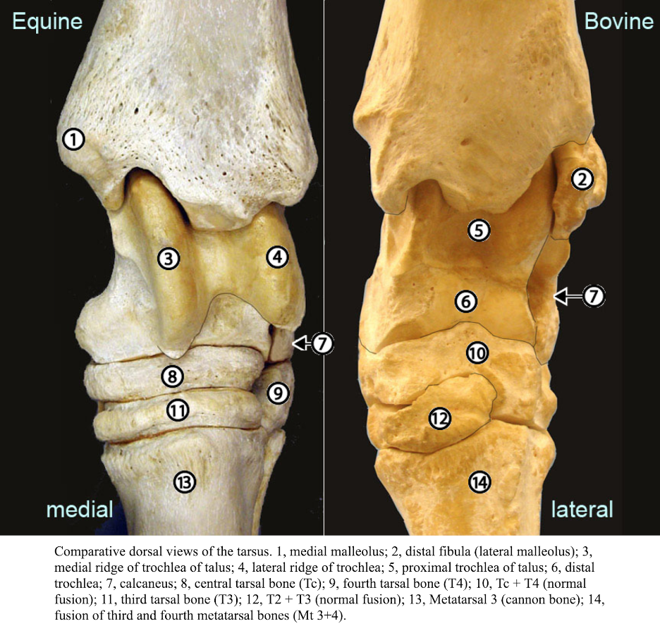
Fig. 5B-6 Comparative dorsal views of the left tarsus. 1, medial malleolus; 2, distal fibula (lateral malleolus); 3, medial ridge of trochlea of talus; 4, lateral ridge of trochlea; 5, proximal trochlea of talus; 6, distal trochlea; 7, calcaneus; 8, central tarsal bone (Tc); 9, fourth tarsal bone (T4); 10, Tc + T4 (normal fusion); 11, third tarsal bone (T3); 12, T2 + T3 (normal fusion); 13, Metatarsal 3 (cannon bone); 14, fusion of third and fourth metatarsal bones (Mt 3+4). (http://vanat.cvm.umn.edu/ungDissect/Lab07/Img7-1.html)

Figure 5B-8 Equine right tarsus, medial view. Labeled: calcaneus, talus, S – sustentaculum tali, T4, T1, T2, T3 Tc, Mt4, Mt2, Mt3. Commonly T1 and T2 are fused to form a T1+2 bone. Note the fusion of the medial splint bone (Mt2) to the cannon bone (Mt3).
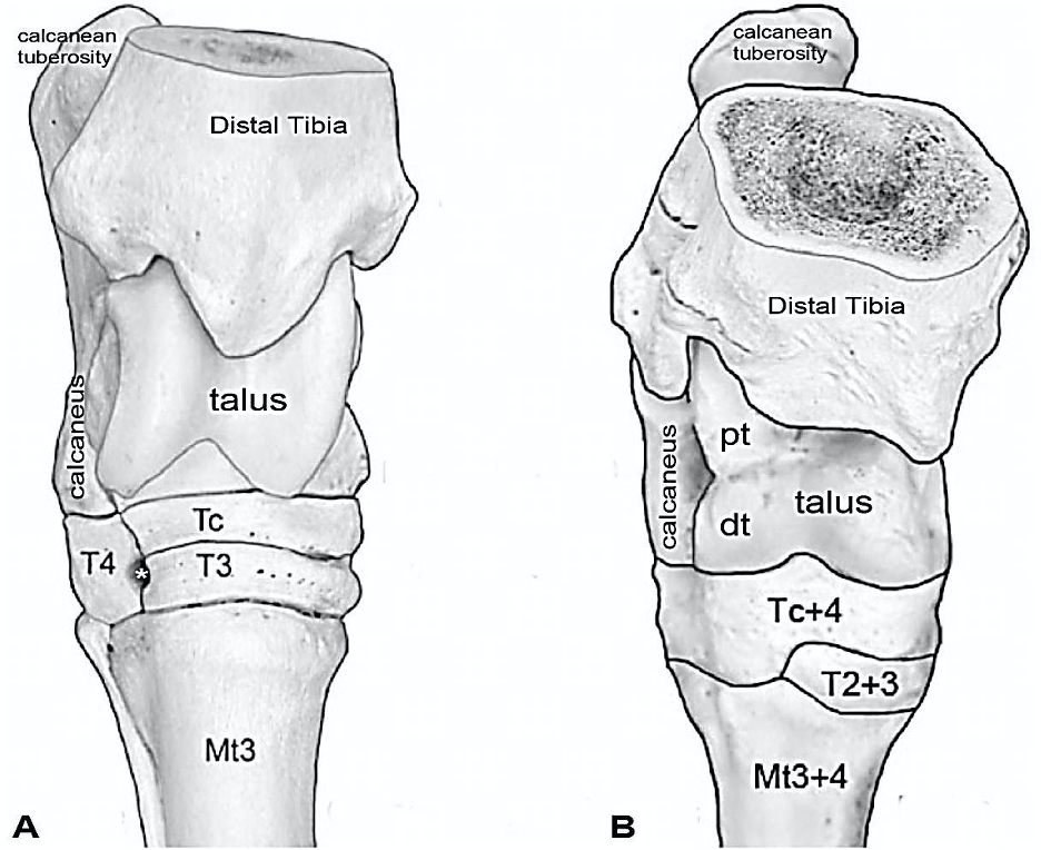
Figure 5B-7 Comparative right tarsal anatomy, cranial views. A. Equine tarsus: * vascular canal. Labeled: tibia, calcanean tuberosity, talus, calcaneus, Tc, T3, T4, Mt3. B. Bovine tarsus: talus: pt, proximal trochlea; dt, distal trochlea. Labeled: tibia, calcanean tuberosity, talus, calcaneus, Tc+4, T2+3, Mt3+4.
OX
- Calcaneus and talus: (Figs. 2.60, 5B-6, 5B-7)
-
- The talus has two trochleae: a proximal trochlea, which corresponds to the only trochlea of the HORSE, and a distal trochlea (Fig. 5B-7/B, pt, dt).
- On the calcaneus, there is a calcanean tuberosity (projecting caudally and lateral) and the sustentaculum tali (medially).
- Clinical Note: Both RUMINANTS and CAMELIDS have two trochleae, which facilitate greater tarsal flexion (i.e., greater range of motion).
- Tarsal bones: (Figs. 2.60, 5B-6, 5B-7)
-
- There is a small first tarsal bone (T1) medially.
- The second and third tarsal bones (T2+3) are combined.
- The central and fourth tarsal bones (Tc+4) are fused, which are the main weight bearing distal tarsal bones.
Metatarsus (Figs. 5B-6 through 5B-8)
HORSE
- The equine metatarsus consists of a large 3rd metatarsal bone (Mt 3) and two smaller metatarsal bones, Mt 2 (2nd metatarsal) and Mt 4 (4th metatarsal).
-
- The Mt 3 has a thick cortex and looks like the barrel of a “cannon” in cross section, hence the common name cannon bone. The cannon bone of the hind limb is more round in cross section, while the forelimb is more oval. The cannon bone of the hind limb is also about one-third longer than that of the forelimb.
-
- The forelimb Mc 3 and hind limb Mt 3 have similar features. The metatarsal tuberosity is on the proximodorsal surface, and the distal end bears a condyle and a prominent sagittal ridge that articulates with a complementary sagittal groove of the proximal phalanx (P1).
- Similar to the forelimb, Mt 2 and Mt 4 are called medial and lateral splint bones, respectively. They are fused tightly to the cannon bone for most of their length by a short interosseous ligament. Don’t confuse the ligament with the interosseous tendon!
- Clinical Note: Splints or splint lesions refers to inflammation and then ossification of the interosseous ligament. The interosseous ligament holds the splint bones to the cannon bone. Shearing forces between the splint bones and the cannon bone results in the initial inflammatory response.
- Clinical Note: The sagittal groove may be the point of origin for a longitudinal/sagittal fracture of P1, which is called a screwdriver fracture. The sagittal ridge of Mt3 is pushed into the sagittal groove of the P1/long pastern bone (see Fig. 7 in A2.1). P1 is also a common site for comminuted fractures (i.e., bone is broken into two or more pieces).
OX
- The BOVINE metatarsus consists of two fused metatarsal bones, the 3rd and 4th metatarsals (Mt 3 and Mt4), of equal size that are fused throughout their entire length, except at the distal end where they separate into two condyles. This fused metatarsal bone in the bovine is also referred to as the cannon bone.
-
- Note that a small, short metatarsal bone is located laterally (Mt 5), on the proximal end of the cannon bone.
Digit(s) – Application 2.1, 2.3
The digit(s) (phalanges, sesamoids and hoof/claw) components of the hind limb are similar to those in the forelimb, replacing palmar terms with plantar. EQUINE bear weight on one digit (3rd) in their forelimbs and hind limbs, while BOVINE bear weight on two digits (3rd and 4th) in their forelimbs and hind limbs. The medial (3rd) fore digit and lateral (4th) hind digit are the most common sites for bovine lameness.
- Proximal sesamoid bones (2 in each equine foot, 4 in each bovine foot).
- Proximal phalanges/long pasterns/P1 (1 in each equine foot, 2 in each bovine foot).
- Middle phalanges/short pasterns/P2 (1 in each equine foot, 2 in each bovine foot).
- Distal phalanges/coffin bones/P3 (1 in each equine foot, 2 in each bovine foot called pedal bones).
- Navicular bones/distal sesamoid bones (1 in each equine foot, 2 in each bovine foot).
Objective
A3.2 Identify and describe the joints, joint angles, joint actions, and muscle groups of the pelvic limb.
Joints of the pelvic (hind) limb
- Clinical Notes: joint pouches are extensions of the synovial capsule and cavity past the joint surface. In more mobile joints these pouches can be more expansive/extensive. When you “tap a joint” (i.e., aspirate) you will not always remove fluid right at the bony union of a joint, but rather near the extension of these synovial cavities. These pouches may also bulge outwards and can visibly been seen due to inflammation. Additionally, anesthesia or medications can be injected in and near the joint capsules. (more Supplemental Clinical in LAS Nerve and Joint Blocks)
- Sacroiliac (SI) joint: there is very little movement at this joint as there is a firm attachment between the pelvic limb and sacrum. During standing, the joint transfers the weight of the trunk to the hind limbs. During motion, the joint transfers the thrust of the hind limbs to the trunk.
- This joint is formed by union of the wings of the sacrum and part of the wings of the ilium on each side.
- Clinical Notes: dislocation of the sacroiliac joint can be diagnosed by observing unilateral elevation of the tuber sacrale on the side of dislocation.
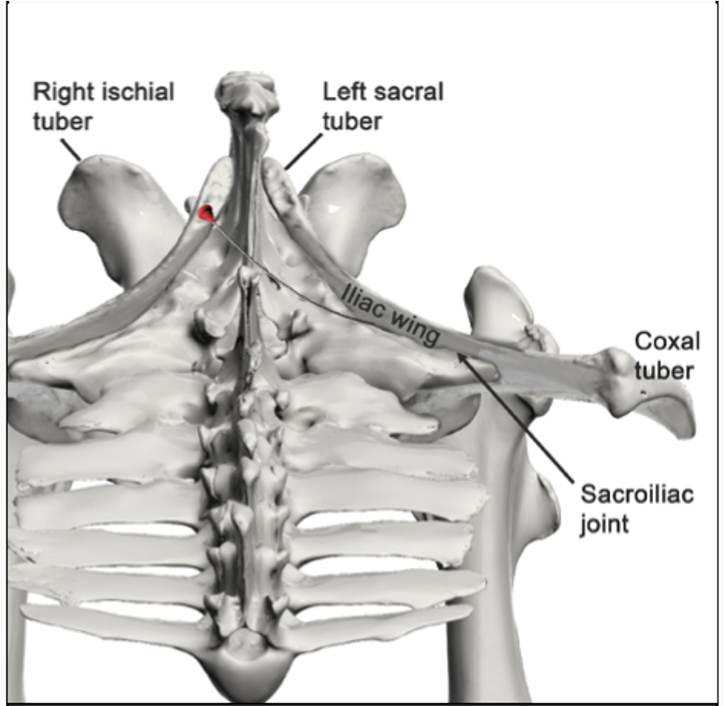
Fig 2-3: Left equine sacroiliac (SI) joint. Craniodorsal view. The periarticular craniomedial approach is intended to place the needle tip near the medial periarticular region of the SI joint. A long needle is advanced from the dorsal midline with the clinician standing on the opposite side of the joint to be treated. See Moyer et al (2011) for details. (from Equine Anatomy Guide: The Forelimb; Mansour, Steiss, Wilhite)
- Hip (aka coxofemoral) joint: is formed by the articulation of the head of the femur with the acetabulum. The stability of the hip joint is due to the depth of the acetabulum and its encircling rim of cartilage.
- Accessory ligament of the femoral head: this is unique to the HORSE and DONKEY. It arises from the prepubic tendon and reaches the joint by passing through the acetabular notch and inserts on the head of the femur (Fig. 4-7).
- Ligament of the femoral head (eq and bov): passes from the acetabulum to the fovea capitis (a divot in the head of the femur) and is relatively short. (Fig. 5B-1)
- There is also a transverse acetabular ligament (eq and bov) that spans the acetabular notch.
- Clinical Notes: subluxation or luxation (i.e., dislocation) of the hip joint is more common in BOVINE, since equine have two ligaments holding the femoral head in place. Equine may also have a reduced ability to sidekick due to this extra ligament.
Figure 4-6 Equine left acetabulum, ventral lateral view. A, Articular surface of the acetabulum (acetabular fossa); g, shallow groove for the accessory ligament of the femoral head; double headed arrow, acetabular notch and the location of the transverse acetabular ligament.
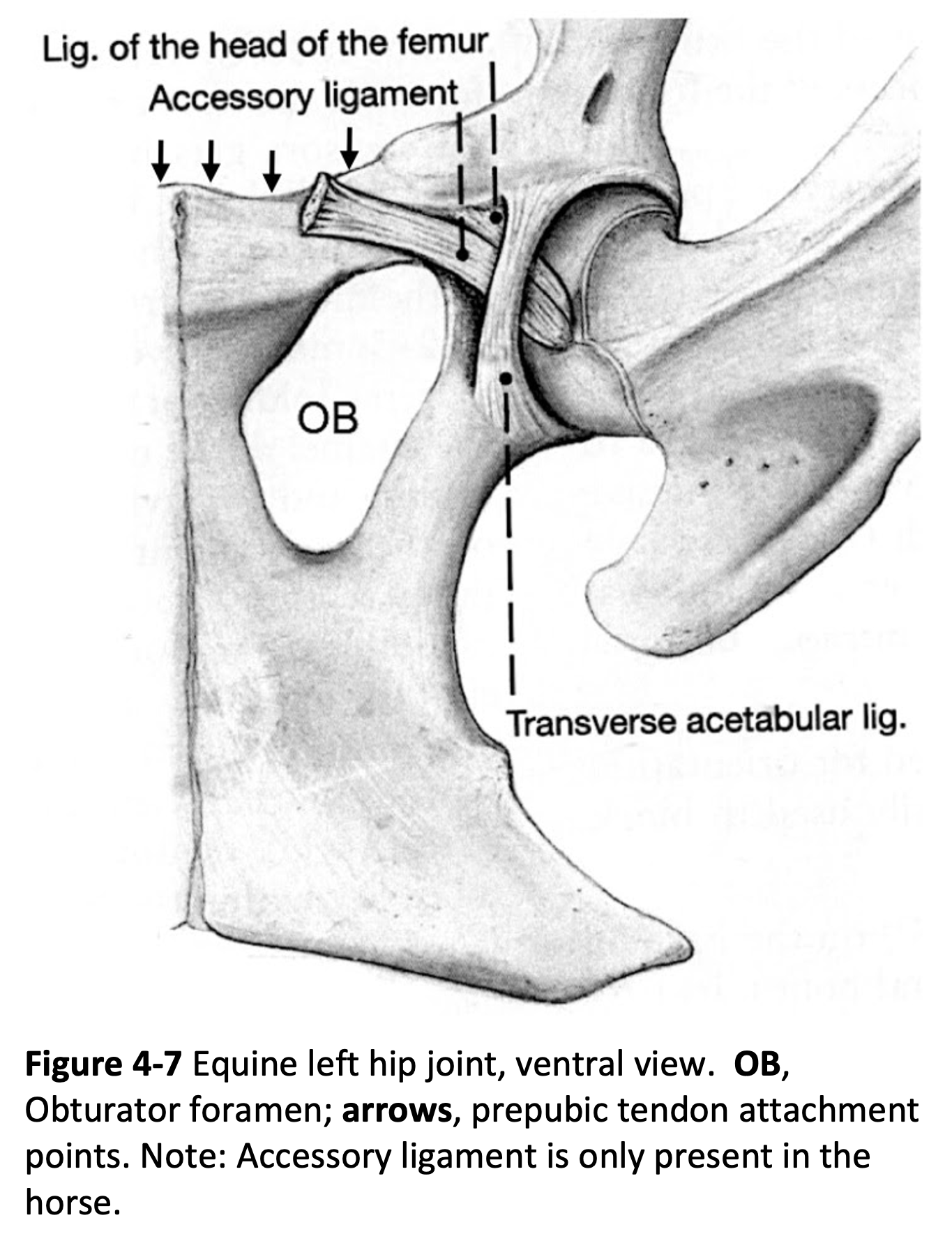
Fig. 4-7 Equine left hip joint, ventral view. OB, obturator foramen; arrows, prepubic tendon attachment points. Note: accessory ligament is only present in the horse. Labeled structures: ligament of the head of the femur, transverse acetabular ligament.

Fig 2-4: Left equine hip joint capsule (in green). Caudolateral approach. Using a lateral approach, the needle is inserted between the cranial and caudal parts of the greater trochanter, approximately 1/2 inch above the level of the cranial part of the greater trochanter. (from Equine Anatomy Guide: The Forelimb; Mansour, Steiss, Wilhite)
- Stifle joint: is formed by the articulation of the femoral condyles with the condyles of the tibia.
- There are 3 patellar ligaments (medial, middle/intermediate and lateral) that help in supporting the joint and each attaches to the tibial tuberosity.
- The stifle joint is also stabilized by medial and lateral collateral ligaments. (Fig. 5B-3)
-
The cranial and caudal cruciate ligaments pass between the intercondylar fossa of the femur and the intercondylar eminence of the tibia. They are named for their attachment on the tibia. (Fig 2-11) (Supplemental Clinical LAS Tendon and ligament issues, Cruciate injuries)
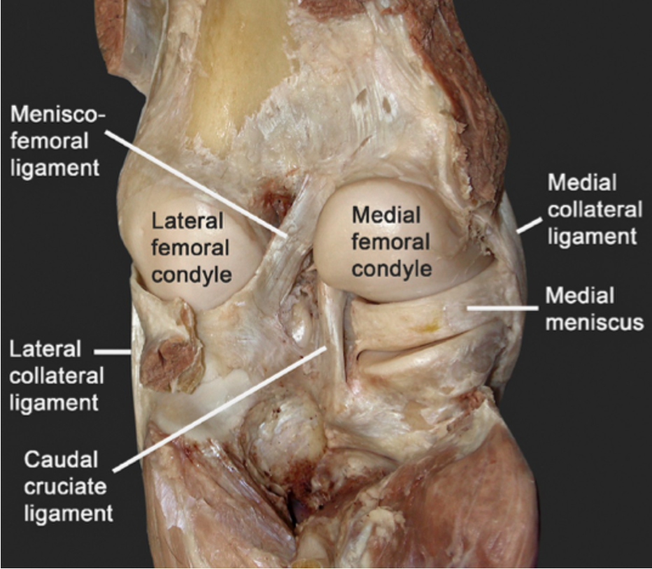
Fig 2-11: Left equine hind limb. Caudal view, deep dissection showing the ligaments of the stifle joint: lateral collateral ligament, medial collateral ligament, caudal cruciate ligament, medial meniscus. (from Equine Anatomy Guide: The Forelimb; Mansour, Steiss, Wilhite)
-
- Patellar locking mechanism: The trochlea of the femur, especially in the HORSE, has a large medial ridge and a smaller lateral ridge. The medial and middle patellar ligaments, together with the patella and para-patellar fibrocartilage, form a loop around the hook-like medial trochlear ridge. This mechanism locks the stifle and allows the horse to bear weight without muscular effort. Although the patellar ligaments are similarly divided in the OX, they do not have a well-developed patellar locking mechanism.
- There are three associated joint compartments or sacs of the stifle: medial femorotibial, femoropatellar, and lateral femorotibial. The femoropatellar and medial femoral tibial sacs usually communicate. The lateral femorotibial sac communicates with the femoropatellar sac in one out of four equine stifle joints.

Fig 2-6: Left equine hind limb. Stifle region. Cranial view of the femoropatellar joint capsule (dark green), medial femorotibial joint capsule (light green) and lateral femorotibial joint capsule (blue). Note the communication between the femoropatellar and medial femorotibial joint capsules. Labeled structures: tendons of long digital extensor and fibularis (peroneus) tertius mm., patellar ligaments (lateral, middle/intermediate, and medial), fibula, tibia, femur, patella. (from Equine Anatomy Guide: The Forelimb; Mansour, Steiss, Wilhite)
- Tarsal/hock joint: located between the distal tibia and the 2nd, 3rd, and 4th metatarsals distally. (Fig. 2.60 below to see equine and bovine) The tarsus includes four joints (listed from proximal to distal).
- Tibiotarsal (aka tarsocrural, talocrural): is the largest of the four joints and has the largest range of motion, while the other three tarsal joints are relatively low motion (proximal and distal intertarsal, tarsometatarsal).
- Clinical Notes: A common site of osteochondrosis in the horse is the distal intermediate ridge of the tibia (often called DIRT lesions).
- There are four tarsocrural joint pouches, two dorsal and two plantar (dorsomedial, dorsolateral, medioplantar, lateroplantar) and all communicate with each other.
- Proximal intertarsal (PIT): is the articulation between the talus and calcaneus proximally and the articulation between the talus, Tc, and T4 distally.
- Distal intertarsal (DIT): is the articulation of Tc with T1+2 and T3.
- Tarsometatarsal: is the articulation of T1+2, T3, and T4 with all three metatarsal bones.
- Clinical Notes: There is a single fibrous joint capsule that wraps around the four synovial compartments. The tibiotarsal and proximal intertarsal joints communicate. Each of the four synovial compartments can be accessed for injection or aspiration.
- Clinical Notes: “spavin” is a generic term for equine hock/tarsal joint pathology (i.e., synovitis/capsulitis, arthritis; see more in the last objective).
- Tibiotarsal (aka tarsocrural, talocrural): is the largest of the four joints and has the largest range of motion, while the other three tarsal joints are relatively low motion (proximal and distal intertarsal, tarsometatarsal).
FIG. 2.60 The bones of the tarsal skeleton in the carnivores (Car), horse (eq), cattle (bo), and pig (su) schematic. Roman numerals identify the metatarsal bones, Arabic numerals the distal tarsal bones. C, calcaneus; c, central tarsal bone; F, fibula; T, talus; Tib., tibia. (TVA)
- Fetlock/metatarsophalangeal: formed by articulations between the Mt3 (cannon) bone, P1, and the paired proximal sesamoid bones.
- Pastern/proximal interphalangeal/PIP: formed between P1 and P2 and is a low motion joint.
- Coffin/distal interphalangeal/DIP/pedal: formed between P2 and P3.
Clinical issues at each joint are listed in the table below:
| Joint | Feature | Clinical |
| Sacroiliac jt. | Forms firm attachment between hind limb and trunk. | Dislocation possible |
| Hip jt. | Deep socket; accessory ligament (equine) | Bovine – can see luxation; Equine – not likely to luxate due to two ligaments holding femoral head within socket |
| Stifle jt. | Equine, bovine: 3 patellar ligaments, three joint compartments with variable connections; Equine: patellar lock mechanism | Patellar fixation |
| Tarsus | Four levels of joints; proximal has largest degree of motion | Equine tarsal joint problems are common |
| Digit(s) | Similar to forelimb | Similar clinical problems to forelimb. |
Extrinsic Muscle of the hind limb:
- The iliopsoas m., similar to carnivores, consists of the psoas major and iliacus mm.. It originates on the lumbar vertebrae and iliac crest and inserts on the lesser trochanter of the femur. It can assist in flexion at the hip (aka protraction of the hind limb).
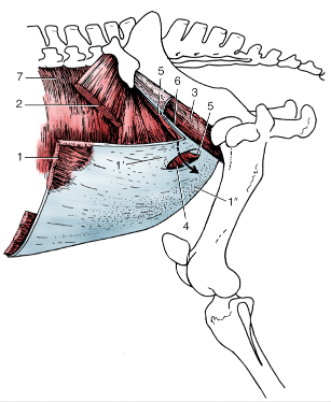
FIG. 21.4 The muscles of the inguinal region. The arrow passes through the inguinal canal. 1, External abdominal oblique; 1′ and 1″, pelvic and abdominal tendons of external oblique aponeurosis, respectively; 2, internal abdominal oblique; 3, iliopsoas partly enclosed by iliac fascia; 4, superficial inguinal ring; 5, cranial border of deep inguinal ring; 6, attachment of pelvic tendon of external oblique aponeurosis on iliopsoas and sartorius (“inguinal ligament”); 7, transversus abdominis. (TVA)
Intrinsic Muscles, Joint Actions, and Muscle Groups (with Innervation) of the hind limb:
- Extensors of Hip: biceps femoris, semitendinosus, semimembranosus, middle gluteal mm. (innervation – sciatic/tibial n., or cranial gluteal n.)
- Biceps femoris m. (eq) acts to extend the hip; when the limb is weight bearing, it can extend the stifle; when the hoof is raised, it can flex the stifle; via its connection to the common calcanean tendon it can extend the hock/tarsus.
- Semitendinosus m. acts to extend the hip; when the limb is weight bearing, it can extend the stifle; when the hoof is raised, it can flex the stifle; via its connection to the common calcanean tendon it can extend the hock/tarsus.
- Semimembranosus m. acts to extend the hip; when the limb is weight bearing, it can extend the stifle; when the hoof is raised, it can flex the stifle.
- Middle gluteal m. acts to extend the hip and can also abduct the thigh.
- Accessory gluteal m. is considered to be a deep portion of the middle gluteal m. The accessory gluteal m. has a tendon that crosses over the cranial part of the greater trochanter of the femur and attaches between the greater trochanter and lesser trochanter. A bursa is situated deep to the tendon and is called the trochanteric bursa. (Fig. 3-3)
- Clinical Note: the trochanteric bursa can become inflamed. Standardbred horses sometimes suffer from a unilateral inflammation of this bursa called trochanteric bursitis (whirl/whorl bone disease). Horses with this bursitis often stand with the affected limb abducted, and adopt a gait where the limb swings in an arc. This type of inflammation may also be seen in cattle.

Fig 3-3: Equine left hind limb. Lateral view of the hip region. The trochanteric bursa is injected by inserting a needle deep to the tendon of the accessory gluteal m and superficial to the cranial part of the greater trochanter. (from Equine Anatomy Guide: The Forelimb; Mansour, Steiss, Wilhite)
- Flexors of Hip: tensor fasciae latae, superficial gluteal, rectus femoris mm. (innervation – gluteal nn., femoral, or saphenous nn.) (Figs. 24.2, 31.2)
- Tensor fasciae latae m. acts to flex the hip and can also advance the hind limb. The fascia lata is a wide aponeurosis that attaches to the tensor fasciae latae m. cranially, runs along the cranial and lateral edge of the thigh, and blends into the fascia near the stifle and the crus.
- Superficial gluteal m. (eq) acts to flex the hip and can also abduct the thigh.
- Rectus femoris m. can act to flex the hip due to its origin on the ilium and its insertion on the patella (through the quadriceps m. tendon).
- The sartorius and pectineus mm. may also act to flex the hip.
- Deep gluteal m. acts to abduct the thigh.
- Gluteobiceps m. (bov) is the combination of the superficial gluteal m. and the biceps femoris m. in BOVINE.
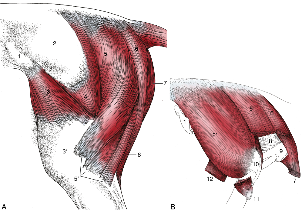
FIG. 24.2 (A) Equine muscles of the croup and thigh, lateral view. (B) Croup muscles, resected to expose the ischial tuber; lateral view. 1, tuber coxae; 2, deep gluteal fascia; 2′, middle gluteal; 3, tensor fasciae latae; 3′, fascia lata; 4, superficial gluteal; 5, vertebral head of biceps femoris; 5′, the three distal divisions of the biceps femoris; 6, semitendinosus; 7, semimembranosus; 8, sacrosciatic ligament; 9, tuber ischii; 10, caudal part of greater trochanter; 11, third trochanter; 12, stump of rectus femoris. (TVA)
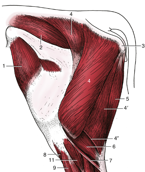
FIG. 31.2 Muscles of the bovine left hindlimb, lateral view. 1, Tensor fasciae latae; 2, middle gluteal; 3, tuber ischii; 4, 4′, and 4″, gluteobiceps, transected at 4″; 5, semitendinosus; 6, lateral head of gastrocnemius; 7, rudimentary soleus; 8, tibialis cranialis; 9, fibularis (peroneus) tertius; 11, fibularis (peroneus) longus. TVA
- Muscles of the rump (aka croup) (Figs. 24.2, 31.2, see images below)
- In EQUINE, normally the rump is rounded and smooth due to the middle gluteal m., which is covered by the superficial gluteal m. and its broad fascia. The middle gluteal m. is a powerful extensor of the hip and it propels the body forward during running.
- In the caudal rump, the semitendinosus and semimembranosus mm. travel over the tuber ischii and attach more dorsally near the vertebrae. From a caudal view of the rump, the semitendinosus is lateral while the semimembranosus is medial.
- In BOVINE, there is less muscle filling the rump, leaving dips in along the ilium of the pelvic bone. This main muscle in this region is the gluteobiceps m. (superficial gluteal + biceps femoris m.).
- In the caudal rump, the semitendinosus and semimembranosus mm. attach to the tuber ischii, which reduces the amount of muscle filling in the region between the ischium and vertebrae.
- In EQUINE, normally the rump is rounded and smooth due to the middle gluteal m., which is covered by the superficial gluteal m. and its broad fascia. The middle gluteal m. is a powerful extensor of the hip and it propels the body forward during running.
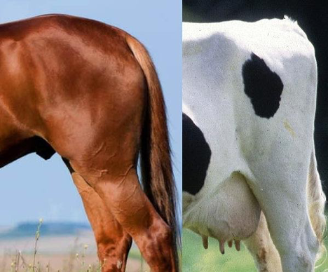
Croup region in equine (left) and bovine (right).
- Internal obturator m. tendon (eq) – this tendon emerges from the lesser sciatic foramen. (innervation – sciatic n.)
- Extensors of Stifle: quadriceps femoris m. group (rectus femoris, vastus lateralis, vastus medialis, vastus intermedius mm.) (innervation – femoral n.)
- Quadriceps femoris m. group acts to extend the stifle.
- Flexors of Stifle: biceps femoris, semitendinosus, semimembranosus mm. (innervation – sciatic/tibial n.) (Figs. 24.2, 31.2)
- When the hoof is raised, these three muscles can flex the stifle.
- Adductors of the Thigh: gracilis, adductor, pectineus mm. (innervation – obturator n.)
- Extensors of Tarsus: gastrocnemius (medial and lateral heads) m. (innervation – tibial n.)
- Gastrocnemius mm. act to extend the tarsus (aka hock) via its attachment to the calcanean tuberosity via the common calcanean tendon.
- Clinical Notes: rupture of the gastrocnemius m. occurs when the stifle is forced into overextension while the tarsus is flexed. Foals may develop gastrocnemius rupture during dystocia and with assisted delivery, while adult horses may develop gastrocnemius rupture during exercise like strenuous stopping. Clinical signs includes a dropped tarsus with extended stifle and inability to straighten the hind limb. (Supplemental Clinical LAS Gait abnormalities, Gastrocnemius rupture)
- Flexors of Tarsus: cranial tibial, fibularis (aka peroneus) tertius, fibularis longus (bov) mm. (innervation – common fibular nn.)
- Cranial tibial m. acts to flex the tarsus (aka hock).
- The cunean tendon (of cranial tibial m.) (eq) is the medial insertion of the cranial tibial m. tendon on the tarsal bones (first and second).
- Fibularis (aka peroneus) tertius m. acts to flex the tarsus, and to link the hock/tarsus and stifle joints as a part of the reciprocal apparatus.
- Fibularis longus m. (bov) originates near the lateral aspect of the stifle and inserts near the plantar aspect of the hock/tarsus. It is superficial to the lateral digital extensor m.
- Cranial tibial m. acts to flex the tarsus (aka hock).
- Extensors of the Digits: long digital extensor (medial and lateral heads (bov)), lateral digital extensor mm. (innervation – common fibular n.)
- The extensor retinacula are three bands of fascia extending across the dorsal aspect of the tarsus that bind down the long digital extensor tendon that crosses the dorsal aspect of the tarsus.
- Flexors of the Digits: superficial digital flexor (SDF), deep digital flexor (medial and lateral heads) mm. (innervation – tibial n.)
- The flexor retinaculum is a thick band of fascia extending across the medial aspect of the tarsus (covering the tarsal canal) that binds down nerves and flexor tendons that pass through the tarsal canal.
- Actions at Joints
- Note: Some muscles that cross the hock/tarsus may also act on the tarsus, but the primary action is listed at the digits. The muscles that extend the digit(s) also flex the hock. The muscles that flex the digit(s) also extend the hock.
- Annular ligaments (EQUINE): note palmar terms are now plantar in the distal hind limb.
- Plantar annular ligament: holds superficial and deep digital flexor tendons (plantar surface near fetlock) in place superficial to the proximal sesamoid bones (see A2.4 for Fig. 23.29/5).
- Proximal digital annular ligament: superficial to the digital flexor tendons on the plantar aspect of P1, makes an “X” shape in EQUINE (see A2.4 for Fig. 23.29/6).
- Distal digital annular ligament: superficial to the deep digital flexor tendons on the plantar aspect of P2 and P3, makes a “U” shape in EQUINE (see A2.4 for Fig. 23.29/7).
- Bovine have a plantar annular ligament and digital annular ligaments, as well as the distal (and proximal) interdigital ligaments (see A2.4 for Fig. 3-7/5,8,10).
Objective
A3.3 Identify and describe all components contributing to the equine suspensory apparatus and hind limb stay apparatus; identify components that are similar to the forelimb stay apparatus.
- HORSES have a special connective tissue system that allows them to conserve muscle energy while standing for long periods of time. Both the forelimb and hind limb have a passive stay apparatus. The passive stay apparatus in the hind limb is composed of several ligamentous mechanisms that prevent collapse of the joints. The hind limb passive stay apparatus includes structures spanning from the patella to the distal phalanx and a reciprocal and suspensory apparatus. The hind limb stay apparatus includes the:
- The patellar locking mechanism is formed by a loop of structures including: the middle patellar ligament, patella and parapatellar fibrocartilage, and medial patellar ligament.
- The medial patellar ligament loops over the large medial trochlear ridge and results in fixation or locking of the resting surfaces of the patella and the trochlea.
- The temporary upward displacement of the patella prevents flexion of the stifle. When the stifle is locked in extension, the hock/tarsus will also extend because of the reciprocal mechanism. Due to the increased weight of the animal while standing, there is increased tension on the superficial digital flexor tendon (SDFT) and also the common calcanean tendon.
- The quadriceps femoris mm. can act to unlock and lock the patella.
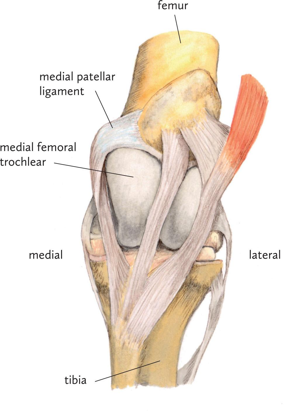
Fig above – Left equine stifle region. Labeled structure: femur, tibia, medial patellar ligament, medial trochlear ridge of the femur.
- The reciprocal apparatus includes the fibularis (aka peroneus) tertius and superficial digital flexor (SDF) mm., which act together and link the actions of the stifle and tarsal (aka hock) joints. Flexion or extension of one joint leads to the corresponding action in the other joint. (Fig. 5B-4)
- The SDF originates in the supracondylar fossa, then crosses the calcanean tuberosity, and attaches distally to the proximal and middle phalanges.
- The common calcanean (aka Achilles) tendon is a collective term for a group of tendons, most notably the superficial digital flexor m. and the gastrocnemius m., that attach to the calcaneus bone. This tendon has an associated subtendinous calcaneal bursa between it and the underlying calcanean tuberosity. There is also a subcutaneous calcaneal bursa between the calcaneus and the skin.
- Clinical Notes: bursitis of the hock/tarsus is when one of the calcaneal bursae is traumatized and becomes inflamed, causing a “capped hock.” (Supplemental Clinical LAS Hock joint problems, capped hocks)
- Fibularis tertius originates in the extensor fossa of the femur and attaches distally near the tarsus and proximal metatarsus.
- The fibularis tertius and SDF mm. have lost most of their muscular bellies and consist of mainly tendon.
- The SDF originates in the supracondylar fossa, then crosses the calcanean tuberosity, and attaches distally to the proximal and middle phalanges.
Figure 5B-4 Equine left hind limb, lateral view of the reciprocal apparatus, where active flexion or extension of the stifle joint results in similar passive actions of the tarsal joint. Labeled structures: supracondylar fossa, extensor fossa, extensor groove, fibula, lateral malleolus, calcanean tuber(osity); superficial digital flexor m., fibularis tertius m.
-
- Clinical Notes: If the hind limb is caught in or under something, the result can be hyperextension of the limb. As an animal tries to pull its foot out of trouble, tension can be put on the fibularis tertius tendon causing it to rupture. A clinical test to check if the tendon is intact includes flexing the stifle and seeing if you can extend the hock/tarsus joint. Normally you would not be able to do this. If the fibularis tertius tendon is ruptured, the hock joint can be extended when the stifle is flexed. (Supplemental Clinical LAS Gait abnormalities, Fibularis tertius rupture)
- The structures that make up the distal portion of the passive hind limb stay apparatus are similar to those of the forelimb. Hyperextension of the fetlock joint is prevented by the suspensory apparatus and SDF and deep digital flexor (DDF) tendons and their acting check ligaments. The SDF does not have a true check ligament in the hind limb. Instead, the attachment of the SDF to the calcaneus, as the SDF courses distally to the digit, acts as a check ligament. The check ligament of the DDF is typically not as well developed in the hind limb.
- The suspensory apparatus spans the plantar aspects of the cannon (Mc3), long pastern (P1), and short pastern (P2) bones.
- The suspensory apparatus includes the suspensory ligament (aka interosseous tendon), proximal sesamoid bones, and the distal sesamoidean ligaments (superficial/straight and oblique/middle, see images in Fig. 3-32, 3-36 in Application 1.3).
- Extensor branches of the suspensory ligaments join the common digital extensor m. tendon on the dorsal surface of the foot. These help counteract the pull of the DDF on the coffin joint.
- The distal most extent of the SDF and DDF tendons also contribute.
- The suspensory apparatus includes the suspensory ligament (aka interosseous tendon), proximal sesamoid bones, and the distal sesamoidean ligaments (superficial/straight and oblique/middle, see images in Fig. 3-32, 3-36 in Application 1.3).
- Clinical Notes: Digital tendon sheaths surround the long and lateral digital extensor tendons, as well as the superficial and deep digital flexor tendons near the fetlock and pastern as in the forelimb. The sheaths can become inflamed and swell, called tenosynovitis. Tendons (and sheaths) can become inflamed or infected (tendonitis) due to ascending foot infections (in both EQUINE and BOVINE). (Supplemental Clinical LAS Tendonitis and bowed tendons)
Objective
A3.4 For each region of the hind limb, provide the name and describe the general location of the nerves innervating flexor and extensor muscle groups and identify clinical signs of nerve damage.
Nerves of the equine hind limb (from the lumbosacral plexus):
- Clinical Notes: Injury to the nerves of the lumbosacral plexus can be seen in ungulates due to the proximity of some of these nerves to bone. They can also be seen when ungulates accidentally get their hind limb(s) stuck or hung and they subsequently pull and stretch the nerves. Stretching of the nerves can also occur if the ground/floor is slippery or wet.
- Note: nerves carry motor and sensory information to and from target regions, respectively. In the most distal extent of the pelvic limbs, the nerves are mostly carrying sensory information, as there are no muscle bellies in the distal pelvic limb.
- There are several nerves we will not fully discuss, and some that will appear later with the pelvis. These include:
- The cranial and caudal gluteal nn. primarily innervate the gluteal mm., which are powerful extensors of the hip (they also innervate: tensor fasciae latae).
- The obturator n. innervates muscles that adduct the limb (pectineus, gracilis, external obturator, adductor).
- The femoral n. innervates the quadriceps femoris m. group (rectus femoris, vastus lateralis, vastus medialis, vastus intermedius mm.).
- The saphenous n. carries sensory information from the skin on the medial aspect of the limb from the mid-crural region to the fetlock.
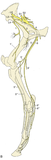
FIG. 24.18 The nerves of the equine hindlimb. (B) The principal nerves, medial view. 1, Femoral nerve (n.); 1′, saphenous n.; 2, obturator n.; 3, cranial gluteal n.; 4, sciatic n.; 5, common fibular (peroneal) n.; 5′, lateral cutaneous sural n.; 5″ and 5′″, superficial and deep peroneal nerves, respectively; 6, tibial n.; 6′, caudal cutaneous sural n.; 6″ and 6′″, medial and lateral plantar nerves, respectively (the lateral nerve gives rise to the plantar metatarsal nerves); 7, caudal gluteal n.; 8, caudal cutaneous femoral n.; 9, pudendal n.; 10, pelvic n.; 11, caudal rectal n. TVA
- Sciatic n. provides innervation to the hamstrings (biceps femoris, semitendinosus, semimembranosus mm.), which extend the hip and have multiple actions on the stifle. Proximal to the stifle the sciatic n. splits into the common fibular and tibial nerves.
- The common fibular n. distributes to the craniolateral muscle group of the crus and innervates the long digital extensor, lateral digital extensor, cranial tibial, fibularis tertius, and fibularis longus (bov) mm. which flex the tarsus and extend the digits.
- On the lateral side of the proximal crural region, the common fibular n. divides into superficial and deep fibular nn. The deep fibular n. courses with the cranial tibial a. At the tarsus, the deep fibular n. divides into medial and lateral dorsal metatarsal nn. These nerves provide sensory innervation of the tarsus joint capsule, metatarsus, and fetlock.
- The tibial n. distributes to the caudal muscle group of the crus and innervates the SDF, DDF, and gastrocnemius mm. which extend the tarsus and flex the digits. (Figs. 4-12, 24.19)
- Near the calcanean tuberosity, the tibial n. divides into the medial and lateral plantar nn. Distal to the hock/tarsus, the lateral plantar n. gives off a deep branch that gives rise to the medial and lateral plantar metatarsal nn. These nerves course along the axial side of Mt 3 and Mt 4 and innervate the deep structures of the metatarsus and digit including the interosseus tendon.
- Both plantar nn. continue distally adjacent to the flexor tendons in the metatarsal region. The communicating branch connects them in the mid-cannon region and crosses over the SDF.
- At the level of the proximal sesamoid bones, the plantar nn. change names and become the medial and lateral plantar digital nn.
- The common fibular n. distributes to the craniolateral muscle group of the crus and innervates the long digital extensor, lateral digital extensor, cranial tibial, fibularis tertius, and fibularis longus (bov) mm. which flex the tarsus and extend the digits.
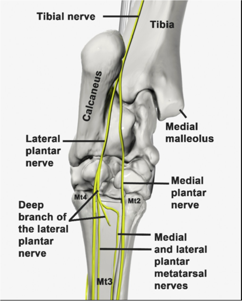
Fig 4-12: Left hind limb. Plantar view showing the division of the tibial nerve into the medial and lateral plantar nerves at the hock/tarsus. (from Equine Anatomy Guide: The Forelimb; Mansour, Steiss, Wilhite)

FIG. 24.19 The nerves (nn.) of the equine right hindfoot. 1 and 2, Medial and lateral plantar nn. (from tibial), respectively; 1′, communicating branch; 2′, deep branch (for plantar metatarsal nn.), cut; 3 and 3′, medial and lateral dorsal metatarsal nn. (from deep peroneal), respectively; 4 and 4′, medial and lateral plantar metatarsal nn. (from lateral plantar, 2′), respectively; 5 and 5′, medial and lateral digital nn., respectively; 6, dorsal branch of digital nerve; 7, branch to digital cushion. TVA
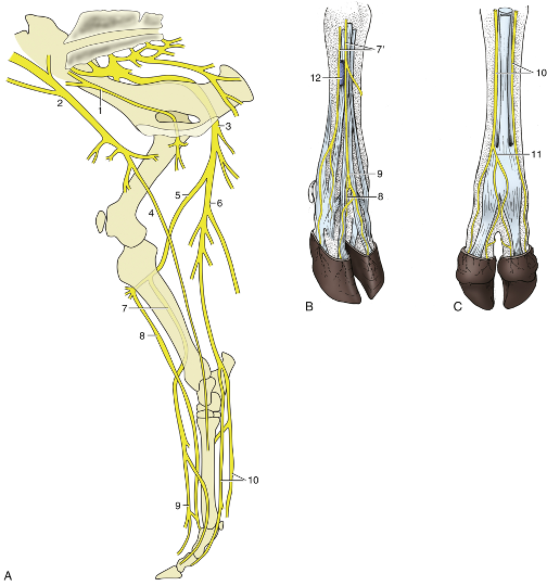
FIG. 31.12 Nerves of the right bovine hindlimb. (A) Medial view. (B) Right hindfoot, dorsolateral view. (C) Right hindfoot, plantar view. 1,Obturator nerve (n.); 2, femoral n.; 3, sciatic n.; 4, saphenous n.; 5, common fibular (peroneal) n.; 6, tibial n.; 7, superficial peroneal n.; 7′, lateral and middle branches of superficial peroneal n.; 8, deep peroneal n.; 9, dorsal common digital n. III; 10, medial and lateral plantar nerves; 11, plantar common digital n. III; 12, cranial tributary of lateral saphenous vein. TVA
Nerve Damage in Equine and Bovine (for images of these conditions see Supplemental Clinical LAS Nerve damage, hind limb nerve damage)
- The obturator n. innervates muscles that adduct the hind limb and damage results in the inability to adduct the limb or prevent splaying of the hind limbs. Once down it is difficult to rise up.
- The femoral n. innervates the quadriceps femoris m. group and damage causes loss of stifle extension, collapse of the hind limb, and inability to bear weight.
- The sciatic n. innervates several muscles of the thigh, crus, and digits. Severe damage would cause the hind limb to hang loose. The sciatic n. can be damaged due to hip fracture or an improper injection site. Once animals are down, it is difficult for them to rise again when the sciatic or tibial nn. are damaged.
- Tibial n. damage includes over-flexion of the tarsus and overextension of the fetlock, resulting in a vertical pastern.
- Common fibular n. damage includes overextension of the tarsus and over-flexion of the joints distal to the tarsus, with the foot resting on the dorsal surface. (Fig. 31.13)
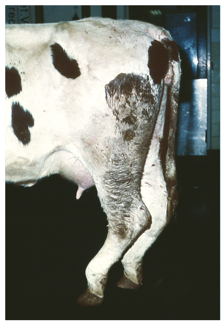
FIG. 31.13 Cow with peroneal paralysis – overextension of the hock and over-flexion of the more distal joints; the limb rests on the dorsal surface of the flexed digits; The animal eventually learns to compensate for this defect by flicking the foot forward before placing it on the ground. TVA
Objective
A3.5 For each region of the hind limb, identify the pathway/distribution and components of the major arterial trunk and clinically relevant veins.
Arteries
- In the limbs, arteries are in more protected locations: medially in the proximal portion of the pelvic limb, and caudally (and on the plantar aspect) in the more distal portions of the pelvic limb.
- The cranial and caudal gluteal aa. supply the muscles of the rump and arise from the internal iliac a. and its branches. These arteries exit the pelvic cavity through the sacrosciatic ligament.
- Main arteries that feed the pelvic limbs begin with the external iliac aa. and continue as the femoral aa. into the thigh. (Figs. 24.17, 31.8)
- The femoral a. and v. are found within the boundaries of the femoral triangle, which is created by the sartorius and pectineus mm. and the abdominal body wall. The femoral a. becomes the popliteal a. caudal to the stifle and then divides into the cranial and caudal tibial aa. in the proximal crus.
- The cranial tibial a. passes between the tibia and fibula. On the dorsal surface of the tarsus, the cranial tibial a. becomes the dorsal pedal a.
- The dorsal pedal a. gives off the perforating tarsal a. at the tarsus and then becomes the dorsal metatarsal a. in the metatarsal region (lateral along the cannon bone in EQUINE and more “dorsal” in BOVINE).
- The dorsal metatarsal a. is the main blood supply to the foot. It divides into the medial and lateral plantar digital aa. at the fetlock.
- The cranial tibial a. passes between the tibia and fibula. On the dorsal surface of the tarsus, the cranial tibial a. becomes the dorsal pedal a.
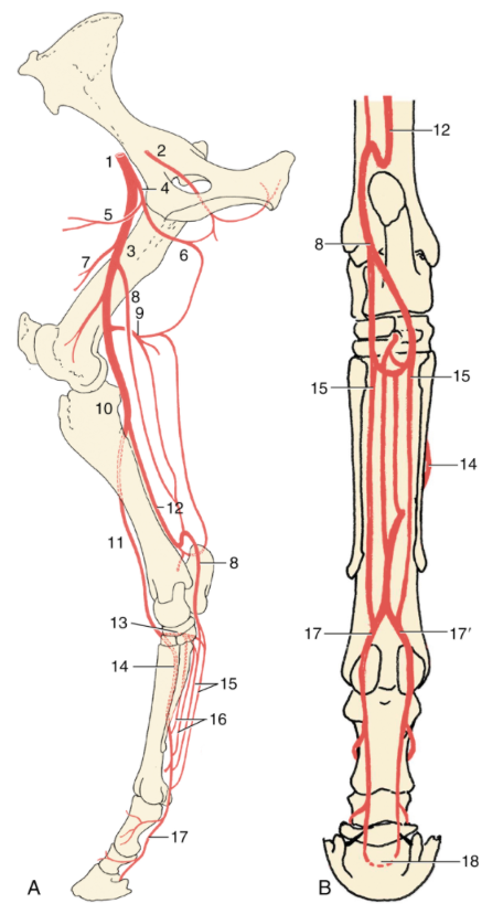
FIG. 24.17 The principal arteries (aa.) of the equine right hindlimb: (A) medial view; (B) caudal view. 1, External iliac artery (a.); 2, obturator a.; 3, femoral a.; 4, deep femoral a.; 5, pudendoepigastric trunk; 6, medial circumflex femoral a.; 7, lateral circumflex femoral a.; 8, saphenous a.; 9, caudal femoral a.; 10, popliteal a.; 11, cranial tibial a.; 12, caudal tibial a.; 13, perforating tarsal a.; 14, dorsal metatarsal a.; 15, medial and lateral plantar aa.; 16, medial and lateral plantar metatarsal aa.; 17 and 17′, medial and lateral plantar digital aa., respectively; 18, terminal arch, anastomosis of digital aa. within the distal phalanx. TVA
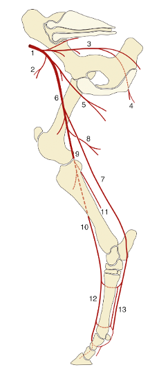
FIG. 31.8 The principal arteries of the bovine right hindlimb, medial view. 1, External iliac artery (a.); 2, deep circumflex iliac a.; 3, internal iliac a.; 4, caudal gluteal a.; 5, deep femoral a.; 6, femoral a.; 7, saphenous a.; 8, caudal femoral a.; 9, popliteal a.; 10, cranial tibial a.; 11, caudal tibial a.; 12, dorsal metatarsal arteries; 13, medial and lateral plantar and metatarsal (closer to the bone) arteries. TVA
Veins
- The femoral a. and v. are found within the boundaries of the femoral triangle, which is created by the sartorius and pectineus mm. and the abdominal body wall. The saphenous vv. drain to the femoral vv.
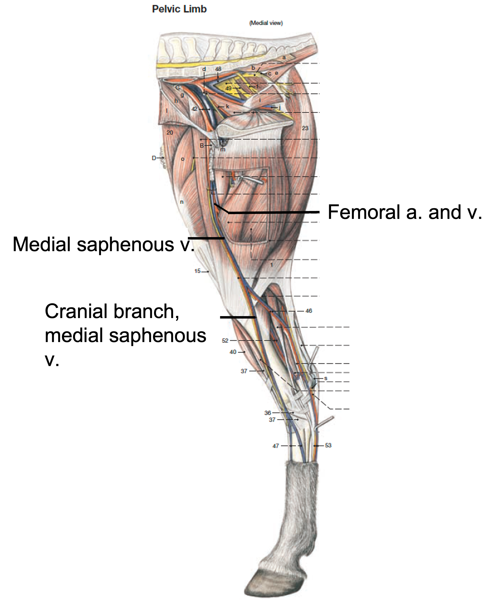
Fig. (above) Equine right pelvic limb, medial view. Labeled veins: femoral v., medial saphenous v., cranial branch of the medial saphenous v. (Budras, Anatomy of the Horse)
- The medial and lateral saphenous vv. drain the medial and lateral aspects of the hind limb, respectively. Each is more prominent in a particular species, as was seen in carnivore anatomy.
- In EQUNIE, the medial saphenous v. has a cranial branch that crosses the dorsal aspect of the tarsus/hock, and in particular crosses over the cunean (aka medial) tendon of the cranial tibial m. The medial saphenous v. also has a caudal branch, and the lateral saphenous v. is present in EQUINE. (Fig. below)
- In BOVINE, the lateral saphenous v. has a cranial branch that is more prominent. The medial saphenous v. is present in BOVINE.
- In both species, the saphenous vv. run over the hock/tarsus, so any injections or aspirations of this joint need to be carefully placed.
- In BOVINE, to anesthetize the foot for diagnostics or treatment, IV regional anesthesia (IVRA) is often performed. (Supplemental Clinical LAS Bovine Foot Anatomy, Lameness, Approaches)
- Distal veins commonly used for clinical procedures in BOVINE include:
- Dorsal common digital v.: on the dorsal surface of the foot (Fig. 31.9 and below); it drains into the cranial branch of the lateral saphenous v.
- Medial (aka axial) or lateral (aka abaxial) palmar (on forelimbs) or plantar (on hind limbs) digital vv.
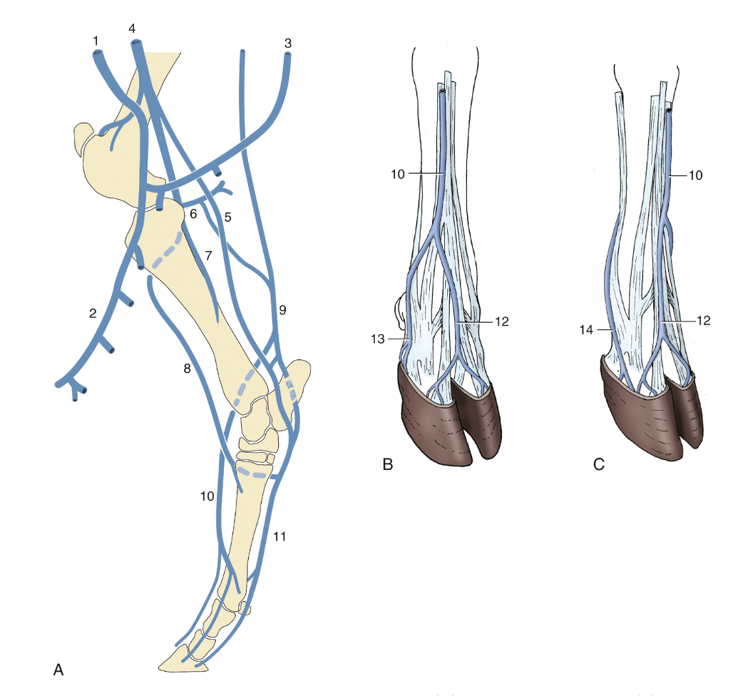
FIG. 31.9 The major veins of the bovine hindlimb. (A) Right limb, medial view. (B) Right hindfoot, dorsolateral view. (C) Left hindfoot, dorsomedial view. 1, External pudendal vein (v.); 2, mammary v.; 3, ventral labial v.; 4, femoral v.; 5, medial saphenous v.; 6, caudal femoral v.; 7, caudal tibial v.; 8, cranial tibial v.; 9, lateral saphenous v.; 10, cranial branch/tributary of lateral saphenous v.; 11, medial and lateral plantar veins; 12, dorsal common digital v. III; 13, plantar v. of lateral digit; 14, plantar v. of medial digit. TVA
Objective
A3.6 Describe clinical syndromes involving the equine hock/tarsus joint.
- The tarsocrural joint has the most mobility of the tarsal joints and has several joint pouches. These pouches can bulge outwards (i.e., swell) if inflammation occurs: dorsomedial, dorsolateral, plantaromedial, plantarolateral.
- Clinical Notes: “spavin“ is a general term for lameness originating in the equine tarsus/hock joint. (see images in Supplemental Clinical LAS Hock joint problems, osteoarthritis)
- Bog spavin: is a condition of the horse that involves fluid distention and swelling of the tibiotarsal (aka tarsocrural) joint. Excess fluid within the joint will bulge out wherever there is an area of ‘least resistance’ in the joint capsule (e.g., between ligaments and tendons) leading to synovitis/capsulitis. This condition is often a sign of joint disease in the horse.
-
- Bone spavin: is an osteoarthritic condition of the horse which mainly involves the medial portions of the distal intertarsal joint and the tarsometatarsal joint.
- The typical treatment includes steroids, but in certain cases transection of the cunean tendon relieves the pressure, and hence, some of the pain in this area. If surgical section (cutting) of the cunean tendon is performed, one must avoid damage to the overlying cranial branch of the medial saphenous v.
- Blood spavin: is a protrusion of the cranial branch of the medial saphenous v. caused by pressure from an enlarged mediodorsal pouch in a horse with bog spavin.
- Bone spavin: is an osteoarthritic condition of the horse which mainly involves the medial portions of the distal intertarsal joint and the tarsometatarsal joint.
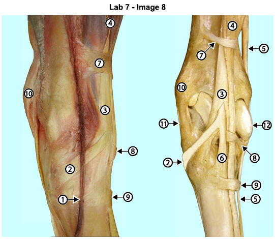
Fig. above – Exposure of the cranial branch of the medial saphenous vein (1) which must be avoided when the cranial tibial cunean/medial tendon (2) aka cunean tendon is cut to lessen the pain of bone spavin. 3, fibularis (peroneus) tertius tendon through which the cranial tibial tendon passes; 4, long digital extensor m.; 5, lateral digital extensor m.; 6, dorsal tendon of cranial tibial m.; 7, 8, and 9 are the proximal, middle and distal extensor retinacula; 10, medial malleolus; 11, medial collateral ligament; 12, lateral ridge of the trochlea of the talus. (http://vanat.cvm.umn.edu/ungDissect/Lab07/Img7-8.html)
