
5 Neck and Thorax
Revised Spring 2024 by T. Clark
Additional Resources for this Chapter:
- Related Supplemental Large Animal Surgery links: LAS Abscesses; LAS Local anesthesia; LAS Esophageal anatomy and surgery; Esophageal obstruction; LAS Temporary tracheotomy; LAS Tracheal disorders; LAS Lymphosarcoma; LAS Pleural space disorders
- Veterinary Gross Anatomy Ungulate Dissection (Cervical and Thoracic Labs 8-10)
- Associated Video Links:
- Cervical Vertebrae (5:30)
- Thoracic Vertebrae and Ribs (2:24)
- Porcine Cervical Region (3:46)
Associated Clinician topics: bovine respiratory disease (BRD), traumatic reticulopericarditis (TRP) – (The most relevant objectives: A5.4, A5.5, A5.6, A7.5)
Objective
A5.1 Identify and describe the structures of a typical vertebra and the distinctive features of the cervical, thoracic, and lumbar vertebrae; compare the general structure of the ribs in equine and bovine; identify and describe the nuchal ligament in the equine and bovine.
- Most vertebrae have a cylindrical body that lies ventral to the arch.
- The arch is formed by pedicles and laminae.
- The arch has a spinous process dorsally and transverse processes laterally.
- The vertebral canal contains the spinal cord; it is formed by the vertebral foramina (space under the arch) of successive vertebrae. A vertebral foramen is formed between the body and arch.
- Vertebral bodies are bound together by fibrocartilaginous intervertebral discs while the arches are bound together by articular processes (cranial and caudal) that form synovial joints.
- The cranial and caudal articular processes are the projections which allow for articulation between adjacent vertebrae. The articulations are synovial joints, while the articular facets are the articular surface of these processes. Joint orientation impacts the directional mobility of a given segment of the vertebral column:
- If there is horizontal orientation of the articular processes (e.g., cervical, cranial thoracic), then lateral flexion can occur.
- If the articular processes are oriented in a dorsal/ventral plane or vertically oriented (e.g., caudal thoracic, lumbar), then dorsal/ventral flexion can occur.
- Between the vertebral bodies, articulation also occurs. There is an intervertebral disc space between vertebral bodies. This is where the intervertebral disc is located. This joint doesn’t have many degrees of movement and is not synovial.
- The cranial and caudal articular processes are the projections which allow for articulation between adjacent vertebrae. The articulations are synovial joints, while the articular facets are the articular surface of these processes. Joint orientation impacts the directional mobility of a given segment of the vertebral column:
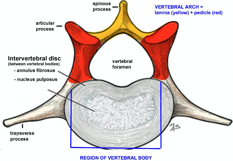
Figure above, cranial view of a typical vertebra. Labeled structures: spinous process, articular process, vertebral foramen, vertebral arch (laminae = yellow, pedicles = red), intervertebral disc, transverse process.
- Clinical Notes: Laminectomy is the partial or complete removal of a lamina. If disc material from the nucleus pulposus pushes into the spinal cord, this pressure can cause impairment of downstream and upstream signals in the nervous system. This procedure is sometimes utilized to decompress the spinal cord.
- Below, a transverse section through a vertebra shows the spinal cord running in the vertebral canal. Dorsal and ventral roots form spinal nerves on each side.
- Most spinal nerves exit between adjacent vertebrae at intervertebral foramina, or spaces, except between the first two cervical vertebrae. Some spinal nerves exit via lateral vertebral foramina, which are complete foramina in certain cervical or thoracic vertebrae (see Figure 8B-4/11).
- Once a spinal nerve exits at an intervertebral foramen or lateral vertebral foramen, the spinal nerve splits into dorsal and ventral branches.
- Dorsal branches of spinal nerves innervate structures located dorsal to the transverse processes of the vertebrae (smaller branches heading in a dorsal direction in the image below). Motor nerve fibers innervate epaxial muscles and sensory nerve fibers innervate the skin in this region.
- Ventral branches of spinal nerves innervate structures located ventral to the transverse processes of the vertebrae (larger branches heading in a ventral direction in the image below). Motor nerve fibers innervate hypaxial muscles and sensory nerve fibers innervate the skin in this region.
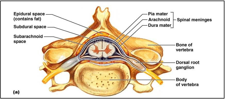
Figure above of cross section of a vertebra and spinal cord: labeled structures: epidural space, subdural space, subarachnoid space, spinal meninges (pia, arachnoid, dura mater), bone of vertebra, dorsal root ganglion, body of vertebra. Structures seen: vertebral canal, intervertebral foramina (right and left) with spinal nerves exiting.
Cervical Vertebrae (Figs. 8B-2 and 8B-3); both HORSE and OX have a total of 7 cervical vertebrae. There are 8 cervical spinal nerves.
- A typical cervical vertebra (Figs. 8B-2, 8B-3) is characterized by a low spinous process, a short but complex transverse process, horizontally oriented cranial and caudal articular processes, and the presence of a transverse foramen that transmits the vertebral a., v., and n.
- With few exceptions, cervical vertebrae have transverse foramina; the exceptions are C1 (OX) and C7 (EQUINE and OX). They can be found on the caudal aspect of each transverse process (or wing of C1).
- The first two spinal nerves (C1 n. and C2 n.) exit through foramina on each side of the vertebral arch called the lateral vertebral foramina. In C1 (atlas) these foramina are on the cranial aspect of the wing. Note the ventral branches of the C1 spinal nerves proceed through bilateral foramina called the alar foramina (alar notch of the DOG), located just ventral to the C1 lateral foramina in Figure 8B-2 (and image below).
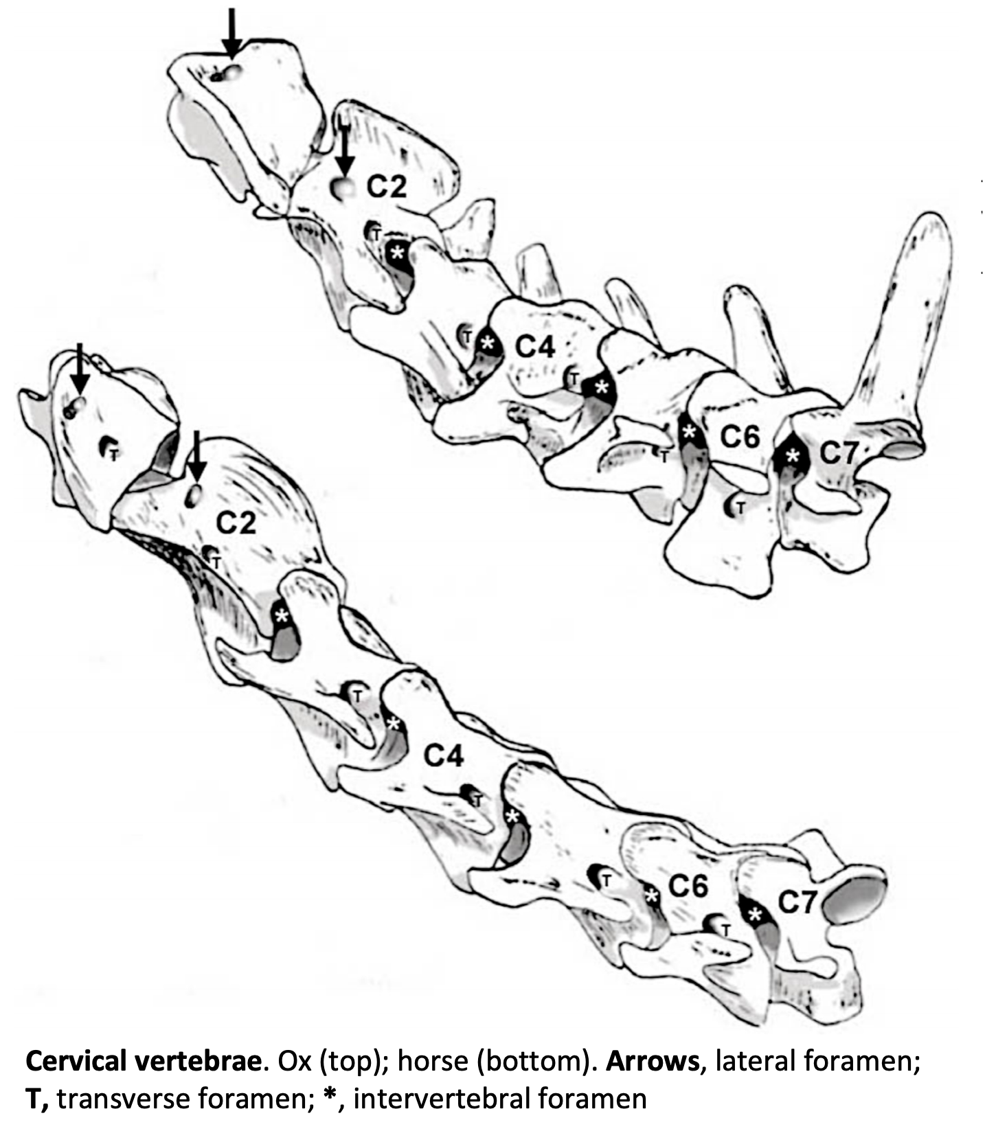
Figure 8B-2 (Above) Cervical vertebrae. Ox (top); horse (bottom). Arrows, lateral foramen (an alar foramen is ventral to the lateral foramen in C1 vertebrae); T, transverse foramen; *, intervertebral foramen. Synovial joints connect the vertebrae (see lab skeletons).
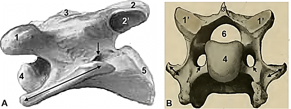
Figure 8B-3 (Above) Typical cervical vertebra. A. lateral view; B. cranial view. Cranial articular process and articular surface: 1, 1′; caudal articular process and articular surface: 2, 2′; 3, spinous process; 4, cranial aspect of vertebral body; 5, caudal aspect of vertebral body; 6, vertebral foramen; double arrows, transverse processes; single arrow, transverse foramina. Modified from Budras (A) and Sisson and Grossman (B).
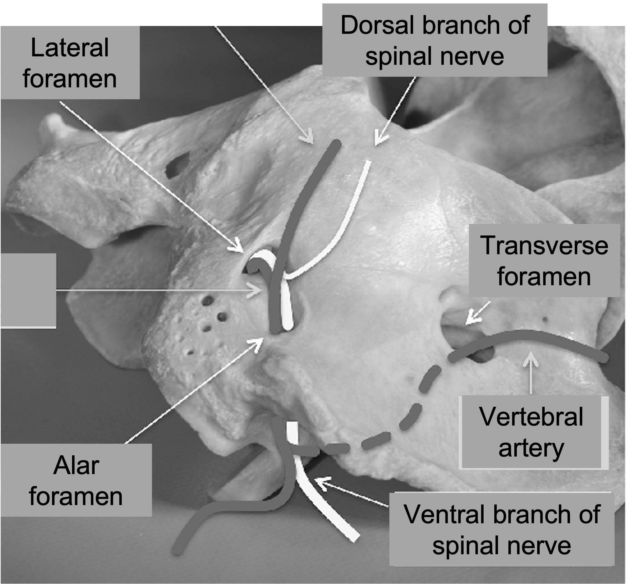
Above: Horse atlas (dorsolateral view), showing route of vessels and nerves. Labeled structures: lateral foramen with spinal nerve exiting and immediately dorsal and ventral branches splitting; alar foramen with ventral branch continuing; transverse foramen with vertebral artery.
- Atlas (C1): The atlas is considered to be a vertebra with dorsal and ventral arches, but a reduced or no body.
-
- The atlas lacks a body and a spinous process. It forms synovial joints with adjacent bones rather than cartilaginous joints that are normally found between the vertebral bodies (i.e., intervertebral discs).
- The broad and long transverse processes of the atlas form the wings of the atlas, making this vertebra wider than all of the other cervical vertebrae.
- In most species, the omotransversarius m. takes its name from its insertion on the wing of the atlas, but in the HORSE this muscle fuses with the brachiocephalicus m. and inserts on the transverse processes of more caudal cervical vertebrae.
- C1 articulates with the occipital condyles creating a synovial joint (and space called the atlanto-occipital space), which allows the head to rock up and down (flexion/extension).
- The atlanto-occipital space is covered by an atlanto-occipital membrane dorsally. (more details in the LAA Dissection eBook)
- Axis (C2): The axis is the longest of the vertebrae.
-
- The axis has a long and wide spinous process (aka spine of the axis) when viewed laterally and a dens (aka odontoid process) protruding cranially. The dens, developmentally, is a component of C1.
- All other vertebral spines are narrow when viewed from the side.
- C3 through C7 vertebrae are more similar to each other.
- These general cervical vertebrae have low spinous processes and relatively short transverse processes, and their general shape is an irregular cube.
- As you proceed to more caudal cervical vertebrae their transverse processes lengthen and the spinous processes can become more developed.
- Clinical Notes: Slight malformations or narrowing of the cervical vertebrae (degenerative processes) or its vertebral canal can result in spinal cord damage leading to gait deficits known as Wobbler syndrome. Symptoms may also include limited dorsiflexion of the neck, depending on the cervical region affected.
- Nuchal ligament: Ungulates have a specialized connective tissue support structure that functions to help support their large heavy head without interfering with their ability to lower their neck when grazing. Muscles help pull the head down, but less muscular effort is needed to bring the head back up due to the springy elastic nature of the nuchal ligament.
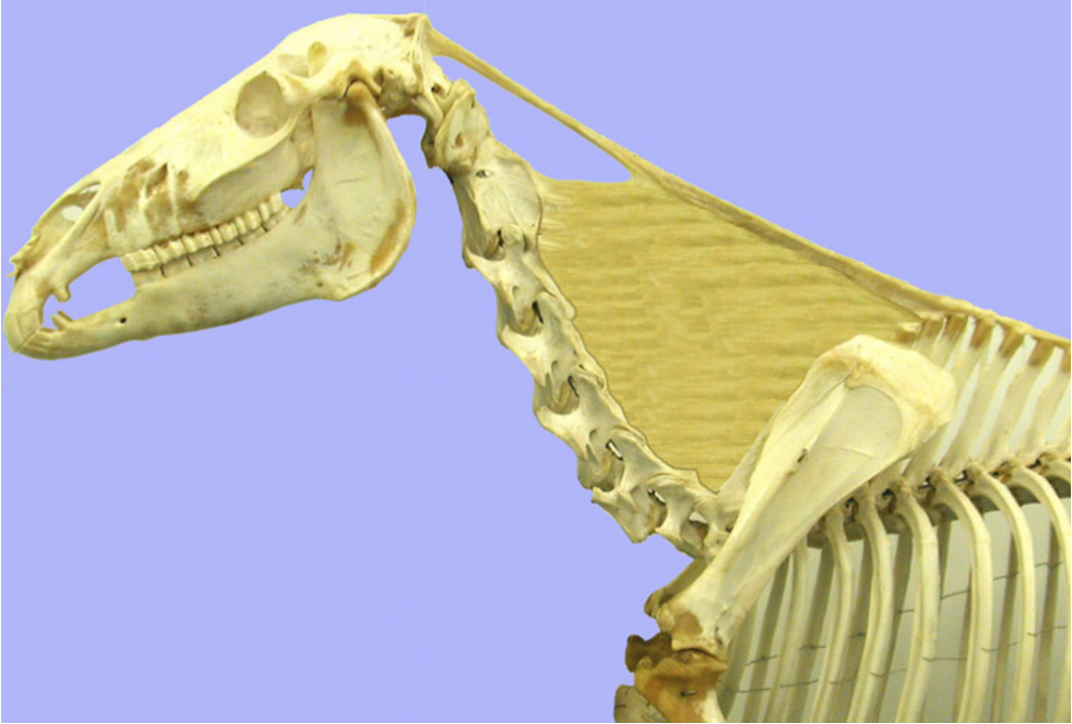
- The nuchal ligament in the HORSE and OX (seen in a horse below) has two parts, is elastic, and is paired. Each side is composed of:
- A funicular part dorsally, which courses between the occipital bone of the skull and the spinous processes of the first few thoracic vertebrae. It is continuous with the supraspinous ligament of the vertebrae. Dogs only have the funicular portion of the nuchal ligament, which attaches to the axis (C2), not to the skull. Remember that the cat does not have a nuchal ligament.
- In the HORSE, deep to the mane lays an interwoven mass of fat and fibrous tissue called the nuchal fatty crest, which is especially prominent in stallions.
- A laminar part is sheet like and fills most of the space between the funicular part and the cervical vertebrae.
- A funicular part dorsally, which courses between the occipital bone of the skull and the spinous processes of the first few thoracic vertebrae. It is continuous with the supraspinous ligament of the vertebrae. Dogs only have the funicular portion of the nuchal ligament, which attaches to the axis (C2), not to the skull. Remember that the cat does not have a nuchal ligament.
- The nuchal ligament in the HORSE and OX (seen in a horse below) has two parts, is elastic, and is paired. Each side is composed of:
- Clinical Notes: There are a few areas where the nuchal ligament is close to bone. In these regions a bursa usually forms. The cranial nuchal bursa forms between the funicular part of the nuchal ligament and the dorsal arch of the atlas (C1). The caudal nuchal bursa forms between the funicular part of the nuchal ligament and the spinous process of the axis (C2). The supraspinous bursa forms between the supraspinous ligament and the most prominent spinous processes of the thoracic vertebrae (known as the “withers”). These bursae may become infected, called bursitis.
- In the poll region this is called “poll evil” aka cranial nuchal bursitis.
- In the withers region this is called “fistulous withers” aka supraspinous bursitis. Brucella can cause fistulous withers, so use caution and culture a sample. (Supplemental Clinical LAS Abscesses)
Thoracic Vertebrae (Fig. 8B-4); HORSES typically have a total of 18 thoracic vertebrae (and 18 sets of ribs), while CATTLE have a total of 13 thoracic vertebrae (and 13 sets of ribs).
- Most of the spinous processes are tall and elongated, and they begin leaning in a caudal direction as they extend caudally through the thorax toward the lumbar region.
- The anticlinal vertebra is the thoracic vertebra with a more perpendicular spinous process, as compared to the typically more angled and caudally directed spinous processes of the cranial thoracic vertebrae and cranially directed spinous processes of the caudal thoracic vertebrae. Each species may have a slightly different thoracic vertebra that is identified as anticlinal: dog/cat ~T11; bovine T11-T13 (see Figure 8B-4); equine ~T16.
- The transverse processes are short and small and attach to ribs.
- Cranial and caudal articular processes – the cervical vertebrae have well developed articular processes while the cranial thoracic vertebrae lack well-defined articular processes, and instead have articular facets on the arch.
- Moving caudally, there is a transition from horizontal to vertical articular processes, as are typical of the lumbar region. This also allows for a change from lateral flexion to dorsal flexion in the spinal column, respectively. The rib cage restricts thoracic vertebral flexion.
- On the caudal edge of each thoracic vertebra note the notch at the base of the arch, just dorsal to the body. This notch forms the basis of an intervertebral or lateral vertebral foramen. If the notch is closed off, a lateral vertebral foramen results. If the notch is not closed off, there is an intervertebral foramen. The spinal nerve that emerges through either foramen will pass laterally along the caudal edge of the adjacent rib.
- There are also costovertebral articulations associated with the ribs (more on these below).
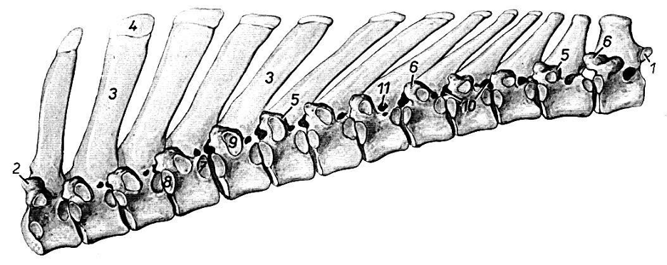
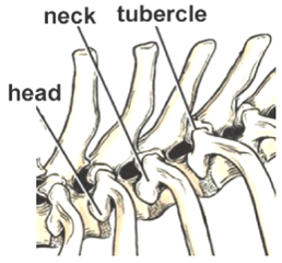
Figure 8B-4 (Above, top) Bovine vertebrae, lateral view. The heads of the ribs articulate with foveae/facets on adjacent vertebral bodies (7, 8); the tuberculum of each rib articulates with an articular fovea/facet (9) on a transverse process (5, 6); caudal and cranial articular processes (1, 2) are present on the ends of the thoracic vertebral series. Intervertebral (10) or lateral vertebral foramina (11) are present at each vertebral level; note the caudal leaning of the spinous processes (3) except for the last shown. (Above, bottom) Image of rib articulation with thoracic vertebrae. (Modified from Guide to Dissection of the Dog, 7th ed. Fig. 2-75)
Ribs HORSES typically have 18 sets of ribs, while CATTLE have 13 sets of ribs.
- There are bony portions of the ribs, and cartilaginous portions. The ribs that are more caudal (“false ribs”), unite and form a costal arch as they connect, while more cranial ribs (“true ribs”) join directly to the sternum via costal cartilages at the costochondral junction. Floating ribs have no connection to the arch or sternum.
- The rib has a head, neck and tubercle (aka tuberculum). The tuberculum and head articulate at two different locations on the thoracic vertebrae (each of these is a synovial joint). Costovertebral articulations: each costovertebral articulation involves the head of a rib and two adjacent vertebral bodies, while a second costovertebral articulation involves the tuberculum of a rib and the transverse process of a thoracic vertebra (Fig. 8B-4 above).
- The head (B in image below) is more ventral and articulates near either one or two vertebral bodies. The articulation surface is called an articular facet.
- The tuberculum (A in image below) articulates more dorsally, along the transverse process of the thoracic vertebra.
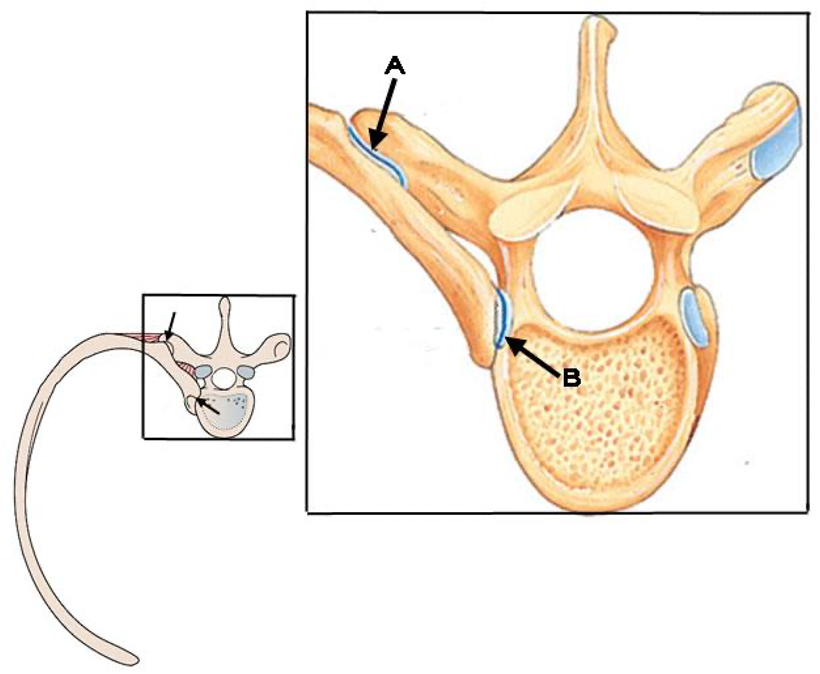
-
- These two proximal articulation points allow the ribs to move craniolaterally and caudomedially.
- As the thoracic body wall moves laterally, the volume of the chest cavity is increased and a small amount of serous fluid in the pleural cavities causes the visceral pleura of the lungs to adhere by capillary attraction to the parietal pleura. Therefore, as the thorax increases in volume, intrathoracic negative pressure draws air into the lungs resulting in inspiration and expansion of the lungs (i.e., the ribs to flare out).
- Comparing HORSES (streamlined thorax, image below on left) and CATTLE (stout thorax, image below on right):
- HORSES have several narrow ribs, while CATTLE have wide and flat ribs.
- In HORSES the last rib is shorter, but in CATTLE all ribs are of similar length and the 13th rib in horses and cattle is of similar length.
- The costal arch is longer and more slanted in HORSES.
- The olecranon in CATTLE is above the sternum, as they have a deeper thorax.
- The depth of thorax is deeper vertically, but shorter in length in CATTLE.
- In HORSES, the sternum is about level with the olecranon. HORSES have a larger thoracic cavity which increases their respiratory capacity.
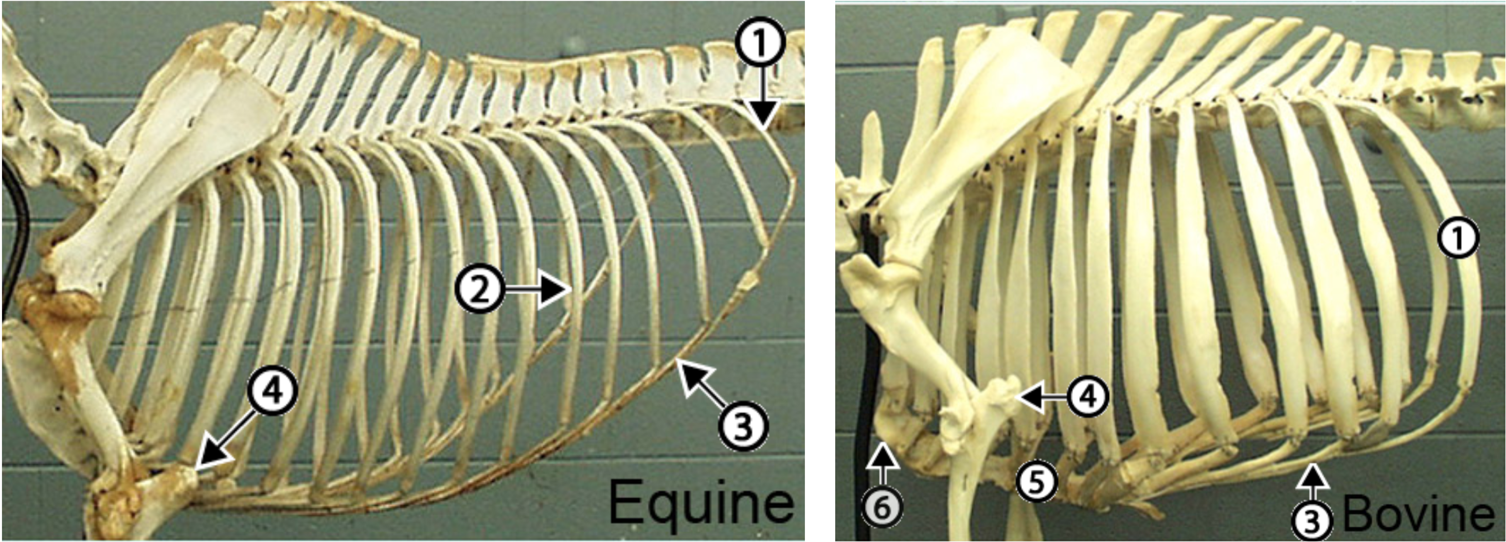
Figure above with equine thoracic skeleton (left) and bovine thoracic skeleton (right). The last rib (1) is shorter in the horse but the 13th rib of the horse (2) is similar in length to the bovine 13th rib (1). The costal arch (3) is much longer and more slanted in the horse. The bovine thorax is deeper than that of the horse and therefore the bovine sternum (5) is ventral to the olecranon (4). The cranial part of the bovine sternum forms the brisket (6). (http://vanat.cvm.umn.edu/ungDissect/Lab09/Img9-1.html)
Lumbar Vertebrae; HORSE and CATTLE typically have a total of 6 lumbar vertebrae.
- The spinous processes are relatively stout and short, while the transverse processes are distinctly long.
- In the EQUINE the transverse processes of L5-6 articulate (Fig. 8B-5).
-
- The cranial and caudal articular processes are vertically oriented.
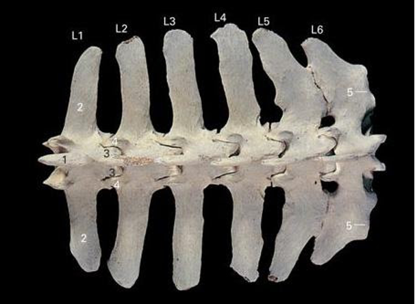
Figure 8B-5 (Above) Lumbar vertebrae (horse), dorsal view. Note the fusion of the transverse processes of L5 and L6. (Image source: Clinical anatomy of the Horse Fig. 3.16)
Sacral Vertebrae/Sacrum; HORSES and CATTLE both have a total of 5 fused sacral vertebrae.
- The sacrum attaches between the wings of the ilium (of the pelvis).
- The sacroiliac joints are formed by the overlapping of the wings of the sacrum (wide modifications of the sacral vertebrae) and the wings of the ilium.
- Fusion of the transverse processes of the sacral vertebrae creates the sacral wings laterally.
- Clinical Notes: Epidurals (in BOVINE) are typically performed in the sacrococcygeal space (the most movable space when the tail is pumped up and down; Supplemental Clinical LAS Local anesthesia).
Caudal/coccygeal Vertebrae
- HORSES can have anywhere from 15-25 caudal vertebrae, with an average of 18.
- CATTLE typically have 18-20 caudal vertebrae.
- Caudal vertebrae reduce in size going distally along the tail.
Objective
A5.2 Identify and describe the muscles, vessels, and associated structures in the equine, bovine, and porcine neck; relate these structures to venipuncture or other clinical procedures in the neck.
Muscles of the Neck and Thorax (Figs. 19.2, 1-2, 32.11, 18.38, 32.12)
- The cervical part of the trapezius m. courses between the funicular part of the nuchal ligament and the spine of the scapula.
- The brachiocephalicus m. can have different named portions depending on the species. In EQUINE the upper cleidocephalicus portion only contains the cleidomastoideus m. (mastoid part of cleidocephalicus m.). In BOVINE, the cleidocephalicus portion includes the cleidomastoideus m. (mastoid part of cleidocephalicus m.) and the cleido-occipitalis m. (occipital part of cleidocephalicus m.). Remember, the lower portion is called the cleidobrachialis m.
- In EQUINE, the omotransversarius m. is fused to the cleidocephalicus m.
- In BOVINE, the omotransversarius m. is a separate muscle, and is deep and between the brachiocephalicus m. and cervical part of the trapezius m.
- Deep and running between the brachiocephalicus m. and cervical part of the trapezius m. are (from most dorsal to most ventral):
- the cervical part of rhomboideus m.
- the splenius m. (part of transversospinalis m. system)
- the serratus ventralis cervicis m.
- On the ventral neck, the superficial “V” shaped muscle is the cutaneous colli and is found in EQUINE and PORCINE.
- Deep to the cutaneous colli m. are paired muscles running from the sternum cranially and include the (cross section of these muscles in equine Fig. 18.38):
- The sternocephalicus m. is cranial to the brachiocephalicus m.
- In EQUINE, there is only a sternomandibularis m. (mandibular part of sternocephalicus) portion.
- In BOVINE, there is a sternomandibularis (mandibular part of sternocephalicus) and sternomastoideus m. (mastoid part of sternocephalicus) (shown in Fig. 3-2, LAA Dissection eBook)
- In PORCINE, there is only a sternomastoideus m. (mastoid part of sternocephalicus) portion (Fig. 32.11,12).
- Moving cranially again, there is another muscle, the sternothyroideus m. running from the sternum to the thyroid cartilage of the larynx.
- In PORCINE, the sternothyroideus m. is deep and slightly lateral to the sternohyoideus m., but has two distinct and narrow attachment points onto the larynx.
- And finally, the most medial set of muscles cranially, the sternohyoideus m. (sternum to basihyoid bone).
- In EQUINE, there is a prominent omohyoideus m. The omohyoideus m. is lateral to the sternohyoideus m. near its insertion on the basihyoid bone, but deep to the brachiocephalicus and sternocephalicus/sternomandibularis mm. near its origin.
- The sternocephalicus m. is cranial to the brachiocephalicus m.
- In PORCINE, the superficial pectoral mm. (running from the sternum to the point of the shoulder) are important landmarks to assist in venipuncture. (Fig. 32.11,12) More on the neck in pigs: Porcine Cervical Region (3:46).
- Muscles of the Thorax
- Serratus ventralis thoracis is part of serratus ventralis that runs from the scapula to the ribs. It is part of the muscles of the thorax. (Fig. 19.2/2)
- Serratus dorsalis cranialis is deep to the trapezius and cranial portion of latissimus dorsi, while serratus dorsalis caudalis is deep to the caudal portion of latissimus dorsi. Each is connected to the dorsal midline via an aponeurosis and they are superficial to the epaxial muscles.
- Epaxial muscle systems: the epaxial muscles generally lie dorsal to the transverse processes of the vertebrae and are associated with the vertebral column and ribs.
- From lateral to medial, iliocostalis, longissimus, and transversospinalis.
- Intercostal muscles (external and internal), run between the ribs.
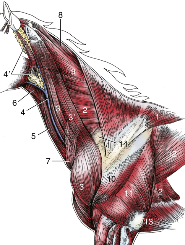
FIG. 19.2 Superficial dissection of the equine neck and shoulder region. 1, Trapezius (cervical, thoracic parts); 2, serratus ventralis cervicis, thoracis; 3, brachiocephalicus; 3′, omotransversarius; 4, external jugular vein; 4′, parotid gland; 5, sternocephalicus/sternomandibularis; 6, omohyoideus; 7, cutaneous colli; 8, rhomboideus, cervical part; 9, splenius; 10, deltoideus; 11, triceps; 12, latissimus dorsi; 13, pectoralis ascendens; 14, subclavius. TVA
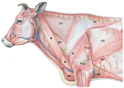
Figure 1-2. Extrinsic muscles of the ox. Extrinsic muscles: CT, Cervical trapezius; TT, thoracic trapezius; OT, omotransversarius, B, brachiocephalicus; SP, superficial pectorals; DP, deep pectorals; LD, latissimus dorsi; SV, serratus ventralis. (Modified from Budras and Habel, 1st ed.)
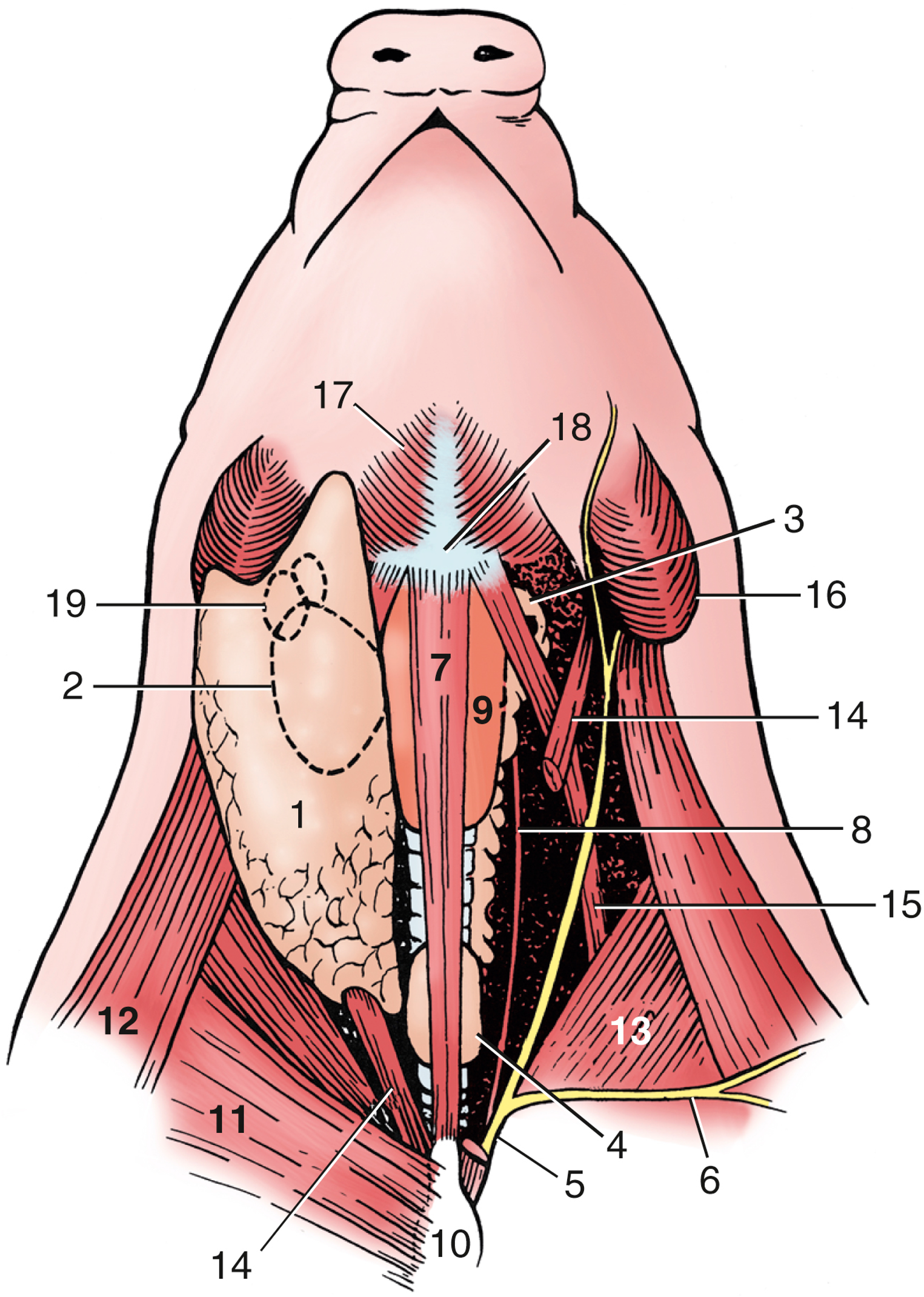
FIG. 32.11 Ventral view of the pig/porcine neck. Deep dissection to the right; superficial dissection, from which the cutaneous colli has been removed, to the left (semischematic). 1, Parotid gland; 2, mandibular gland; 3, thymus; 4, thyroid gland; 5, external jugular vein (yellow); 6, cephalic vein; 7, sternohyoideus (drawn narrower than actual width); 8, internal jugular vein (red); 9, larynx; 10, manubrium sterni; 11, superficial pectoral muscle; 12, brachiocephalicus; 13, subclavius; 14, sternocephalicus/sternomastoideus; 15, omohyoideus; 16, angle of mandible; 17, mylohyoideus; 18, basihyoid; 19, mandibular lymph nodes. TVA
Images above from: Porcine Cervical Region (3:46).
Vessels and Nerves of the Neck and Venipuncture: (Figs. 19.2, 1-2, 32.11, 32.12, 18.38)
- The nerves and vessels of the neck are all relatively closely associated and found running within the carotid sheath, near the trachea, or between adjacent neck muscles.
- In the transverse section of the neck in the HORSE below (Fig. 18.38), the external jugular vv. are found in the jugular furrow which is bordered by the sternomandibularis/sternocephalicus m. medially and the cleidomastoideus/cleidocephalicus m. (of the brachiocephalicus) laterally.
- Clinical Note: The jugular groove (aka jugular furrow) is an important external landmark in the neck when venipuncture and IM (intramuscular) injection is performed. (Image below of live horse, #1; and right side with triangle) The furrow is most prominent in horses, less prominent in bovine, and in camelids (Veterian Key link) you would need to palpate the cervical vertebrae for placement of a jugular catheter.
- The common carotid aa. are found within the carotid sheaths (structures are bilateral), which is deep to the omohyoideus m. in the HORSE. The omohyoideus m. forms a muscular separation between the external jugular v. and the carotid sheath in the cranial half of the neck (Fig. 18.38). In the caudal half of the neck the external jugular v. and the carotid sheath are in apposition.
- Also contained within the carotid sheath is the vagosympathetic nerve trunk (in all species we discuss), while most of the time there is an internal jugular v. within the carotid sheath in BOVINE and PORCINE. In pigs, the internal jugular v. is nearly as large as the external jugular v. and the two veins lie close together, separated by the sternomastoideus m.
- Clinical Note: In EQUNE, there is little separation between the external jugular v. and common carotid a. in the upper neck, and the omohyoideus m. may offer some protection from puncturing the common carotid a. If medications are delivered into the artery versus the vein during intravenous (IV) injection there could be very serious and adverse reactions. Nervous system structures could be impacted including the sympathetic trunk (within the carotid sheath), or the recurrent laryngeal n. (adjacent to the trachea). Sympathetic trunk damage could cause Horner’s syndrome symptoms.
- Clinical Note: In PORCINE, there is a depression (similar to the jugular groove) formed by the borders of the superficial pectoral (caudally), sternocephalicus/sternomastoideus (medially), and brachiocephalicus mm. (laterally) where venipuncture is conducted. In this depression is where the internal and external jugular vv. would join to form the short brachiocephalic v. that drains into the cranial vena cava. There would be slightly different approaches when aiming for the jugular vv. versus the cranial vena cava, as well as technique used for adult pigs versus piglets. See images below for more information (Fig. 32.12). You will also learn about this procedure in pigs in LAS and Swine courses.
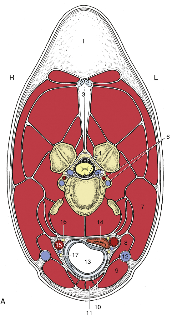
FIG. 18.38 (A) Transection of the equine neck at the level of the fourth cervical vertebra. L, Left side; R, right side. 1, Nuchal fatty crest; 2 and 3, funicular and laminar parts of nuchal ligament, respectively; 4, subarachnoid space; 5, internal vertebral venous plexus; 6, vertebral artery and vein; 7, brachiocephalicus; 8, omohyoideus; 9, sternocephalicus; 10, sternothyroideus; 11, sternohyoideus; 12, external jugular vein; 13, trachea; 14, esophagus; 15, common carotid artery; 16, vagosympathetic trunk; 17, recurrent laryngeal nerve. TVA
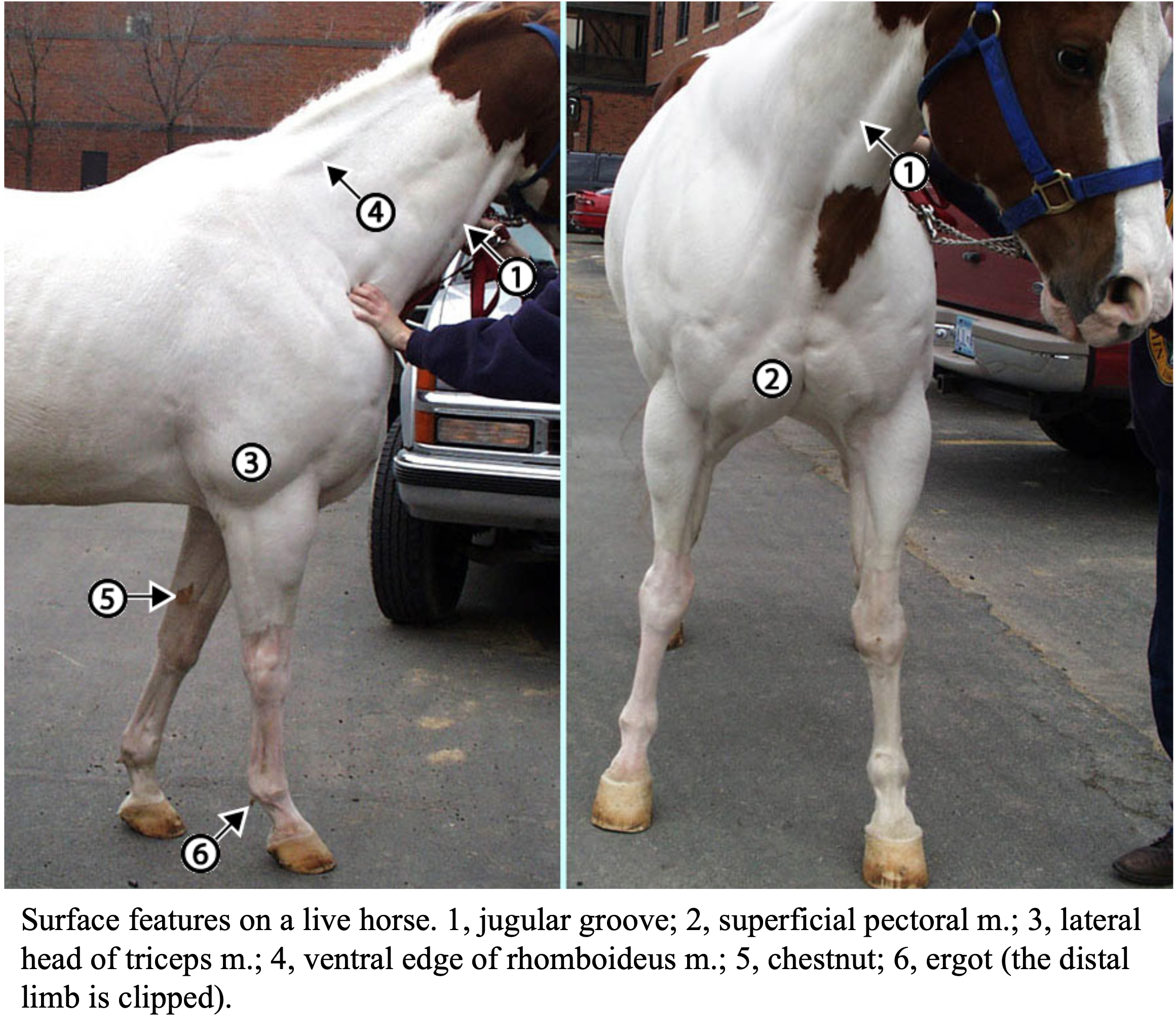
Figure above. Surface features on a live horse. 1, jugular groove; 2, superficial pectoral m.; 3, lateral head of triceps m.; 4, ventral edge of rhomboideus m.; 5, chestnut; 6, ergot (the distal limb is clipped). (http://vanat.cvm.umn.edu/ungDissect/Lab08/Img8-9.html)
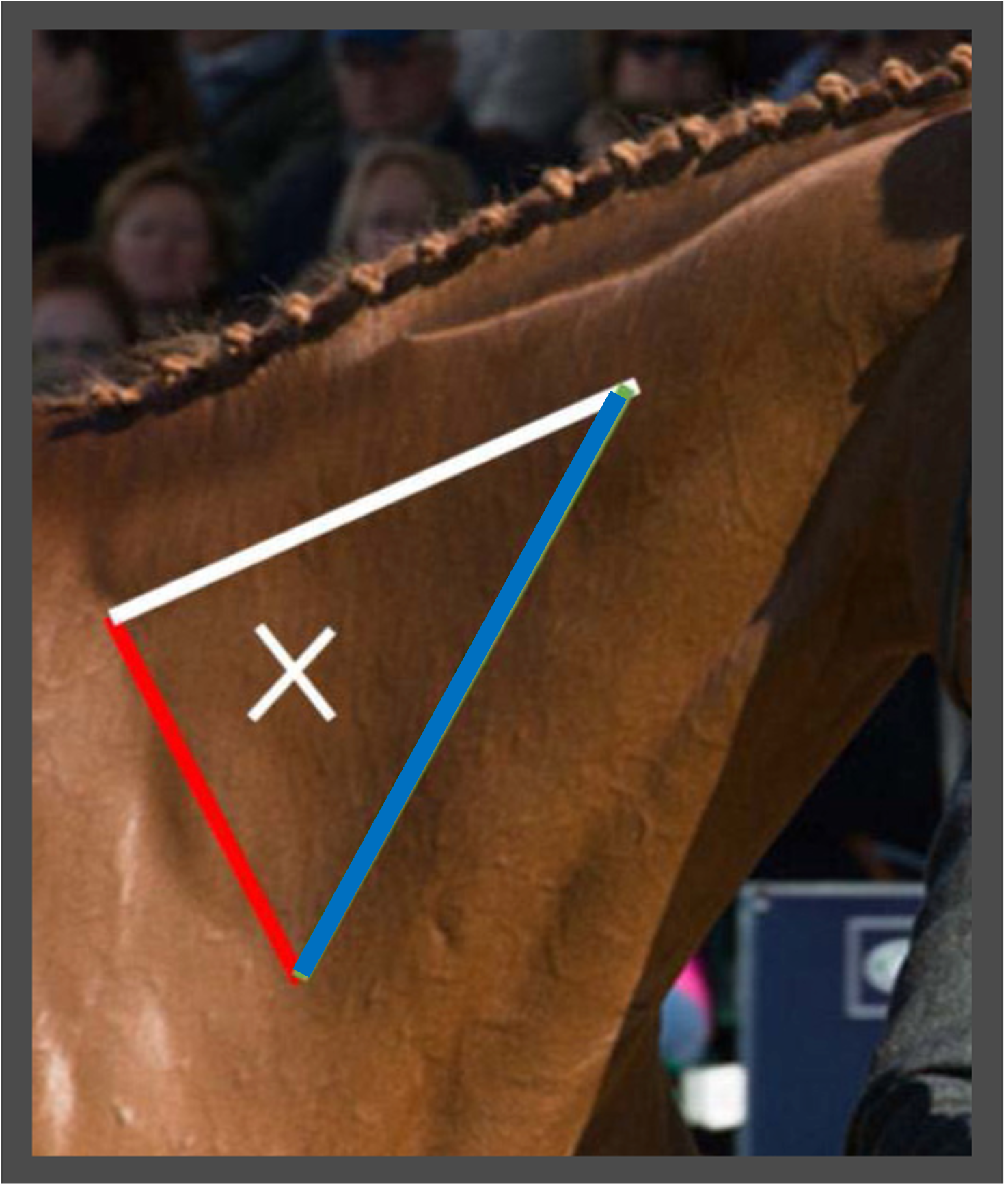
Figure above – The landmarks for IM injecting in the equine neck are: The scapula (shoulder blade) – at the base of the neck (behind the red line). The cervical spine (cervical vertebrae) – at the bottom of the neck (below the blue line and about a hands width up from the jugular groove). The nuchal ligament – at the top of the neck (above the white line, and about a hands width down from the base of the mane). You should inject in the triangle approximately a hands width above the shoulder blade, about halfway between the nuchal ligament and cervical vertebrae. (From https://www.rvc.ac.uk/equine-vet/information-and-advice/fact-files/intra-muscular-injections-in-horses)

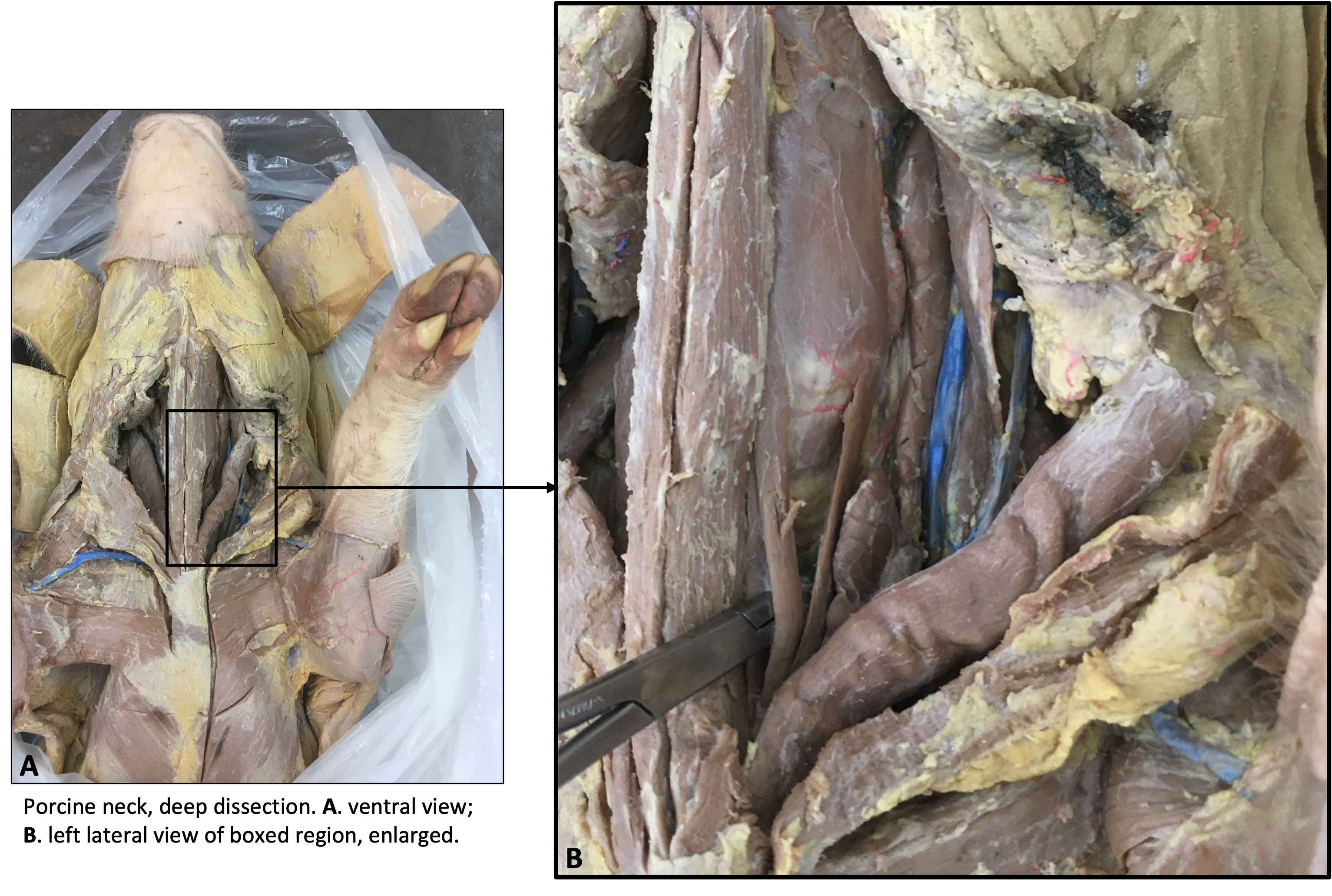
Figure above. Venipuncture landmarks in a pig, inset of veins in the neck of a pig. A surface depression is created by the superficial pectoral (caudally), sternocephalicus/sternomastoideus (medially), and brachiocephalicus mm. (laterally). Labeled structures: CVC – cranial vena cava, E – external jugular v., I – internal jugular v., S – subclavian v., T – thyroid cartilage.
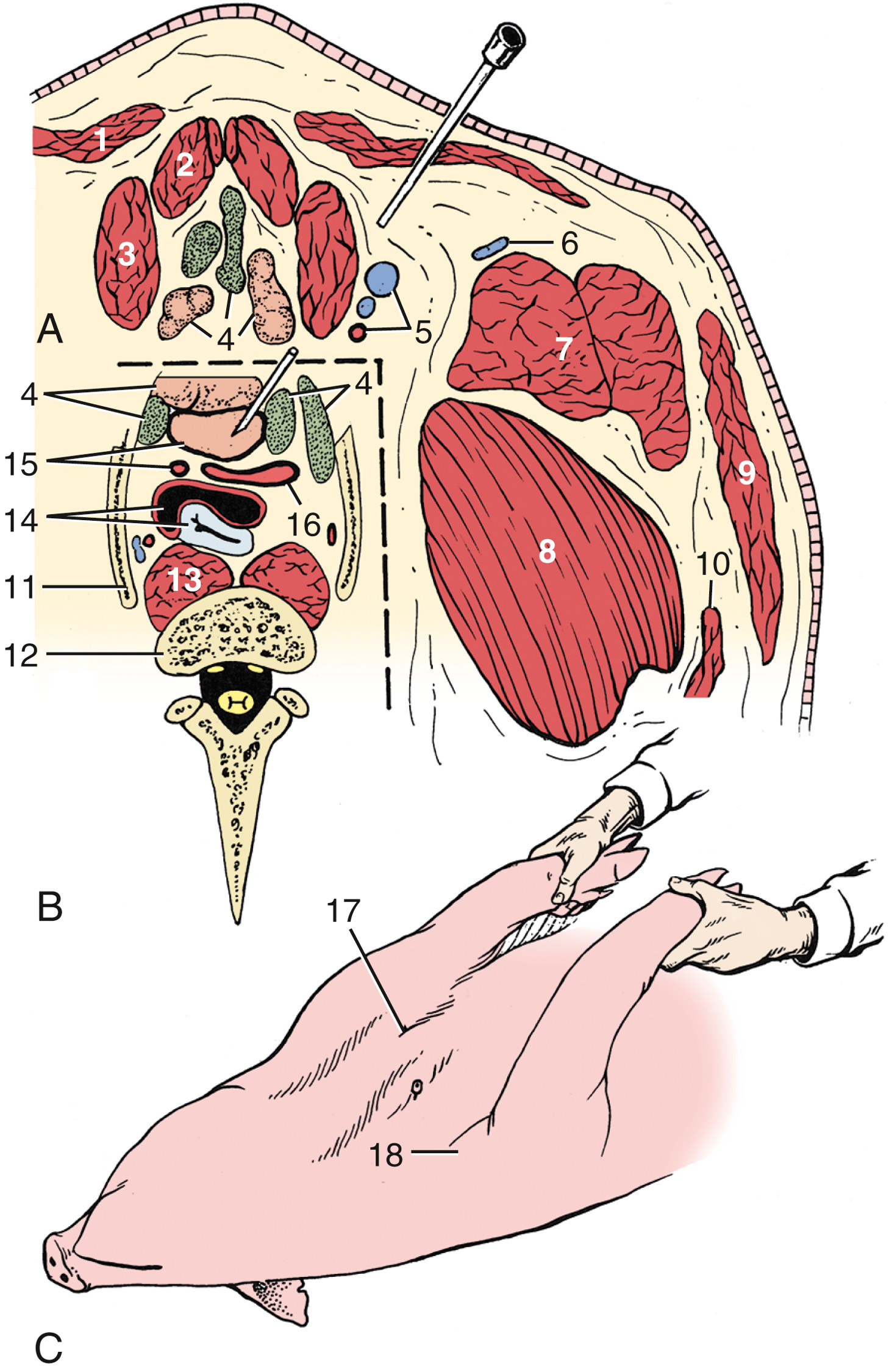
FIG. 32.12 (A) Transverse section of the ventral neck slightly cranial to the manubrium sterni in a pig. (B) The area below and to the left of the broken lines represents the topography at the slightly more caudal level of the first ribs. (C) Pig held on its back for cranial vena cava venipuncture (see needle in position). 1, Cutaneous colli; 2, sternohyoideus; 3, sternocephalicus; 4, lymph nodes and thymus; 5, common carotid artery and external and internal jugular veins; 6, cephalic vein; 7, brachiocephalicus; 8, subclavius; 9, platysma; 10, omotransversarius; 11, first rib; 12, body of C7; 13, longus colli; 14, trachea and esophagus; 15, cranial vena cava and left subclavian artery; 16, bicarotid trunk and right subclavian artery; 17, palpable manubrium sterni; 18, shoulder joint. TVA
Other Structures of the Neck:
- In general, the esophagus is directly dorsal to the trachea at its origin in the upper neck, veers slightly to the left in the mid neck, and then returns to a more dorsal position in the thoracic cavity (Fig. 18.38, 3.29).
- Clinical Notes: it is important to know the location of the esophagus and trachea when performing any scoping procedures or intubations. The esophagus is dorsal to the trachea, and tends to stay just off midline and on the left side of the body. There are several esophageal disorders and procedures to be aware of (especially in equine), including esophagotomy and esophagostomy, to remove foreign objects (e.g., choke or esophageal obstruction) or assist in feeding. (Supplemental Clinical LAS Esophageal anatomy and surgery; Esophageal obstruction)
- Clinical Notes: a tracheotomy can be performed at the midline of the upper neck (below the larynx) by separating the sternohyoideus mm. and cutting between the tracheal cartilages. Temporary tracheotomies are performed when airflow is significantly impaired through the upper airway. The obstruction MUST be proximal to the tracheotomy for it to help. Temporary tracheotomies are also used for intubation for upper airway or dental surgery and when airway access needs are immediate. (Supplemental Clinical LAS Temporary tracheotomy; LAS Tracheal disorders)
- Bovine, equine, and porcine have parotid salivary glands, but in varying amounts. In PORCINE, there is a large amount of parotid salivary gland in the neck and it extends back to the shoulder (Fig. 32.11 and Porcine Cervical Region video). EQUINE may have two portions, near the base of the ear and angle of the mandible (Fig. 19.2), while BOVINE have one region between the angle of the mandible and the base of the ear (Fig. 1-2).
- Bovine, equine, and porcine also have thyroid glands in varying amounts. In PORCINE, the thyroid gland (right and left lobes, often an isthmus between them crossing the trachea ventrally) is caudal in the neck and near the thoracic inlet (32.11,12). In EQUINE and BOVINE, the thyroid glands are cranial in the neck and the lobes lie on the lateral surface of the trachea near the larynx (on both left and right sides). As EQUINE age, their thyroids may enlarge (and this is a normal process of aging).
- In young BOVINE and PORCINE there may be a well-developed cervical thymus present, which can look like a chain of glandular tissue (Fig. 32.11) and can be confused for the thyroid gland.
- In young BOVINE cervical thymus tissue continues into the cranial mediastinum as well as the neck. Young cattle can suffer from thymic lymphosarcoma in the neck or mediastinum. (Supplemental Clinical LAS Lymphosarcoma)

FIG. 3.29 Lateral view of the bovine neck. In the mid-neck the esophagus lies on the left dorsolateral aspect of the trachea. 1, Esophagus; 2, trachea; 3, pharyngeal musculature; 4, sternocephalicus muscle; 5, nuchal ligament. TVA
Viborg’s triangle:
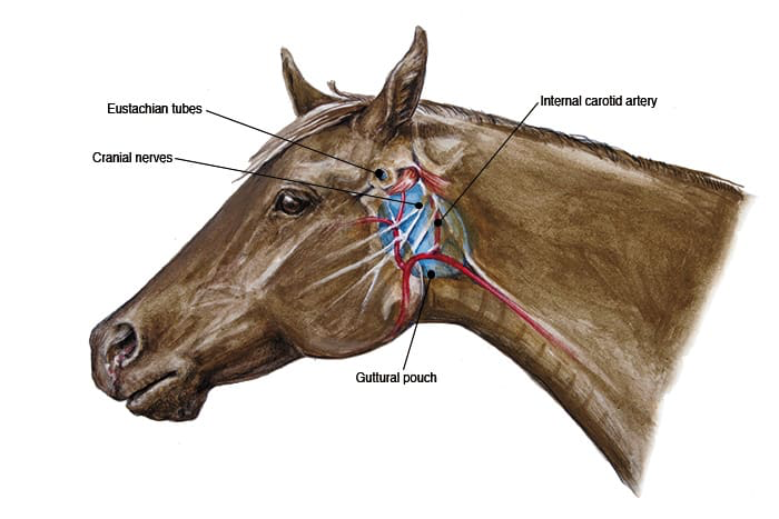
- Clinical Notes: In EQUINE, a region in the neck that is of clinical significance is called Viborg’s triangle. It is an access point, where you can avoid major vessels and nerves, to drain fluid from the guttural pouch. This pouch is a dilatation of the auditory tube in the horse (see image below). There is a medial and lateral pouch on each side of the head. Streptococcus equi can infect this region and the condition is called “strangles” (a highly contagious upper respiratory infection). This infection can grow and put pressure on the airway or pharynx, which is ventral to these pouches.
- The boundaries of Viborg’s triangle:
- Dorsal/caudal – tendon of the sternocephalicus/sternomandibularis m.
- Rostral/cranial – angle of mandible.
- Ventral – linguofacial vein. The linguofacial joins with the maxillary to form the external jugular v.
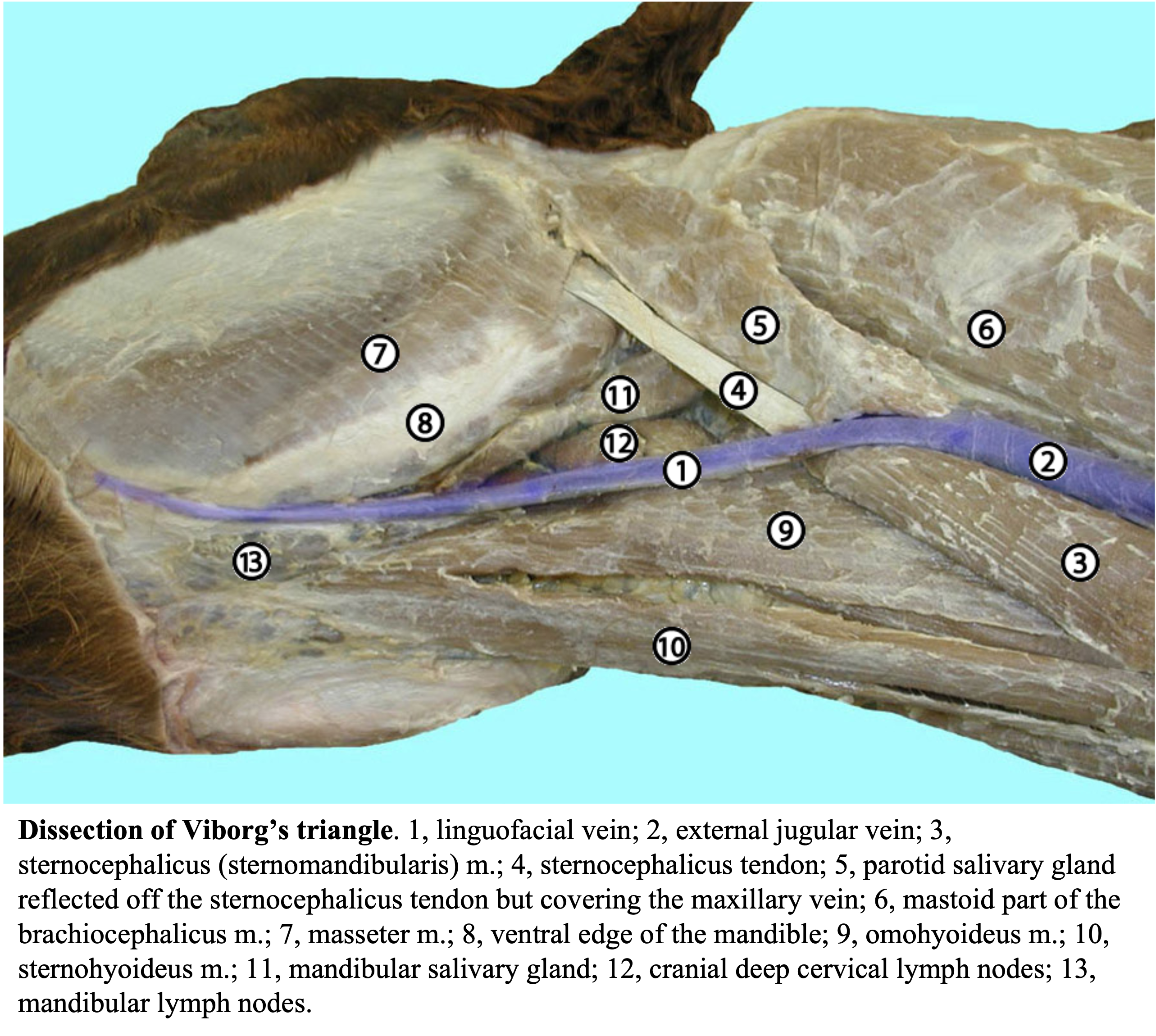
Figure above. Dissection of Viborg’s triangle. Labeled structures: 1, linguofacial vein; 2, external jugular vein; 3, sternocephalicus/sternomandibularis m.; 4, sternocephalicus tendon; 5, parotid salivary gland reflected off the sternocephalicus tendon but covering the maxillary vein; 6, mastoid part of the brachiocephalicus m.; 7, masseter m.; 8, ventral edge of the mandible; 9, omohyoideus m.; 10, sternohyoideus m.; 11, mandibular salivary gland; 12, cranial deep cervical lymph nodes; 13, mandibular lymph nodes.
Lymph structures in the Neck:
- There are several lymph nodes in the neck and head, as well as in the thorax. The images below show the superficial cervical lymph nodes or lymphocenter (in equine).
- In the BOVINE, you may also see small dark spots that look like lymph nodes. These are hemal nodes, and are well developed in most ruminants and are considered to be modified vascular structures. They can be found throughout the body.
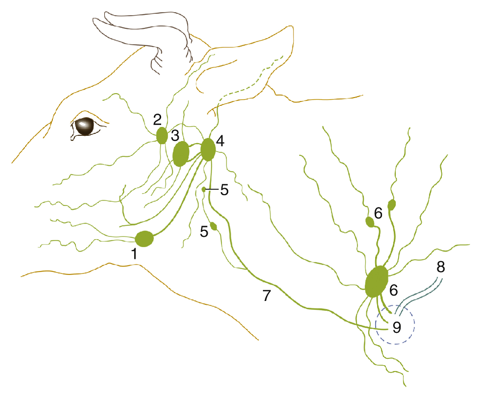
FIG. 25.26 The lymph drainage of the head and neck in bovine. 1, Mandibular lymph node; 2, parotid lymph node; 3, medial retropharyngeal lymph node; 4, lateral retropharyngeal lymph node; 5, deep cervical lymph nodes; 6, superficial cervical lymph nodes; 7, tracheal duct; 8, thoracic duct; 9, area within which lymphatic vessels enter veins. TVA
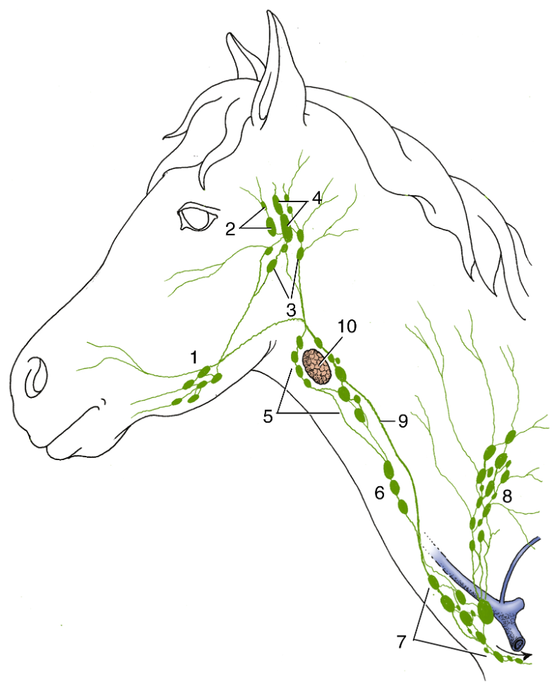
FIG. 18.41 Lymphatic structures of the head and neck of the horse (schematic). 1, Mandibular lymph nodes; 2, parotid lymph nodes; 3, medial retropharyngeal lymph nodes; 4, lateral retropharyngeal lymph nodes; 5, 6, and 7, cranial, middle, and caudal deep cervical lymph nodes, respectively; 8, superficial cervical lymph nodes/lymphocenter; 9, tracheal duct; 10, thyroid gland. TVA
Objective
A5.3 Identify and describe the somatic and autonomic nervous system components found within the neck and thorax.
Autonomic Nervous System Structures: (all bilateral structures)
- The vagosympathetic trunk is in the neck region, within the carotid sheath. This includes sympathetic and parasympathetic nerves (via the vagus aka cranial nerve [CN] X) going to the thoracic and abdominal viscera.
- Similar to small animal anatomy, there are cervical sympathetic ganglia near the thoracic inlet including the cervicothoracic and middle cervical ganglia, which are connected by the ansa subclavia (that wraps around the subclavian a.) to the sympathetic trunk running along the dorsal body wall near the vertebrae in the thoracic cavity.
- The vagus sends off parasympathetic branches to the heart and lungs before continuing caudally in the thorax. As it descends towards the abdominal cavity, the vagus splits into dorsal and ventral branches (one from each side), the right and left branches join either on the ventral aspect of the esophagus or dorsal aspect of the esophagus, forming dorsal and ventral vagal trunks. These trunks then proceed through the diaphragm along with the esophagus to reach the celiacomesenteric ganglia and plexus of the abdominal viscera. These vagal branches are carrying parasympathetic innervation to the abdominal viscera.
- Vagal parasympathetic innervation to the heart, slows the heart. Sympathetic innervation from other nerves would speed up the heart.

Figure (above) Left equine thorax. 1, apex of heart (left ventricle); 2, right ventricle; 3, circumflex branch of left coronary a.; 4, paraconal branch of left coronary a.; 5, left atrium; 6, right auricle; 7, pulmonary trunk; 8, aorta; 9, brachiocephalic trunk; 10, left subclavian artery, in order from caudal, branches are: costocervical, deep cervical and vertebral; 11, phrenic n.; 12, left vagus n.; 13, esophagus; 14, ventral vagal nerve trunk; 15, dorsal vagal nerve trunk; blue asterisk, thoracic duct; black asterisk, rib 7. (http://vanat.cvm.umn.edu/ungDissect/Lab10/Img10-5.html)
Somatic Nervous System Structures:
- The R and L vagus nerves have recurrent laryngeal branches which carry somatic motor to the skeletal muscles of the larynx. Each side has a path around a vessel (aortic arch – left; right subclavian a. – right) and then the continues cranially along the trachea to reach the larynx via the caudal laryngeal n. branches.
- There are also the R and L phrenic nerves, which innervate the diaphragm (a skeletal muscle).
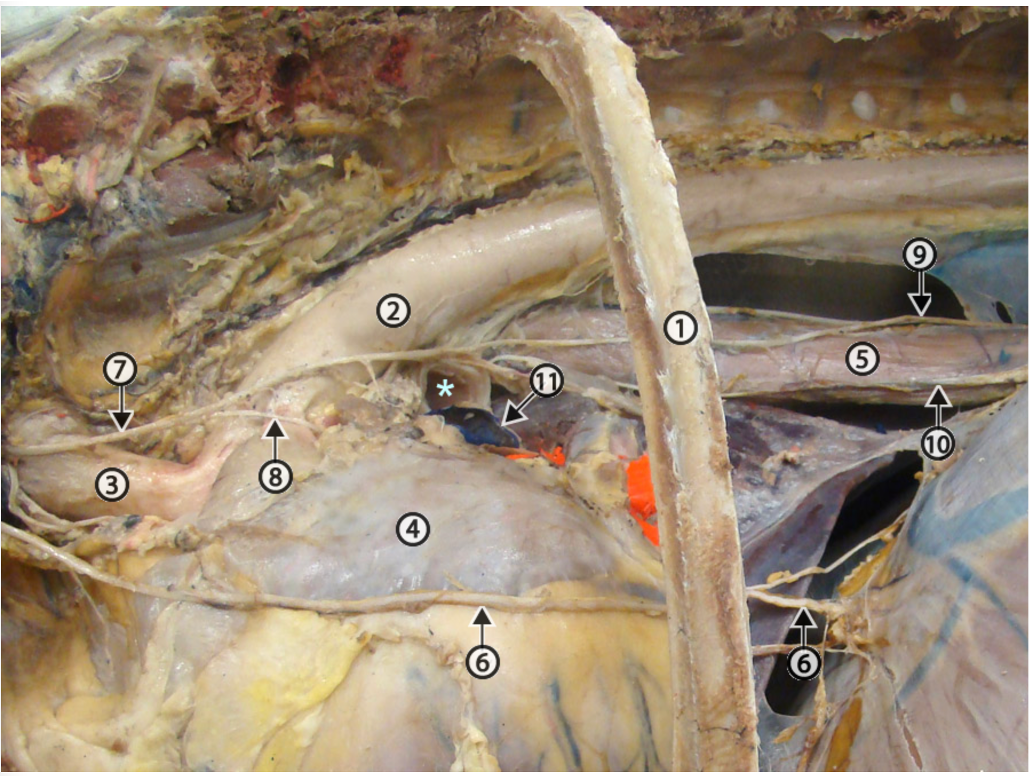
Figure (above) Left side, equine mediastinal dissection. 1, rib 7; 2, aorta; 3, brachiocephalic trunk; 4, left atrium; 5, esophagus; 6, phrenic n.; 7, vagus n.; 8, recurrent laryngeal branch of the vagus n. crossing the ligamentum arteriosum; 9, dorsal vagal nerve trunk; 10, ventral vagal nerve trunk; 11, left pulmonary artery; asterisk, lumen of left primary bronchus. (http://vanat.cvm.umn.edu/ungDissect/Lab10/Img10-3.html)
Objective
A5.4 Identify and describe structures of the mediastinum and diaphragm.
Mediastinum
- The mediastinum is the space between the pleural cavities. The mediastinum includes: thymus, thoracic lymph nodes, heart, aorta, trachea, esophagus, vagus nerves, as well as other nerves and vessels.
- The thymus fills most of the cranial part of the mediastinum, while the heart sits in the middle portion.
- The esophagus and aorta are in the dorsal and caudal parts of the mediastinum, while the caudal mediastinal lymph nodes are also in the caudal mediastinum.
- The tracheobronchial lymph nodes are at the boundary of the mediastinal and pleural cavities, at the branching of the trachea into bronchi.
- Clinical Notes: Caudal mediastinal lymph nodes can be found in the dorsal aspect of the caudal thoracic cavity, near the esophagus and aorta. These may get infected and swell (sometimes due to pneumonia), and could potentially impair function of nearby structures, including the dorsal vagal trunk which innervates the rumen in BOVINE, and could lead to bloat, “vagal indigestion,” and weight loss.
- Clinical Notes: the mediastinum differs in bovine and equine.
- BOVINE have a fibrous and tough connective tissue and pleura creating the boundaries of the mediastinum. This reduces the likelihood of conditions spreading from the mediastinum to the pleural cavities, or spreading from one pleural cavity to the other. This also allows for standing thoracic and pericardial surgery in BOVINE.
- EQUINE, however, have a more fragile mediastinal boundary (i.e., there are perforations that allow communication) and conditions can move from one pleural cavity to the other or from within the mediastinum to the pleural cavities.
- Wounds in the axillary region can lead to pneumomediastinum. (Supplemental Clinical LAS Pleural space disorders)
- Clinical Note: “plucks” are heart plus lung specimens that have been “plucked” out of the chest cavity. Plucks are a common tissue set that is examined during meat inspection. In this situation the evaluation of lymph nodes associated with the pluck (mediastinal and tracheobronchial), and those associated with the carcass (aortic and sternal) are of considerable importance in the detection of diseased animals.
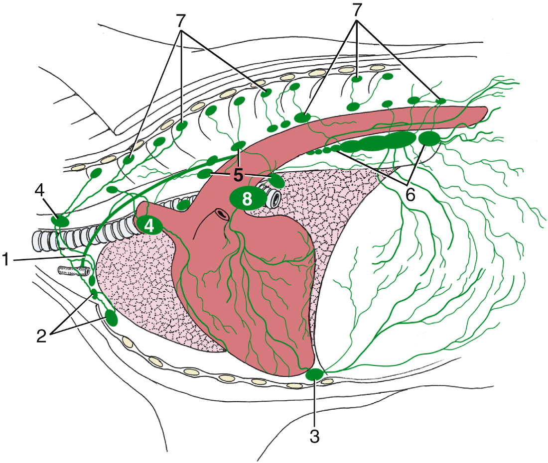
FIG. 27.8 Lymph drainage of the bovine thoracic wall and mediastinum. 1, Thoracic duct; 2, cranial sternal lymph nodes; 3, caudal sternal lymph node; 4, cranial mediastinal lymph nodes; 5, middle mediastinal lymph nodes; 6, caudal mediastinal lymph nodes; 7, intercostal and thoracic aortic lymph nodes; 8, tracheobronchial node. TVA
Diaphragm
- The diaphragm is the muscular partition between the thoracic and abdominal cavities.
- Tendinous center (aka central tendon): a V-shaped tendinous sheet in the middle portion of the diaphragm.
- The muscular parts of the diaphragm (attached to the tendinous center) are the lumbar, costal, and sternal parts; these are named according to their regions of attachment.
- The lumbar part forms the diaphragmatic crura: a left crus and a right crus. The crura (plural) are somewhat triangular slips of muscle and tendinous tissue that attach the diaphragm to the vertebral column and surround structures passing through the diaphragm.
- Passageways through the diaphragm:
-
Aortic hiatus: located dorsally in the central diaphragm, between the muscular diaphragmatic crura. It allows for the passage of the aorta, azygous vein(s), and thoracic duct through the diaphragm.
-
Esophageal hiatus: centrally located in the diaphragm, between the muscular diaphragmatic crura. It allows for the passage of the esophagus, esophageal vessels, and vagal nerve trunks through the diaphragm.
-
Caval foramen: usually located slightly to the right side, passing through the central tendon of the diaphragm. It allows for the passage of the caudal vena cava through the diaphragm.
-
Objective
A5.5 Identify and describe the lungs and associated structures of the equine, bovine, and porcine; summarize notable species differences.
Pleural Cavity – the pleural cavity/space is found between the two layers of pleura (visceral and parietal) within the thoracic cavity.
- Pulmonary (aka visceral) pleura: the serous membrane on the surface of the lungs.
- Parietal pleura: the serous membrane on the “walls” of the thorax, including the boundaries formed by the ribs, diaphragm, and mediastinum and has costal, diaphragmatic, and mediastinal pleura, respectively.
- Line of pleural reflection: the line along which diaphragmatic pleura reflects off the diaphragm and onto the ribs as costal pleura. This is a clinically important landmark as it is the caudoventral extent of the pleural cavities. (Figs. 20.1, 27.2A)
- Costodiaphragmatic recess: the narrow angled space (lined by pleura) between the convex surface of the diaphragm and the thoracic body wall. Lung tissue usually doesn’t extend into this space, and therefore makes it a place where you can collect a sample of pleural fluid without entering/damaging lung tissue; aka thoracocentesis/thoracentesis (see Clinical Notes below).
- In large animals, the edge of the rib cage (i.e., costal arch) is more caudal while the lung field is more cranial (especially in ruminants, see image below on right Fig. 27.2A). In bovine, the abdominal contents push cranially into the thoracic cavity and are found deep to the caudal-most ribs.
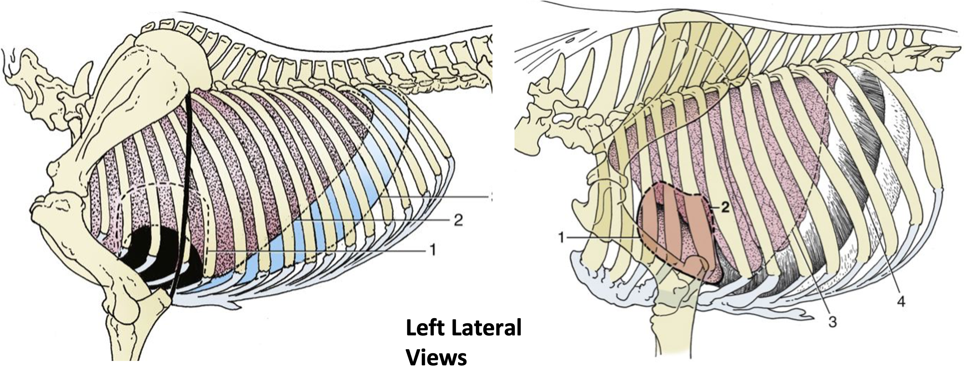
Figures above, Equine (left, Fig. 20.1) outline of thorax cavity structures from a left lateral view: 1, outline of heart; 2, basal border of lung, 3, line of pleural reflection. Bovine (right, Fig. 27.2A): 1,2, cranial and caudal extents of the heart; 3, basal border of lung; 4, line of pleural reflection. TVA
- Clinical Notes: when conducting a physical exam, you will need to know where the lung fields can be auscultated in each species. Auscultation of the lungs, especially in bovine, should be conducted more cranially. Equine have a much more extended rib cage and increased expansion of their lung field in the caudal and dorsal directions. Also note the heart fields and cardiac notches on the left and right sides in large animals. (see images above [Figs. 20.1, 27.2A]; below, only left sides shown)
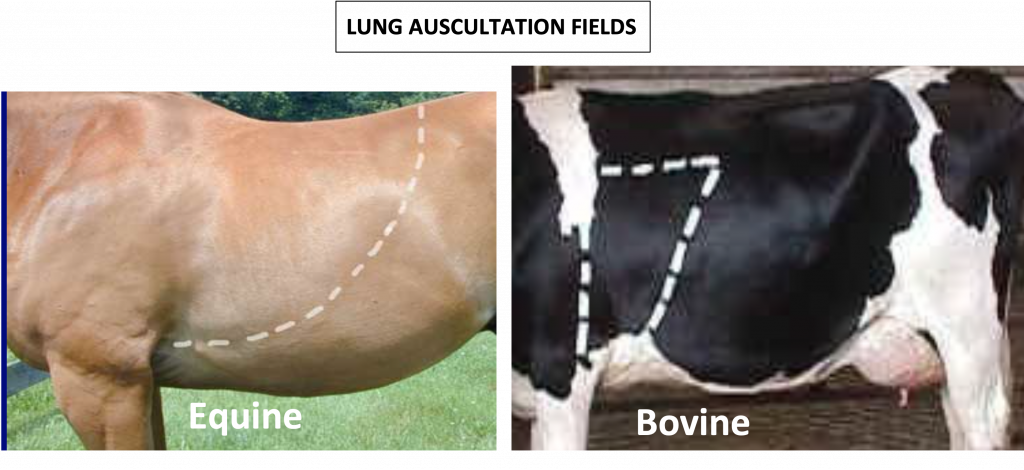
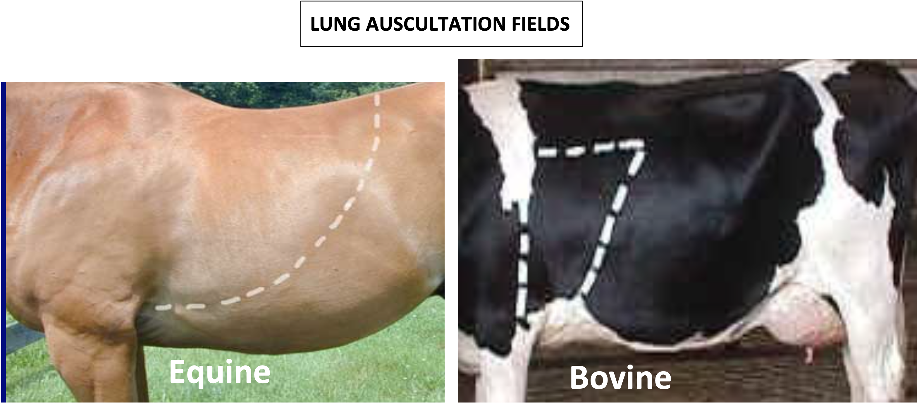
Figures above, Equine (left) and bovine (right) left lateral view of external thorax, with lung fields outlined with dotted lines to indicate where auscultation of the lungs can occur.
- Clinical Notes: to perform a thoracentesis, and remove fluid from the pleural space/pleural cavity, you would insert your needle/trocar as to not pierce the lung tissue. It is likely you would be able to perform this procedure with ultrasound guidance. In horses, the line of pleural reflection (and costal arch) is less steep due to their longer thorax. In bovine, the line of pleural reflection is steeper due to their deeper and shorter thorax. The diaphragm is at a slant and the thoracic and abdominal cavities overlap. Additionally, remember the intercostal a., v., and n. run along the caudal border of the ribs, so be aware of these so as not to damage them when inserting your needle into the intercostal spaces between ribs and through intercostal mm. running in these spaces. Intercostal n. blocks are also relevant to lateral thoracotomy or fractured ribs (DVM 360 link).
Comparative Lungs (equine, bovine, porcine)
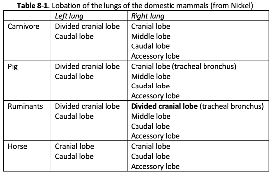
- The lobes of the lungs are named according to the branching of the principal (primary) bronchi from the trachea. Each primary bronchus then branches into lobar bronchi. Bovine and porcine have a separate bronchus called the tracheal bronchus. (See Table 8-1 and Fig. 8-4)
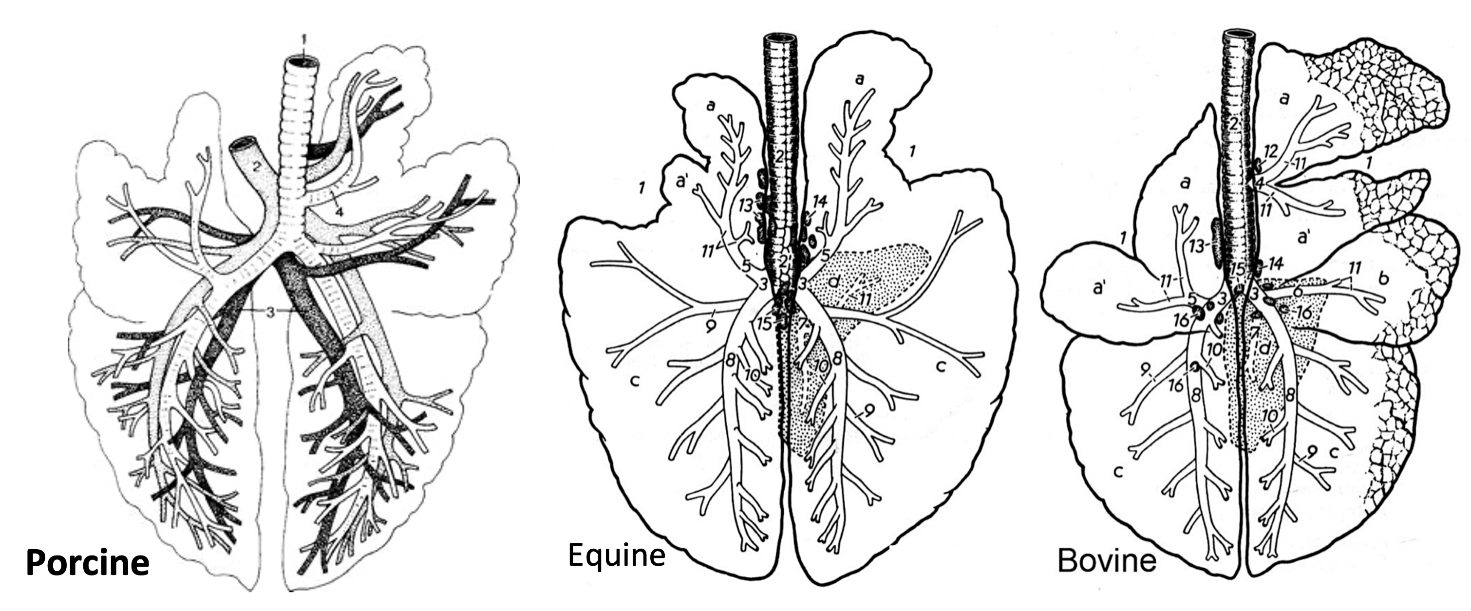
Figure 8-4 (Above) Porcine, bovine, and equine lungs, dorsal schematic view. 1, cardiac notch; 2, trachea; 3, primary bronchus; 4, tracheal bronchus (not in horse); 5, cranial bronchus; 6, middle bronchus (not in horse); 7, accessory bronchus; 8, caudal bronchus; 9-11, segmental bronchi; 12-15, tracheobronchial lymph nodes; 16, pulmonary lymph nodes; a, cranial parts of cranial lobe(s); a’, caudal parts of cranial lobe(s); b, middle lung lobe; c, caudal lung lobes; d, accessory lung lobe.
- EQUINE lungs (Fig. 8-4, middle) lack fissures between the lobes, but the lobes are still based on the branching of the left and right principal bronchi.
- The left lung is divided into cranial and caudal lobes.
- The right lung is divided into cranial, caudal, and accessory lobes. The HORSE lacks the right middle lobe.
- OX and PIG (Fig. 8-4, right and left, respectively) lungs are similar in lobation to the dog and cat.
- The left lung consists of two lobes, a cranial lobe (with cranial and caudal parts separated by a cardiac notch and fissure) and a caudal lobe.
- The right lung consists of cranial (with cranial and caudal parts in the OX), middle, caudal and accessory lobes.
- Species differences to note:
- OX and PIG: have a separate bronchus to the right cranial lobe which arises cranial to the primary bifurcation of the trachea, and is called the tracheal bronchus (Fig. 8-4/4).
- OX: has a subdivided right cranial lobe (cranial and caudal parts).
- HORSES and OX have cardiac notches. Equine have distinct right and left sided cardiac notches, while cardiac notches in bovine are less distinct (Fig. 8-4/1).
- General: Lobulation (distinct lobules separated by fibrous tissue within the lung parenchyma) is obvious in both the OX and PIG. SHEEP and HORSES lack obvious lobulation.
Objective
A5.6 Identify and describe the heart structures and associated major vessels of the thorax; summarize the flow of blood into, through, and out of the heart.
Pericardium
- The heart is surrounded by a membrane called the pericardial sac, which consists of three inseparable components. These are, from outermost to innermost: pericardial mediastinal pleura, fibrous pericardium, and serous parietal pericardium.
- The serous visceral pericardium (aka epicardium) is the serous membrane tightly adhered to the surface of the heart itself.
- The endocardium is a membrane that lines the internal surfaces of the heart.
- Clinical Notes: Hardware disease or traumatic reticuloperitonitis can be with or without pericarditis (more on this in the Application 5 live session and Application 7 eBook chapter).
- Cows eat all sorts of stuff. Any foreign bodies usually end up in the reticulum (e.g., nails, wire, ropes, etc.) Sharp objects can be pushed through the reticular wall, due to the churning in the rumen and reticulum. A hole in the reticulum can leak ingesta and cause peritonitis and abscesses. Further migration can occur, penetrating through the diaphragm, and then the pericardial sac of heart. This can lead to pericarditis and pleuritis.
Heart Structures
- The apex (ventral pointing tip of the heart) is associated with the left ventricle (LV). The right ventricle (RV) is cranial to the LV in a left lateral view (see Figure below).
- The ear-like appendages sitting upon each atrium (right atrium [RA], left atrium [LA]), near the dorsal aspect of the heart, are the right and left auricles. Both can be seen from the left lateral (auricular) view (see Figure below).
- The coronary groove contains fat and is between the atria and ventricles. The right and left coronary aa. are embedded in the fat of this groove between the right and left atria and ventricles, respectively.
- Arteries:
-
- The RV ‘cones down’ (aka conus arteriosus) to terminate at the base of the pulmonary trunk. Deoxygenated blood flows through the pulmonary/pulmonic valve (with semilunar cusps) into the pulmonary trunk and then splits into right and left pulmonary aa. to enter the right and left lungs, respectively.
- Cranial to the pulmonary trunk is the base of the aorta where the right and left coronary aa. originate. The coronary aa. provide oxygenated blood to the heart muscle. The ligamentum arteriosum (fetal ductus arteriosus) is a fibrous connection between the pulmonary trunk and the aortic arch. During development it was patent and allowed blood to directly enter the aorta, bypassing the fetal lungs.
-
- The left coronary a. traverses under the pulmonary trunk and immediately branches.
-
-
- The paraconal (interventricular) branch runs ventrally between the right and left ventricles. The paraconal name is derived from its “parallel” relationship to the conus arteriosus.
- The circumflex branch lays in the coronary groove and circles around the base of the heart, between the left auricle/atrium and ventricle.
- In RUMINANTS (and canines) the left (via the circumflex) coronary a. terminates as the subsinuosal (interventricular) a.
-
-
- The right coronary a. arises from the base of the aorta and travels in the coronary groove between the right auricle/atrium and ventricle.
- In the HORSE and PIG, the right coronary a. terminates as the subsinuosal (interventricular) branch (Fig. 8B-7/2′). This name is in reference to its relationship to the coronary “sinus,” as it is “below” the coronary sinus and between the ventricles.
- The right coronary a. arises from the base of the aorta and travels in the coronary groove between the right auricle/atrium and ventricle.
- Aorta
- The ascending aorta is quite short and gives rise to the right and left coronary aa.
- In the HORSE and RUMINANT, the aortic arch gives rise to:
- A large brachiocephalic trunk. (Fig. 4 below)
- The left and right subclavian aa. are branches of the brachiocephalic trunk, and give off several branches including the internal thoracic aa. (which run alongside the inside of the sternum). The subclavian aa. eventually become the axillary aa. once they exit the thoracic body wall.
- The bicarotid trunk is another branch of the brachiocephalic trunk, and gives rise to the right and left common carotid aa.
- A large brachiocephalic trunk. (Fig. 4 below)
- The descending aorta (thoracic and abdominal) gives rise to vessels supplying thoracic structures, abdominal organs, and terminates in the caudal abdomen as iliac aa.
Figure (above) Left equine thorax. 1, apex of heart (left ventricle); 2, right ventricle; 3, circumflex branch of left coronary a.; 4, paraconal branch of left coronary a. (great cardiac v. alongside); 5, left atrium; 6, right auricle; 7, pulmonary trunk; 8, aorta (descending); 9, brachiocephalic trunk; 10, left subclavian artery, in order from caudal, branches are: costocervical, deep cervical and vertebral; 11, phrenic n.; 12, left vagus n.; 13, esophagus; 14, ventral vagal nerve trunk; 15, dorsal vagal nerve trunk; blue asterisk, thoracic duct; black asterisk, rib 7. (http://vanat.cvm.umn.edu/ungDissect/Lab10/Img10-5.html)
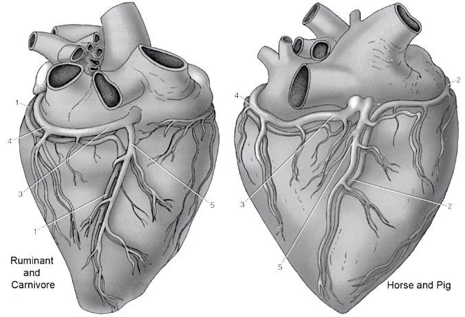
Figure 8B-7 (Above) Cardiac vessels in ruminants/carnivore and horse/pig, right-sided view. 1, circumflex branch of the left coronary a.; 1’, subsinuosal a.; 2, right coronary a.; 2’, subsinuosal a.; 3, 4, great cardiac v.; 5, middle cardiac v.

Fig 4. The aortic arch branching pattern found in bovine (and equine) has a single brachiocephalic trunk originating from the aortic arch and eventually splits into the bilateral subclavian arteries and a bicarotid trunk. AJNR Am J Neuroradiol 27:1541– 42
- Veins:
- The great cardiac v. is a major route for venous drainage of the left ventricular muscle mass. It begins at the paraconal interventricular groove and ends at the coronary sinus (which empties into the RA). The coronary sinus sits within the coronary groove.
- The cranial vena cava and caudal vena cava empty into the RA.
- The cranial vena cava collects blood from the external jugular vv., internal jugular vv. (bov, por), and subclavian vv.
- The right azygous v. (found in equine and some bovine) drains into the RA, sometimes via the cranial vena cava. (see image below).
- The left azygous v. (found in bovine and porcine) often drains into the great cardiac v. and then to the coronary sinus.
- Pulmonary vv. enter the LA bringing oxygenated blood from the lungs into the LA.

Figure (above) Right equine mediastinal dissection, the right lung is removed, the left is present. 1, first rib; 2, rib 7; 3, right ventricle; 4, left ventricle; 5, caudal vena cava; 6, cranial vena cava; 7, right atrium; 8, right coronary a. (great cardiac v. and coronary sinus cranial to); 9, subsinuosal vessels, 10, (right) azygous v.; 11, costocervical v.; 12, supreme intercostal v.; 13, longus colli m.; 14, lumen of right primary bronchus; 15, thymus. http://vanat.cvm.umn.edu/ungDissect/Lab10/Img10-9.html
Inside the Atria and Ventricles
- The cranial and caudal vena cavae enter into the RA. Inside the RA there are several internal structures. (Figs. 7.8, 8B-8 and equine fresh heart)
- The intervenous tubercle is a ridge of tissue on the inside wall of the atrium that helps direct the flow of blood into the RA. Near the intervenous tubercle you may see a depression called the fossa ovalis (fetal foramen ovale), which once allowed blood to pass from the RA to the LA.
- Separating the termination of the cranial vena cava from the inner chamber of the right auricle is a muscular ridge called the terminal crest (crista terminalis), so named because it marks the end of the cranial vena cava but also the entrance to the right auricle.
- The coronary sinus is the entry point for venous blood from the heart and has a small orifice/entrance close to the caudal aspect of the right atrioventricular (AV) orifice. The great cardiac v. collects blood from the left and right sides of the heart and directs it into the coronary sinus.
- The right atrioventricular (AV) orifice (i.e., the opening between right atrium and ventricle) and right AV (aka tricuspid) valve are at the junction of the RA and the RV.
- Blood flows through the right AV valve and into the RV which eventually narrows to a region called the conus arteriosus (cone-shaped area). From here blood flows through the pulmonary valve, pulmonary trunk, right and left pulmonary aa., and then lobar pulmonary aa. to the lungs.
- The RV internally has: chordae tendineae (connective tissue “heart strings” that connect the AV valve cusps to papillary muscles), papillary muscles (muscular bumps projecting from the wall of the ventricle and attached to chordae tendineae), and a trabecula septomarginalis (aka moderator band; a band of connective tissue connecting the interventricular septum to the ventricle wall). The moderator band will have conduction fibers to help with the stimulation of the heart muscle. Based on observations, this structure is often found in sheep, horse, pigs, and carnivores. It may also be variable in size and location. (Figure below, equine fresh heart)
- The pulmonary vv. carrying oxygen-rich blood from the lungs enter the LA. There is a left AV orifice, left AV (aka mitral, aka bicuspid) valve, and chordae tendineae attaching to papillary muscles and AV valve cusps. Blood exits the LV by passing through the aortic valve located at the aortic root of the aorta. The aortic valve also has semilunar cusps, and deep to these cusps are where the right and left coronary aa. arise.

Figure 8B-8 (Above) Interior of the right atrium of the dog (similar to other species). Labeled structures: cranial and caudal vena cava, azygous v., aortic arch, coronary sinus opening, intervenous tubercle, crista terminalis, right atrium, right auricle, right AV valve cusp, right ventricle, left ventricle, asterisks indicate pulmonary vv. and aa. (Modified from Guide to Dissection of the Dog, 7th ed. Fig. 2-23)
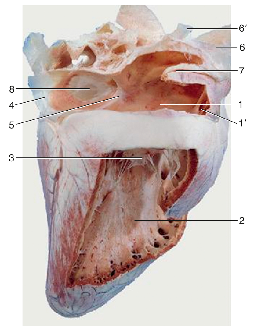
FIG. 7.8 Overview of the interior of the right atrium and right ventricle of the equine heart. 1, Right atrium; 1′, right auricle; 2, right ventricle; 3, right atrioventricular valve; 4, caudal vena cava; 5, intervenous tubercle; 6, cranial vena cava; 6′, right azygos vein; 7, terminal crest; 8, fossa ovalis. TVA
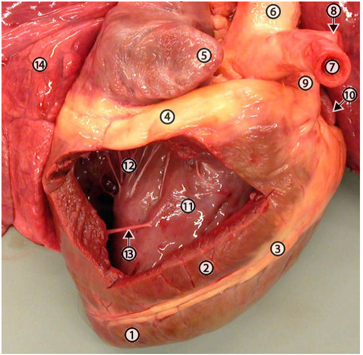
Figure (above) Equine fresh heart, inside view of right ventricle. 1, apex of heart (always marks the left ventricle); 2, right ventricle; 3, paraconal groove filled with fat; 4, coronary groove filled with fat; 5 auricle of right atrium; 6, aorta; 7, lumen of brachiocephalic a.; 8, left subclavian a.; 9, pulmonary trunk; 10, auricle of left atrium; 11, interventricular septum; 12, clump of five chordae tendineae; 13, trabecula septomarginalis; 14, lung. http://vanat.cvm.umn.edu/ungDissect/Lab10/Img10-11.html
Clinical Notes: the auscultation points for heart valves are important to remember. The pattern of valves and regions for auscultation are similar to what was described in carnivore anatomy.
- On the left side of the thorax, you can hear the pulmonic, aortic, and mitral valves (P A M) in the 3rd, 4th, and 5th intercostal spaces (ICS), respectively.
- On the right side of the thorax, you can hear the tricuspid valve (T) in the 4th intercostal space (ICS). There can be some variability. (see images below)
- The toughest part is to get the stethoscope up under the triceps brachii mm. and along the body wall to hear the heart in these spaces. Use the point of the elbow (olecranon) as a landmark and then go deep to the forelimb and triceps m., shoving the stethoscope forward/cranial.
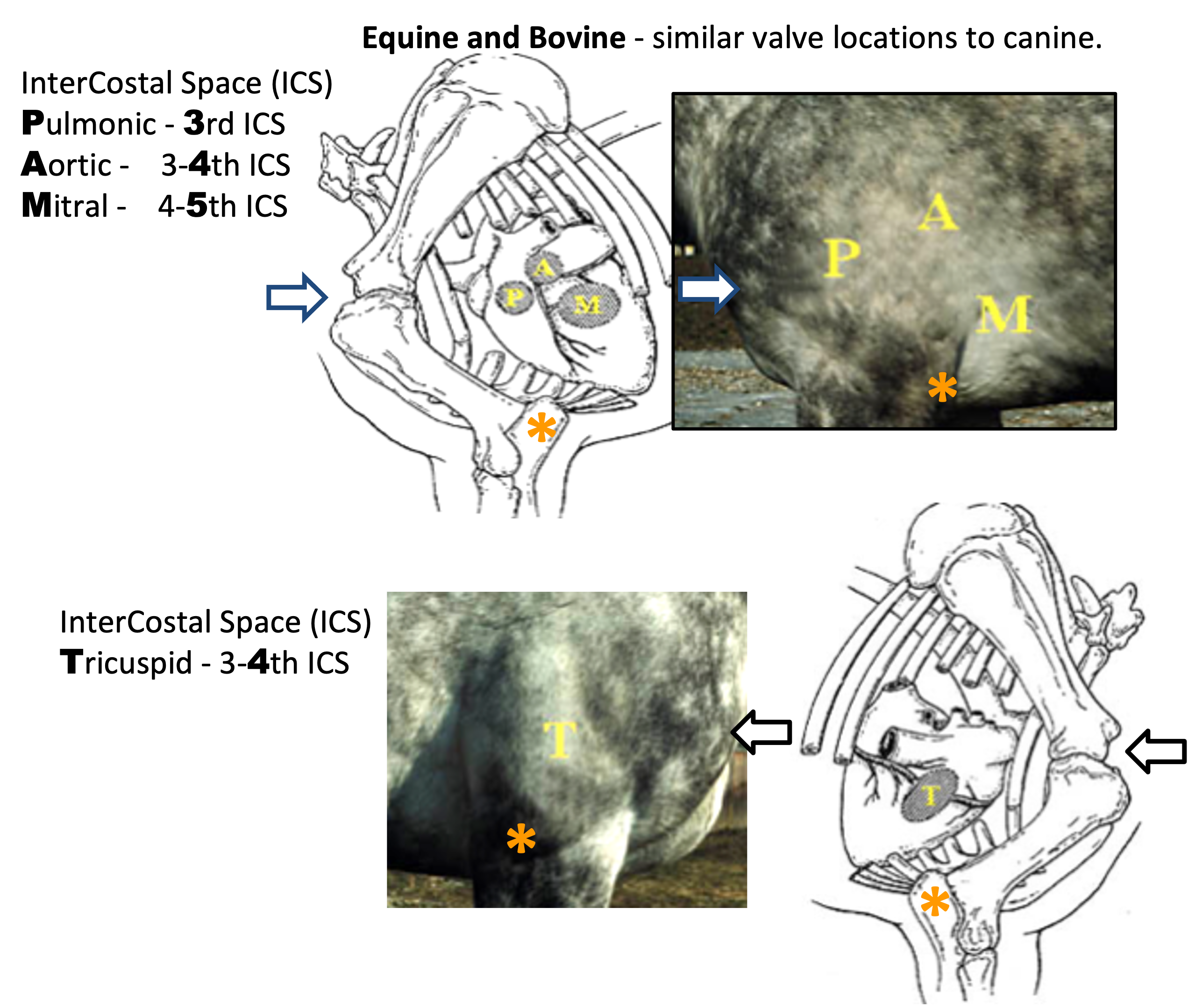
Figures (above) right (bottom) and left (top) sided views of heart auscultation points in large animals. P, pulmonic; A, aortic; M, mitral; T, tricuspid; asterisk, point of ulna the olecranon.
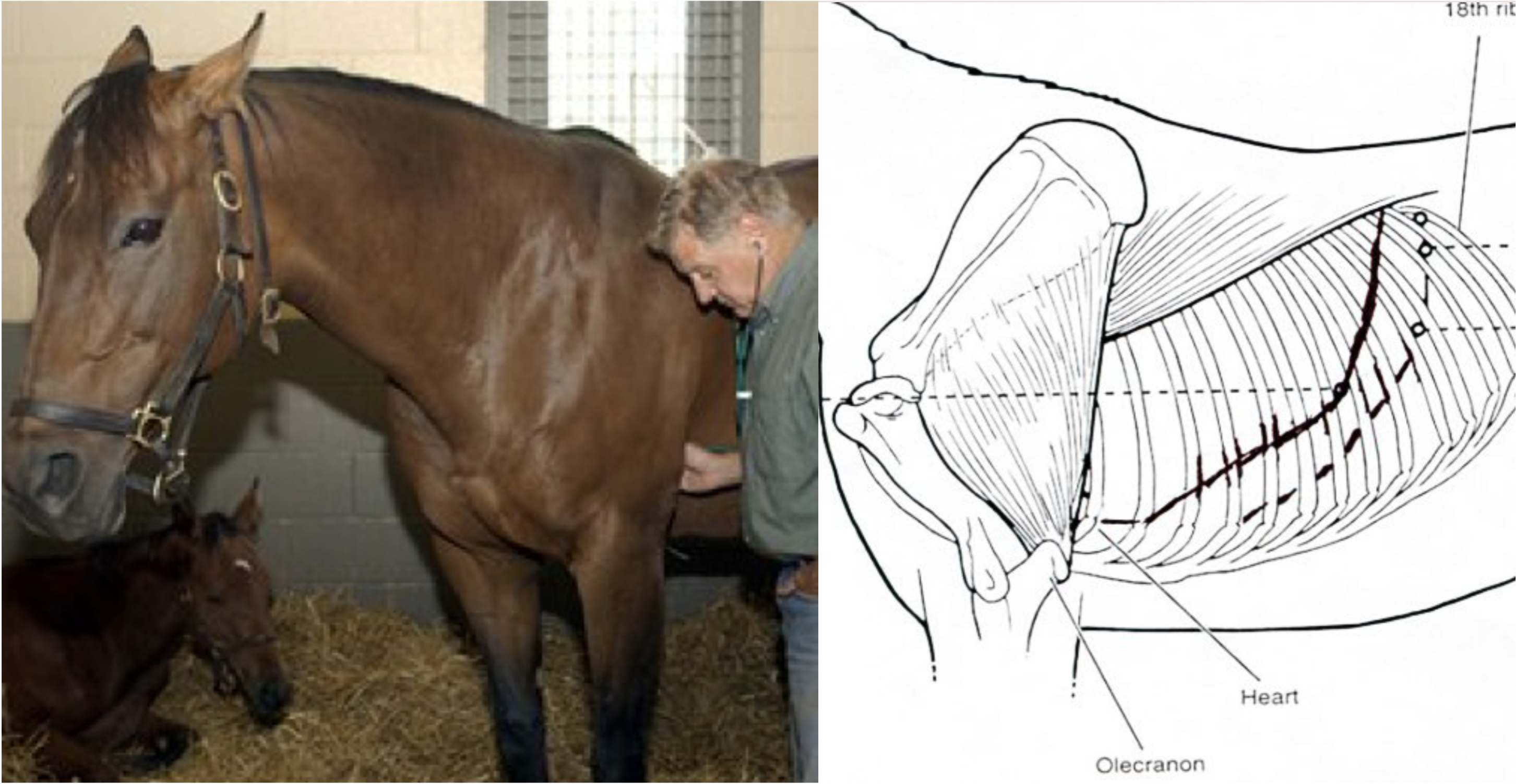
Figure (above): Heart sounds are deep to the forelimb (i.e., dig the stethoscope into the axillary region).
Be able to list the normal route of blood flow into, through, and out of the heart:
right atrium > right AV valve > right ventricle > pulmonic/pulmonary valve > pulmonary trunk > pulmonary aa. > lungs > pulmonary vv. > left atrium > left AV valve > left ventricle > aortic valve > aorta > out to the body > right atrium > repeat
- Oxygen-poor blood from the body and heart: cranial and caudal vena cava, and the coronary sinus (from great cardiac vein) empty into the RA. From the RA, blood flows through the right AV orifice and valve into the RV. From the RV, blood flows through the pulmonic valve and the pulmonary trunk, and exits via the right and left pulmonary aa. to reach the lungs. At the lungs, blood is re-oxygenated.
- Oxygen-rich blood to the body and heart: The pulmonary vv. collect the oxygen rich blood and deliver it to the LA. From the LA the blood flows through the left AV orifice and valve into the LV. From the LV, blood flows through the aortic valve, and to the heart itself via the right and left coronary aa. (the first branches of the aorta) and to the rest of the body via the other portions and branches of the aorta (arch, descending).
