
Part 1: Neck
Abby Brown
IMPORTANT NOTE: For this part of the chapter, the Guide directions will refer to either “ALL specimens” (meaning dissect pony and calf specimens in the same way) or to one species in particular, i.e., “ALL (PONY) specimens” or “ALL (CALF) specimens.” This added instructional note is needed because the dissection may differ slightly according to species.
Terminology Notes: Four anatomic adjectives, nuchal, cervical, jugular, and colli all refer to the neck. (The cervix is the “neck” (narrow part) of the female genital tract. Colli is the Latin root of the English word ‘collar’. Be careful to avoid confusion with coli which refers to the colon, as in E. coli, a bacterium found in the colon.) The term ‘poll’ refers to the region between the ears and should not be confused with ‘polled’ which refers to a bovid animal without horns.
Skin Reflection and the Nuchal Ligament
DISSECTION NOTE: You will be dissecting both sides of the neck on your hanging specimens in the same manner unless otherwise noted (i.e., RIGHT side or LEFT side notation) for this lab. Some structures are midline structures and will only be dissected once, but should be viewed by all group members.
- ALL specimens: If needed, incise, reflect, and remove any remaining skin in the neck region. (Be careful to preserve the vessels in the throat region.)
- Pony skin reflection: If needed, on both sides of your specimen, incise any remaining skin of the neck region along the ventral border of the mane.
- Make a rostral incision that extends from just caudal to the ears, moving toward ventral midline, following the angle of the mandible (see Fig. 3-1).
- Reflect the skin ventrally. Use care in this reflection so that you preserve the underlying vessels in the throat region.
- Discard the reflected skin.
- Calf skin reflection: If needed, extend a dorsal midline incision from the region of the poll to the withers.
- Make vertical incisions from the poll (region between the ears) along the caudal border of the mandible. (Make this cut as far cranially as the halter on the calf’s head will allow.)
- Carefully reflect and remove the cervical skin (and discard it).
- Pony skin reflection: If needed, on both sides of your specimen, incise any remaining skin of the neck region along the ventral border of the mane.
- ALL (PONY) specimens: Reflect the nuchal fatty crest caudally. Be careful not to damage the underlying nuchal ligament.
-
- Deep to the mane lays an interwoven mass of fat and fibrous tissue called the nuchal fatty crest, which is especially prominent in stallions. Identify the nuchal fatty crest in the pony specimen.
- Important Dissection Note: Ventral to the nuchal fatty crest lays an important two-part elastic tissue called the nuchal ligament, which you will identify in the next step.
- Reflect and remove the remaining mane skin + the nuchal fatty crest as far caudally as the withers; take care not to damage the underlying nuchal ligament tissue!
- Deep to the mane lays an interwoven mass of fat and fibrous tissue called the nuchal fatty crest, which is especially prominent in stallions. Identify the nuchal fatty crest in the pony specimen.
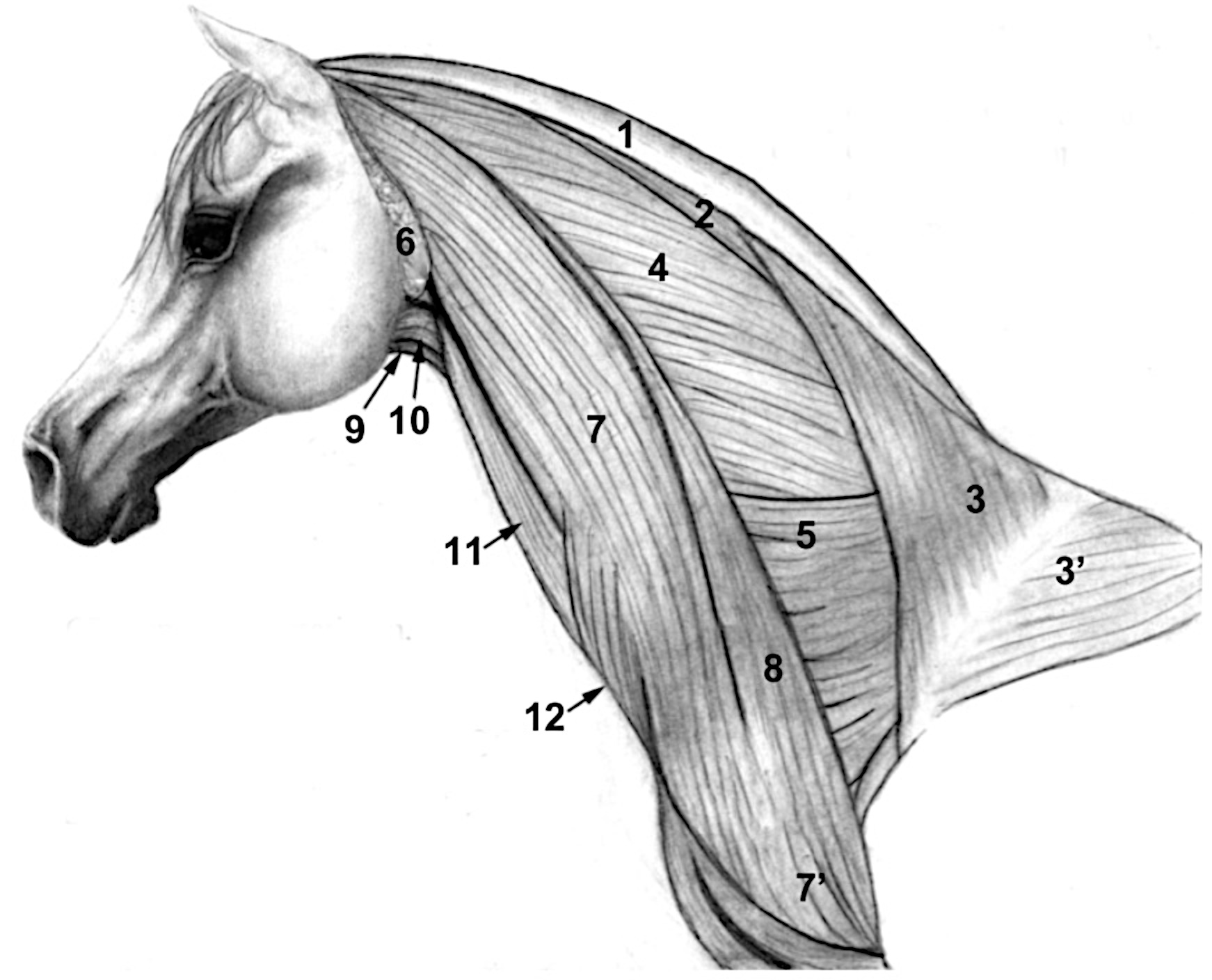
Figure 3-1. Pony neck dissection, lateral view. 1, nuchal fatty crest; 2, rhomboideus m.; 3, 3′, trapezius m.; 4, splenius m.; 5, serratus ventralis m.; 6, parotid salivary gland; 7 cleidocephalicus m. (cleidomastoideus m.); 7′ cleidobrachialis m.; 8, omotransversarius m. fused to cleidocephalicus m.; 9, sternohyoideus m.; 10, omohyoideus m.; 11, sternomandibularis m.;12, cutaneous colli m. Note: The cutaneous colli m. is a V shaped muscle that originates from the sternum and fans out laterally over the ventral edge of the brachiocephalicus m. (see Fig. 3-5 for a cranial view). The external jugular vein is black, lying between 7 and 11.
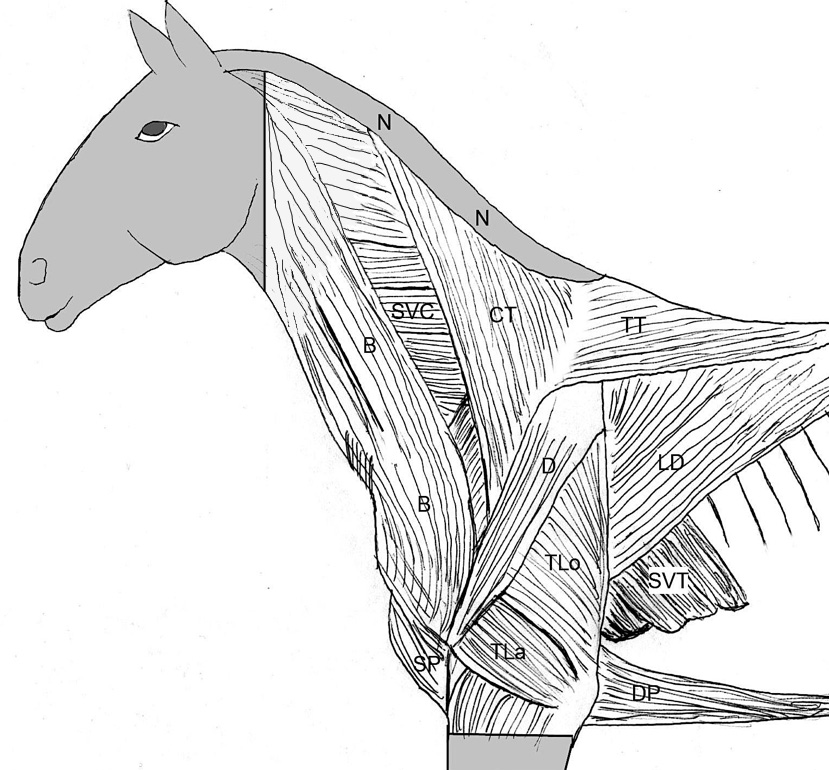
(Duplicate) Figure 1-2. Extrinsic and intrinsic muscles of the horse (upper) and ox (lower). (N) Indicates the location of the nuchal fatty crest present only in the horse. Extrinsic muscles: CT, Cervical trapezius; TT, thoracic trapezius; B, brachiocephalicus;SP, superficial pectorals; DP, deep pectorals; LD, latissimus dorsi;SVC, serratus ventralis cervicis; SVT, serratus ventralis thoracis. Labeled intrinsic forelimb muscles: D, Deltoideus; TLo, long head of the triceps brachii; TLa, lateral head of the triceps brachii. (Not shown is the cutaneous trunci m. which may come off when the skin is reflected. If present, it will cover the upper forelimb and structures caudal to it.) (Modified from Budras and Habel, 1st ed.)
3. ALL specimens: Identify and separate the two parts of the nuchal ligament: funicular and laminar.
-
- The nuchal ligament is made up of two parts: a dorsally positioned, round, cord-like funicular part and a deeper, sheet-like laminar part. Identify both parts of the nuchal ligament.
- Calf Dissection Note: In the calf (and dog), the rhomboideus m. extends over the nuchal ligament, but in the horse this muscle lies lateral to it.
-
- In the calf, reflect the rhomboideus m. as needed to see the nuchal ligament.
- To best visualize the nuchal ligament of the calf, approach from the left side by reflecting the muscle stumps/remnants of the cervical part of the trapezius and rhomboideus mm.
-
- Calf Dissection Note: In the calf (and dog), the rhomboideus m. extends over the nuchal ligament, but in the horse this muscle lies lateral to it.
- Dissection Note: The nuchal ligament attaches to the caudal skull region (an area that was exposed on your pony when your specimen was suspended with a wire loop).
- ALL (PONY) specimens: Explore the caudal region of the skull for remnants of this ligamentous attachment site.
- ALL (CALF) specimens: The attachment of the nuchal ligament to the caudal skull will be more ventral than in the pony. (This is due to the caudal extension of the large frontal bone of the ox.) If possible, look for the attachment point of the nuchal ligament on the caudal aspect of the skull. (Note that the halter suspending the calf specimen may prevent this in some specimens.)
- ALL specimens: Differentiate the two parts of the nuchal ligament by locating the midline groove on the dorsal side of the (more superficially located) funicular nuchal ligament and carefully separate it into right and left halves throughout its length.
- In the middle of the neck, retract the funicular nuchal ligament laterally to expose the underlying (deeper) laminar nuchal ligament. Note that the laminar part extends cranially only as far as the caudal edge of the spine of the axis (TVA 535).
- The nuchal ligament is made up of two parts: a dorsally positioned, round, cord-like funicular part and a deeper, sheet-like laminar part. Identify both parts of the nuchal ligament.
4. ALL (PONY) specimens: Attempt to identify the atlanto-occipital space and any remaining parts of the atlanto–occipital membrane.
-
- At the base of the skull, identify the atlanto-occipital space. The wire loop suspending your pony passes around the dorsal arch of the atlas, penetrating this space between the atlas and the occipital bone of the skull.
- Attempt to identify any visible remnants of the atlanto-occipital membrane which is the membranous covering of the atlanto-occipital space. (This membrane was cut/reflected to allow passage of the wire loop to suspend each pony.)
NECK MUSCLES AND ASSOCIATED STRUCTURES
5. Hypaxial muscles generally lie ventral to the transverse processes of the vertebrae; in this chapter, we will dissect some hypaxial muscles associated with the neck and thorax. Epaxial muscles generally lie dorsal to the transverse processes of the vertebrae and are associated with the vertebral column and ribs. Later in this chapter (during the dissection of the external thorax), we will dissect some of these muscles that are divided into three parallel muscle systems running longitudinally: iliocostalis system, longissimus system, and transversospinalis system.
6. ALL specimens: Re-identify the cervical part of the trapezius m. and the cervical part of the rhomboideus m. on the right side (previously identified in Chapter 1); find the cut portions of these muscles on the left side.
-
- Re-identify the cervical part of the trapezius m. and the cervical part of the rhomboideus m. that were dissected in Chapter 1.
- On the right side, identify the muscles.
- On the left side, identify the cut edge of the muscles where the limb was removed.
- Recall that the rhomboideus m. lies deep to the trapezius m. and is composed of cervical and thoracic parts (there is no capitis part of rhomboideus as is seen in the carnivore).
- Re-identify the cervical part of the trapezius m. and the cervical part of the rhomboideus m. that were dissected in Chapter 1.
7. ALL specimens: Identify the splenius m. and serratus ventralis cervicis m. on both sides.
-
- On both left and right sides, ventral to the rhomboideus m., isolate the large flat splenius m.
- Separate the cervical rhomboideus m. from the underlying splenius m.
- Ventral to, and overlapping the splenius m., identify and isolate the cranial edge of the serratus ventralis cervicis m. (Figs. 3-1 through 3-3)
- Dissection Note: The rhomboideus m. attaches to the dorsal border of the scapula while the serratus ventralis m. attaches to the deep face of the scapula.
- On both left and right sides, ventral to the rhomboideus m., isolate the large flat splenius m.
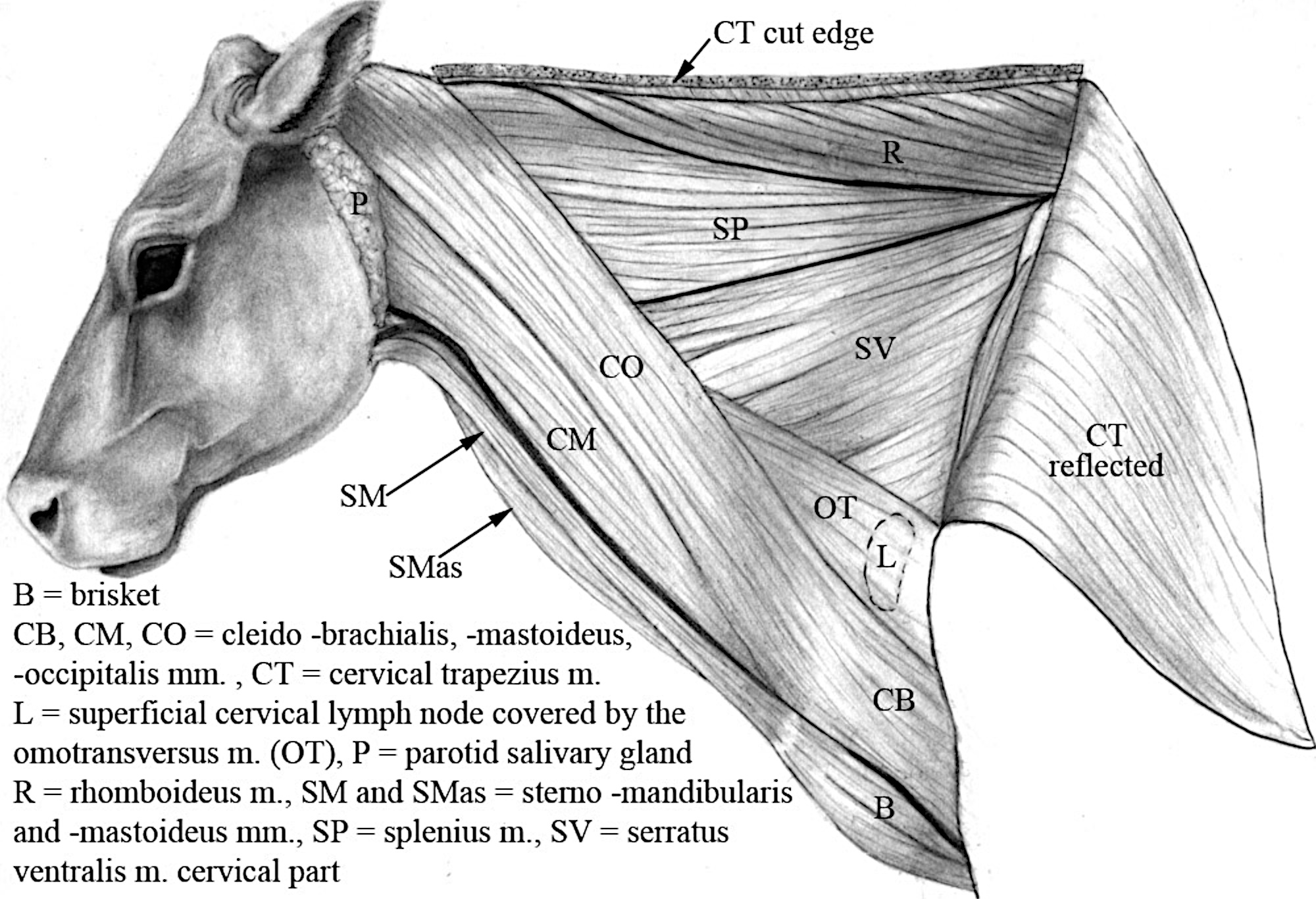
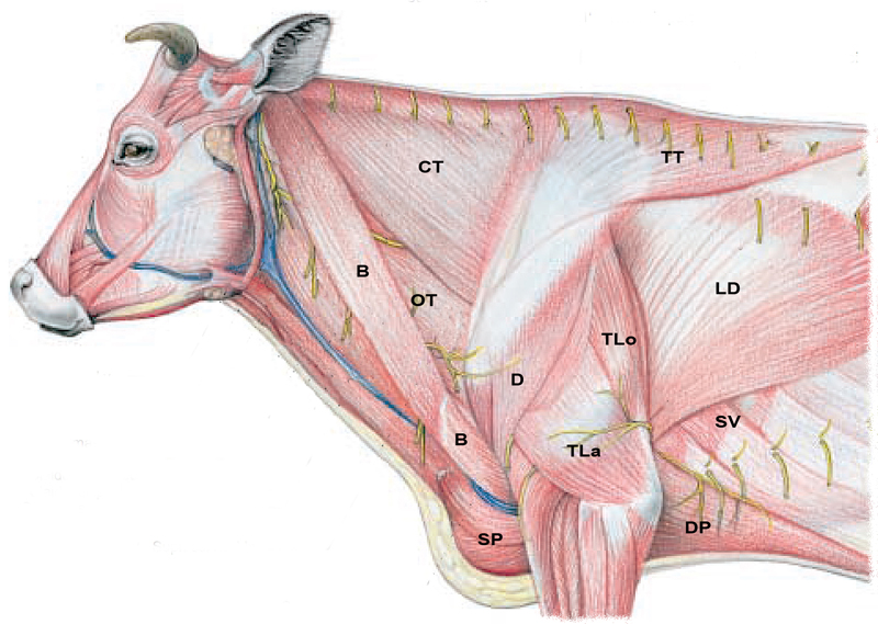
Figure 3-2. Neck dissection of the ox, lateral view. The external jugular vein lies between SM and CM. The brisket (ventral chest region), B, is well developed in cattle but not in small ruminants.
8. ALL (PONY) specimens: On both sides, identify the beginning of the external jugular vein as the confluence of the linguofacial and maxillary veins. (Note that you will need to reflect a portion of the parotid salivary gland to view the maxillary vein.)
-
- On both left and right sides, isolate and trace the external jugular vein in the lateral and cranial neck regions (Fig. 3-3).
- Observe/identify the maxillary vein (upper branch) and linguofacial vein (lower branch) which join to form the external jugular vein. (Fig. 3-4 and Popesko, Eq2R, Fig. 154:17)
- Dissection Note: The maxillary vein will be “hidden” from view by the parotid salivary gland; this region will be fully dissected with the head, but you should reflect the caudal portion of the salivary gland off the maxillary vein to view it.
9. ALL (CALF) specimens: On both sides, identify the external jugular vein.
-
- On both left and right sides, isolate and trace the external jugular vein in the lateral and cranial neck regions (Fig. 3-2).
- In the calf, the halter will obscure your view of the maxillary and linguofacial veins at this time; hence, you do not need to identify these veins now in the calf specimen.
10. ALL specimens, RIGHT side: Re-identify the brachiocephalicus m. (previously identified in Chapter 1). Identify the cleidocephalicus portion of the brachiocephalicus m. and note its parts (pony: cleidomastoideus m. only; calf: cleidomastoideus m. and cleido-occipitalis m.) Note the fused omotransversarius portion (of the brachiocephalicus m.) in the pony and identify the separate omotransversarius m. in the calf.
-
- On the RIGHT side of your specimen, identify the previously dissected brachiocephalicus m. running down the neck and over the shoulder to attach on the humerus (TVA 534). Identify the cleidocephalicus m. portion of brachiocephalicus extending from just cranial to the shoulder (the region where a clavicle would be) toward the head.
-
ALL (PONY) specimens: In the pony, the cleidocephalicus portion of the brachiocephalicus m. is composed of only the cleidomastoideus m.; observe this muscle running parallel and dorsal to the external jugular vein (Fig. 3-4).
-
Also in the PONY, note that the brachiocephalicus muscle is fused to the omotransversarius m. (TVA 534); the only evidence of this fused omotransversarius portion is a series of muscular insertions onto the transverse processes of C2-4. (For this course we will not try to separate out the omotransversarius component.)
-
ALL (CALF) specimens: In the calf, the cleidocephalicus portion of the brachiocephalicus m. has two parts: the cleidomastoideus m. and the cleido-occipitalis m.
-
-
Dorsal and parallel to the external jugular vein, identify the cleidomastoideus m., and dorsal to that muscle identify the cleido-occipitalis m.
-
-
-
Also in the CALF, in the caudal neck, re-identify the omotransversarius m. (previously identified in Chapter 1).
-
-
The omotransversarius m. can be found in the triangle formed by the caudal edge of the cleido-occipitalis m., the cranial edge of the trapezius m., and the scapula (Fig. 3-2). (Cranially, note that the omotransversarius m. is covered by the cleido-occipitalis m.)
-
-
-
- On the RIGHT side of your specimen, identify the previously dissected brachiocephalicus m. running down the neck and over the shoulder to attach on the humerus (TVA 534). Identify the cleidocephalicus m. portion of brachiocephalicus extending from just cranial to the shoulder (the region where a clavicle would be) toward the head.
11. ALL specimens, LEFT side: Identify the cut stump of the brachiocephalicus m. (the cleidocephalicus portion) and be familiar with its contributing parts in both pony and calf (pony: cleidomastoideus m. only; calf: cleidomastoideus m. and cleido-occipitalis m.). Note the fused omotransversarius portion (of the brachiocephalicus m.) in the pony and identify the separate omotransversarius m. in the calf.
-
- On the LEFT side of your specimen, find the transected remnant of the brachiocephalicus m.
- Note that the part left on your specimen is of the cleidocephalicus portion of the brachiocephalicus m. extending from just cranial to the shoulder (the region where a clavicle would be) toward the head.
-
- ALL (PONY) specimens: In the pony, the cleidocephalicus portion of the brachiocephalicus m. is composed of only the cleidomastoideus m.; observe this muscle running parallel and dorsal to the external jugular vein (Fig. 3-4).
- Also in the PONY, note that the brachiocephalicus muscle is fused to the omotransversarius m. (TVA 534); the only evidence of this fused omotransversarius portion is a series of muscular insertions onto the transverse processes of C2-4. (For this course we will not try to separate out the omotransversarius component.)
- ALL (CALF) specimens: In the calf, the cleidocephalicus portion of the brachiocephalicus m. has two parts: the cleidomastoideus m. and the cleido-occipitalis m.
-
-
Dorsal and parallel to the external jugular vein, identify the cleidomastoideus m., and dorsal to that muscle identify the cleido-occipitalis m.
-
-
-
Also in the CALF, in the caudal neck, re-identify the transected remnant of the omotransversarius m. (previously identified in Chapter 1).
-
-
The omotransversarius m. can be found in the triangle formed by the caudal edge of the cleido-occipitalis m., the cranial edge of the trapezius m., and the scapula (Fig. 3-2). (Cranially, note that the omotransversarius m. is covered by the cleido-occipitalis m.)
-
-
-
- Note that the part left on your specimen is of the cleidocephalicus portion of the brachiocephalicus m. extending from just cranial to the shoulder (the region where a clavicle would be) toward the head.
- On the LEFT side of your specimen, find the transected remnant of the brachiocephalicus m.
12. ALL (CALF) specimens, RIGHT side: Transect the omotransversarius m. on the right side and reflect it to expose and identify the large superficial cervical lymph node.
-
- On the right side, transect the omotransversarius m. cranial to the edge of the scapula and reflect it to expose the underlying lymph node.
- Identify the large superficial cervical lymph node.
13. ALL (CALF) specimens, LEFT side: Look for the large superficial cervical lymph node, but note that this lymph node may have been removed with the left forelimb in Chapter 1.
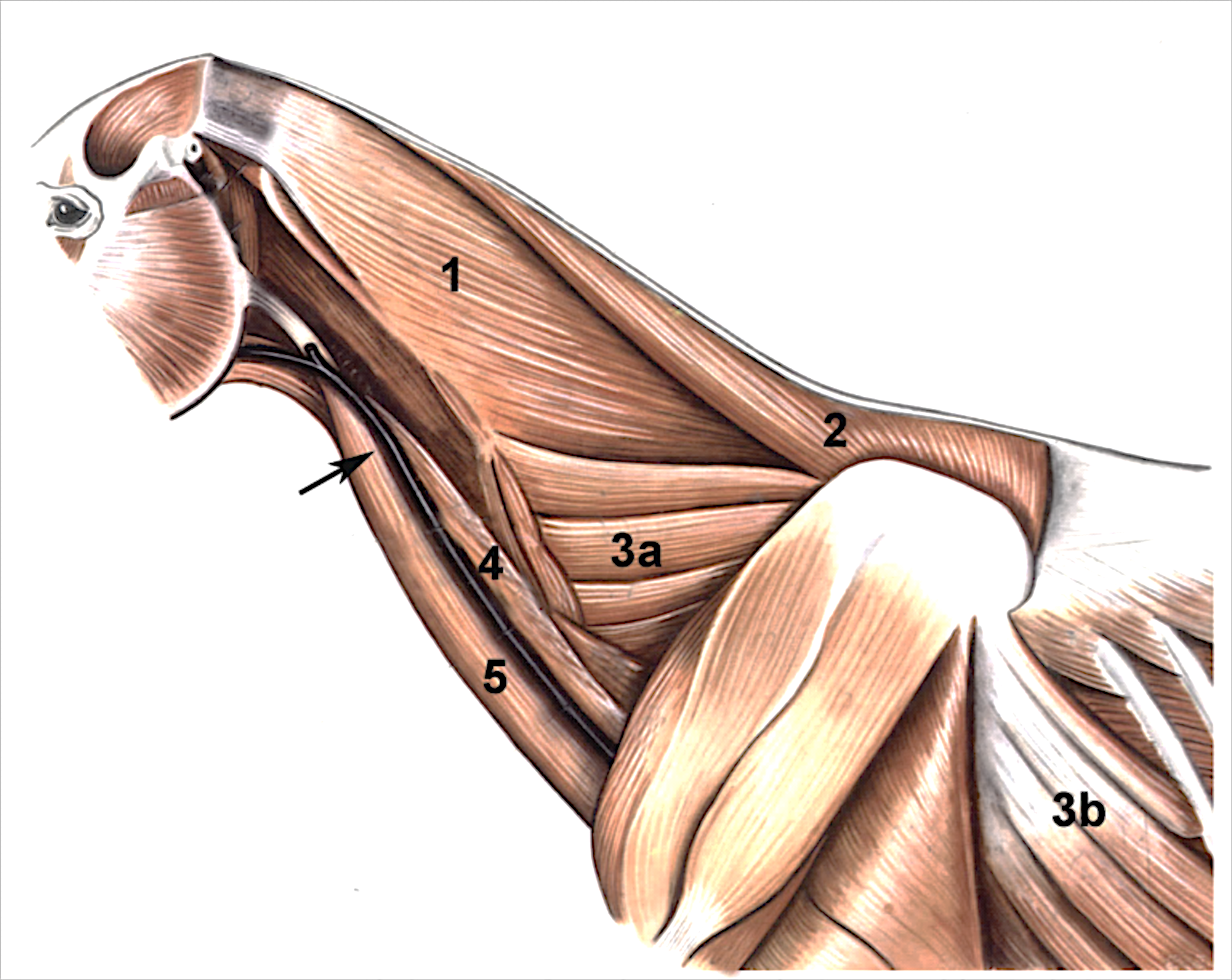
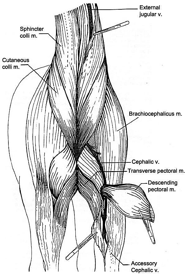
Figure 3-3. Pony neck and forelimb deep dissection, lateral view. 1, splenius m.; 2, rhomboideus m.; 3a, serratus ventralis cervicis m., 3b, serratus ventralis thoracis m.; 4, omohyoideus m.; 5, sternomandibularis m.; arrow, external jugular vein.
14. ALL (PONY) specimens: Identify the cutaneous colli m. (equine only)
-
- In the pony, identify a superficially located muscle in the ventral neck region, the cutaneous colli m. (with a thinner ‘sphincter colli’ subdivision as you move up the neck toward the head).
- The cutaneous colli m. will cover/overlap the ventral edge of the brachiocephalicus muscle and was previously mentioned in Chapter 1 (see Fig. 1-3).
- CALF specimen note: Note that the cutaneous colli m. is virtually non-existent in the calf and other ruminants.
- In the pony, identify a superficially located muscle in the ventral neck region, the cutaneous colli m. (with a thinner ‘sphincter colli’ subdivision as you move up the neck toward the head).
(Duplicate) Figure 1-3. Equine, cranial view of the superficial pectoral muscles and the base of the neck.
15. ALL specimens: On both sides, identify and trace the sternocephalicus m. cranially. This muscle extends from the cranial part of the sternum toward the head, to the angle of the mandible.
-
- ALL (PONY) specimens: In the pony, there is only one part of the sternocephalicus m., the sternomandibularis, which you should identify. (Figs. 3-1 and 3-3)
- Dissection Note: In the equine, the terms sternocephalicus m. and sternomandibularis m. are synonymous and, therefore, interchangeable (i.e., sternocephalicus = sternomandibularis).
- Isolate the sternomandibularis muscle; note how it contributes to the ventral border of the jugular groove.
- Trace the sternomanibularis m. cranially to view its tendon that will insert on the angle of the mandible, deep to the parotid salivary gland.
-
- Important Dissection Note: In the horse, this strong tendon of insertion forms the dorsal border of a clinically significant region called Viborg’s triangle (Fig. 3-4). The ramus of the mandible and linguofacial vein form the triangle’s rostral and ventral borders respectively. Clinically, Viborg’s triangle is an access point, where you can avoid major vessels and nerves, to drain fluid from the guttural pouch. (The guttural pouch will be discussed in more detail in Chapter 7.)
-
- ALL (PONY) specimens: In the pony, there is only one part of the sternocephalicus m., the sternomandibularis, which you should identify. (Figs. 3-1 and 3-3)
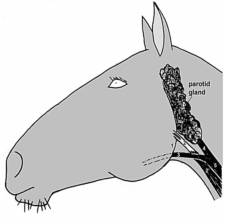
Figure 3-4. Schematic illustration of Viborg’s triangle with boundaries labeled 1-3: 1, angle of mandible; 2, tendon of insertion of 2′ sternomandibularis m.; 3, linguofacial v. 4, maxillary v.; 5, external jugular v. The parotid salivary gland has been reflected since it covers most of the landmarks of the triangle.
-
- ALL (CALF) specimens: Note that the sternocephalicus m. in the calf is divided into 2 components: the sternomandibularis m., which forms the ventral border of the jugular groove, and the sternomastoideus m., which is deep to the external jugular vein. Identify the sternomandibularis and sternomastoideus parts of the sternocephalicus m. in the calf. (Fig. 3-2)
- Identify the sternomandibularis m. and trace it as far cranially as the halter allows.
- Elevate the sternomandibularis m. and the external jugular vein to expose and identify the sternomastoideus m. (which lies more ventrally than the sternomandibularis m.).
- Comparative Dissection Note: Recall that the horse has only the sternomandibularis portion of the sternocephalicus muscle and it also has a well-developed omohyoideus muscle that lies deep to the external jugular vein in the cranial portion of the neck. The omohyoideus muscle in ruminants is insignificant.
- ALL (CALF) specimens: Note that the sternocephalicus m. in the calf is divided into 2 components: the sternomandibularis m., which forms the ventral border of the jugular groove, and the sternomastoideus m., which is deep to the external jugular vein. Identify the sternomandibularis and sternomastoideus parts of the sternocephalicus m. in the calf. (Fig. 3-2)
16. ALL specimens: On both sides, identify the sternohyoideus and sternothyroideus muscles.
-
- Cranially in the ventral neck, identify the narrow sternohyoideus and sternothyroideus mm. (TVA 529(6)) on the ventral midline covering the trachea, arising from a common origin on the sternum.
- Identify and isolate the combined origin of the sternohyoideus and sternothyroideus mm.
-
- Dissection Note: You do not need to try to separate the individual muscle bellies at their point of origin.
-
- Working cranially, trace the more medially positioned sternohyoideus m. to the basihyoid bone, or as far cranially as possible (cranial to the angle of the mandible).
- In the cranial half of the neck, isolate the sternothyroideus m. as it separates laterally from the sternohyoideus m. and runs cranially to insert on the thyroid cartilage of the larynx.
- Comparative Dissection Note: In the equine, these muscles combine to form a tendon midway along their length.
- Identify and isolate the combined origin of the sternohyoideus and sternothyroideus mm.
- Cranially in the ventral neck, identify the narrow sternohyoideus and sternothyroideus mm. (TVA 529(6)) on the ventral midline covering the trachea, arising from a common origin on the sternum.
17. ALL (PONY) specimens: Re-identify the omohyoideus m. and carefully isolate it.
-
- Recall from Chapter 1, that the omohyoideus m. is not present in the dog, is insignificant in ruminants, but is of significance in the equine. The obliquely placed omohyoideus m. forms an incomplete medial (deep) boundary of the jugular groove (Fig. 3-3/4).
- Dissection Note: The omohyoideus m. forms a muscular separation between the external jugular vein and the carotid sheath in the cranial half of the neck. In the caudal half of the neck the external jugular vein and the carotid sheath are in apposition.
- The omohyoideus m. is found lateral to the sternohyoideus m. near its insertion on the basihyoid bone.
- From this point, re-identify the omohyoideus m., isolate it, and trace it caudally, where it lies deep to the jugular vein, sternomandibularis m., and the cleidomastoideus m.
- Elevate the external jugular vein and remove the fascia from the medial wall of the jugular groove to visualize the omohyoideus m.
- Recall from Chapter 1, that the omohyoideus m. is not present in the dog, is insignificant in ruminants, but is of significance in the equine. The obliquely placed omohyoideus m. forms an incomplete medial (deep) boundary of the jugular groove (Fig. 3-3/4).
18. ALL specimens, RIGHT side (attached forelimb): Transect the brachiocephalicus m. in the cranial third of the neck.
19. ALL (PONY) specimens, RIGHT side (attached forelimb): After transecting the brachiocephalicus m., reflect it off of the omohyoideus m. to aid in visualization of the omohyoideus m.
-
- Important Dissection Note: Use caution when making this cut! The omohyoideus m. is closely applied to the deep face of the brachiocephalicus m.
20. ALL (PONY) specimens, LEFT side (detached forelimb): Reflect the previously transected remnant of the brachiocephalicus m. off of the omohyoideus m. to aid in visualization of the omohyoideus m.
-
- Important Dissection Note: Use caution when making this cut! The omohyoideus m. is closely applied to the deep face of the brachiocephalicus m.
CERVICAL VISCERA
21. ALL specimens: In the neck, spread the sterno- muscles apart to identify the trachea and the right and left lobes of the thyroid gland (and connecting isthmus, if possible).
-
- The trachea lies within a deep fascial sheath continuous with the endothoracic fascia; identify the trachea.
- The thyroid gland lies on the lateral surface of the trachea near the larynx (on both left and right sides).
- Just caudal to the larynx, identify the right and left lobes of the thyroid gland lying on the trachea, partially covered by the sternothyroideus muscle.
- Dissection Note: The right and left lobes of the thyroid are connected by a narrow isthmus passing ventral to the first and/or second tracheal rings.
-
- Make a ventral midline incision in the neck to spread the right and left sternohyoideus muscles apart and attempt to observe the isthmus connecting right and left lobes of the thyroid gland on the midline.
-
- Comparative Dissection Note: The thyroid gland is dark brown and ovoid in the horse but flattened in ruminants (TVA 220, 522(18), 522(21).
22. ALL specimens: On the LEFT side of the neck, identify the esophagus. Then, transect the trachea and esophagus caudal to the thyroid gland.
-
- The esophagus begins dorsal to the trachea at the level of the cricoid cartilage of the larynx. From this point, the esophagus inclines to the left so that at the level of C4 the esophagus lies to the left of the trachea. From there the esophagus gradually inclines dorsally so that in the thorax the esophagus is again directly dorsal to the trachea.
- Identify the esophagus in both pony and calf.
- Transect the trachea immediately caudal to the thyroid gland. and note its cross-sectional appearance.
- Observe that the trachea is incomplete dorsally (Fig. 3-5).
- Comparative Dissection Notes: In the dog, the smooth muscle (trachealis m.) that closes the dorsal opening of the tracheal rings is external to the rings, but in ungulates it is internal to the tracheal rings. Additionally, the bovine will have a ‘teardrop’ shape to the trachea as compared to the round shape of the equine trachea.
- Transect the esophagus at the same level as the tracheal transection and note the outer muscular layer and the inner layer of longitudinally folded mucosa.
- The esophagus begins dorsal to the trachea at the level of the cricoid cartilage of the larynx. From this point, the esophagus inclines to the left so that at the level of C4 the esophagus lies to the left of the trachea. From there the esophagus gradually inclines dorsally so that in the thorax the esophagus is again directly dorsal to the trachea.
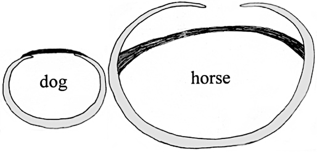
Figure 3-5. Cross-section of the trachea. As noted above, in the dog, the tracheal smooth muscle lies external to the tracheal rings, whereas in the horse (and ungulates in general) the tracheal smooth muscle lies dorsally within the incomplete tracheal rings.
23. ALL specimens: On both sides, identify the carotid sheath. Open the carotid sheath and identify the common carotid a. and vagosympathetic nerve trunk.
-
- On both sides (right and left), dorsolateral to the trachea, expose the carotid sheath by blunt dissection and identify it.
- Recall from Anatomy I that the carotid sheath is the deep cervical fascia surrounding the common carotid artery, vagosympathetic nerve trunk, internal jugular vein, and tracheal lymphatic duct. There is a carotid sheath on both left and right sides of the neck.
- Open the carotid sheath; within it, identify the common carotid artery and the vagosympathetic nerve trunk.
- Also within the carotid sheath, attempt to find a tracheal lymphatic duct (which may be difficult to find unless it has brown tinged lymph in it).
- ALL (CALF) specimens: A small internal jugular vein is also found within the carotid sheath fascia. You should identify this in your calf specimen running alongside your common carotid artery and vagosympathetic nerve trunk, if possible.
- Comparative Dissection Note: The internal jugular vein is small in the bovine, large in the pig, and not present in the equine.
- On both sides (right and left), dorsolateral to the trachea, expose the carotid sheath by blunt dissection and identify it.
24. ALL (CALF) Specimens: Identify the thymus; specifically, identify the cervical thymus.
-
- Look for evidence of thymic tissue in the neck of your specimen.
- The thymus in the young ruminant (and pig) may extend from the thoracic inlet to the cervical region. This is called the cervical thymus (TVA 661(1, 2); Popesko, Bo6L). Identify the cervical thymus in your calf specimen.
- Comparative Dissection Note: Note that the thymus of the horse is typically limited to the chest, so it will (most often) not be visible in the current dissection field.
25. ALL specimens: Re-identify the superficial cervical lymph node(s). In the pony, identify the lymphocenter(s). In the calf, look for hemal lymph nodes. Attempt to identify deep cervical lymph nodes in both species.
-
- Comparative Dissection Note: At any given location, instead of having one or two large lymph nodes as in the dog, ruminant or pig, the horse has a multitude of small lymph nodes known collectively as a lymphocenter. Interestingly, the ruminant has additional dark-colored nodes called hemal lymph nodes that horses lack.
- ALL (PONY) specimens: Re-identify the superficial cervical lymph nodes (lymphocenters) that lie deep to the brachiocephalicus m. and cranial to the subclavius m. (Fig. 7-6, TVA 531(8), 663). Locate and isolate these lymph nodes/lymphocenters on one or both sides of the pony. (LEFT side note: These lymph nodes may have been removed with the left forelimb.)
- ALL (CALF) specimens: Re-identify the large superficical cervical lymph nodes that lie deep to the omotransversarius m. (LEFT side note: This lymph node may have been removed with the left forelimb.)
- Additionally, look for dark-colored hemal lymph nodes during your dissection of the calf.
-
- Hemal lymph nodes are lymphoid structures with blood sinuses instead of lymph sinuses, and are commonly found in cattle and sheep.
-
- Additionally, look for dark-colored hemal lymph nodes during your dissection of the calf.
- In both species, attempt to identify deep cervical lymph nodes scattered along the trachea.
- Dissection Note: The deep cervical lymph nodes are named by their location on the trachea: cranial deep cervical, middle deep cervical and caudal deep cervical. (Attempt to find these nodes, but don’t be disappointed if they are not found as they can be very difficult to find/see!)
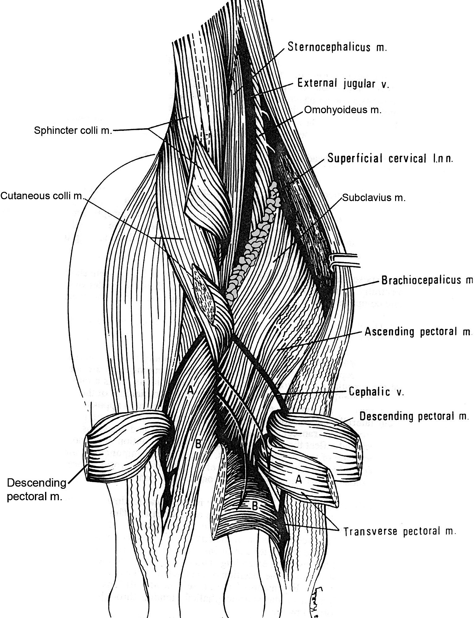
Figure 3-6. Equine, pectoral muscles, deep dissection, cranial view. Sternocephalicus = sternomanibularis in the horse.
PIG CERVICAL REGION
26. Use this brief guide, along with the tissue study video available on Canvas, and the porcine wet specimens available in the lab to identify the listed structures in the neck of the pig.
-
- The neck of the pig, like all domestic mammals, has 7 cervical vertebrae, but proportionately the neck is very short. As a result, the parotid salivary gland extends back to the shoulder and the thyroid gland is near the thoracic inlet.
- Note that the thyroid gland is large and has a broad isthmus which crosses the ventral side of the trachea to join the right and left lobes.
- The larynx is large and extends caudally almost to the thoracic inlet.
- In young animals there may be a well-developed cervical thymus present [TVA 661(1, 2)].
- The external jugular vein lies deep to the parotid salivary gland cranially and the brachiocephalicus m. caudally.
- The mature porcine individual has a thick subcutaneous adipose layer which makes this vein rather inaccessible for venipuncture.
- In pigs, the internal jugular vein is nearly as large as the external jugular vein (Fig. 3-7). The two veins lie close together, separated by the sternomastoideus m.
- In this species, the sternomastoideus m. is the only part of the sternocephalicus m. (therefore sternomastoideus = sternocephalicus in the pig).
- The sternohyoideus m. is wide and covers the sternothyroideus m. but is separate from it.
- The deeper lying sternothyroideus m. is divided into two parts, both of which attach to the large thyroid cartilage.
- The manubrium of the sternum is well developed and serves as the place of origin of the three sterno- muscles and the cutaneous colli m.
- The cutaneous colli mm. are thick at their origin, and the pair forms a V-shaped mass similar to the same muscle in the horse.
- Caudal to the manubrium, the superficial pectoral muscles are directed laterally from the sternum.
- A depression is formed by the superficial pectoral mm. caudally, sternomastoideus m. medially, and the brachiocephalicus m. laterally. This depression is palpated as the external landmark for venipuncture (Fig. 3-7) in the pig.
- The neck of the pig, like all domestic mammals, has 7 cervical vertebrae, but proportionately the neck is very short. As a result, the parotid salivary gland extends back to the shoulder and the thyroid gland is near the thoracic inlet.
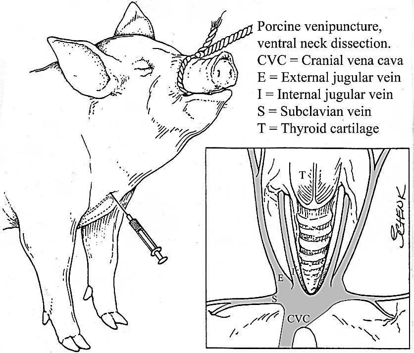
Figure 3-7. Porcine venipuncture and venous structure of the ventral neck.
Dissection Videos for this Section of Material
Neck
