
Part 2: External Thorax
Abby Brown
IMPORTANT NOTE: For this part of the chapter, the Guide directions will refer to either “ALL specimens” (meaning dissect pony and calf specimens in the same way) or to one species in particular, i.e., “ALL (PONY) specimens” or “ALL (CALF) specimens.” This added instructional note is needed because the dissection may differ slightly according to species.
Removal of RIGHT forelimb
On all hanging specimens, you will now remove the RIGHT forelimb in a similar manner to the removal of the left forelimb that was completed in Chapter 1. Please proceed with the RIGHT forelimb removal instructions as outlined below:
- ALL specimens: If needed, transect both cervical and thoracic parts of the trapezius m., as well as both parts of the rhomboideus m.
- ALL (PONY) specimens: Transect the dorsoscapular ligament that lies deep to the rhomboideus m.
- ALL specimens: If the brachiocephalicus m. has not already been transected, transect it approximately one hand width cranial to the shoulder.
- ALL (CALF) specimens: If not already transected, transect the omotransversarius m.
- ALL (PONY) specimens: Transect the omohyoideus m.
- ALL specimens: Transect all parts of the superficial and deep pectoral muslces with a cut adjacent to the sternum.
- ALL specimens: Abduct the forelimb, elevating it away from the body.
- ALL specimens: Transect the cephalic vein as well as all brachial vessels and nerves of the brachial plexus entering the deep face of the limb.
- ALL specimens: Continue to abduct (lift) the limb and transect the serratus ventralis m. close to the scapula to complete the limb removal process.
MUSCLES OF THE THORAX AND ASSOCIATED STRUCTURES
IMPORTANT NOTE: The following procedures should be completed on right and left sides of ALL specimens (unless otherwise specified).
10. ALL specimens: Remove any remaining skin from the thorax and abdomen regions.
-
- If needed, on right and left sides of your specimen, incise any remaining skin of the thorax and abdominal regions along the dorsal midline. Reflect the skin ventrally and remove it.
11. ALL specimens: Identify and reflect/remove the serratus ventralis thoracis m.
-
- Identify the main muscle mass of the serratus ventralis thoracis m. (TVA 590 (1)) on the lateral side of the trunk and reflect it cranially by carefully peeling it away and incising the muscular interdigitations that insert on the ribs.
- Continue reflecting the muscles cranially as you reach the serratus ventralis cervicis portion of the muscle but leave the muscle attached along the cranial edge of the serratus ventralis cervicis.
- Dissection Note: If needed, you may choose to entirely remove the serratus ventralis m. from the trunk, but it is preferred that you leave it attached cranially if possible.
- Dissection Note: Note that you may see a scalenus muscle overlapping part of the serratus ventralis cervicis muscle cranioventrally, but you do not need to dissect or identify it for this dissection.
12. ALL specimens: Identify the serratus dorsalis cranialis and caudalis mm.; transect and reflect both parts.
-
- Dorsally in the external thorax, identify the serratus dorsalis cranialis and caudalis mm. These muscles will be smaller and less defined than the serratus ventralis m. but still have the ‘serrated’ edge and insert on the ribs.
- Note that these muscles are connected to the dorsal midline via an aponeurosis.
- If possible, transect these muscles along their interdigitations inserting on the ribs and reflect them dorsally, leaving them attached by their aponeurotic origins.
-
- Dissection Note: If needed you may choose to entirely remove the serratus dorsalis mm. from the trunk, but it is preferred that you leave them attached dorsally if possible.
-
- Dorsally in the external thorax, identify the serratus dorsalis cranialis and caudalis mm. These muscles will be smaller and less defined than the serratus ventralis m. but still have the ‘serrated’ edge and insert on the ribs.
13. Now we will move on to the dissection of the epaxial mm. Recall that the epaxial muscles generally lie dorsal to the transverse processes of the vertebrae and are associated with the vertebral column and ribs. Here we will dissect some of these muscles that are divided into three parallel muscle systems running longitudinally: iliocostalis system, longissimus system, and transversospinalis system.
14. ALL specimens: Identify and isolate the iliocostalis m. system; transect and reflect it.
-
- Along the dorsal edge of the ribs, identify and isolate the iliocostalis m. system.
- Beginning caudally, reflect the long, slender iliocostalis m. by transecting its attachment at the last rib and reflecting it cranially toward the first rib.
- Note that the iliocostalis m. consists mainly of parallel tendinous bands that insert on the ribs.
- This muscle is the most lateral part of the epaxial muscle systems. Just medial to the iliocostalis m.system is the much thicker longissimus muscle system, which will be identified in the next step.
15 ALL specimens: Identify and isolate the longissiums m. system.
-
- Medial (and slightly dorsal) to the iliocostalis system identify and isolate the longissimus m. system.
- Dissection Note: This muscle system is regionally divided into thoracic and cervical parts. The longissimus cervicis lies deep to the cervical part of the serratus ventralis m. Isolate the longissimus cervicis by separating it from the splenius m. (which will be identified in the next step).
16. ALL specimens: Identify the transversospinalis m. system.
-
- Medial (and slightly dorsal) to the longissimus system identify and isolate the transversospinalis m. system.
- The splenius muscle, which was previously identified during the dissection of the neck in this chapter, is part of the transversospinalis m. system. Re-identify the splenius m. in your specimen.
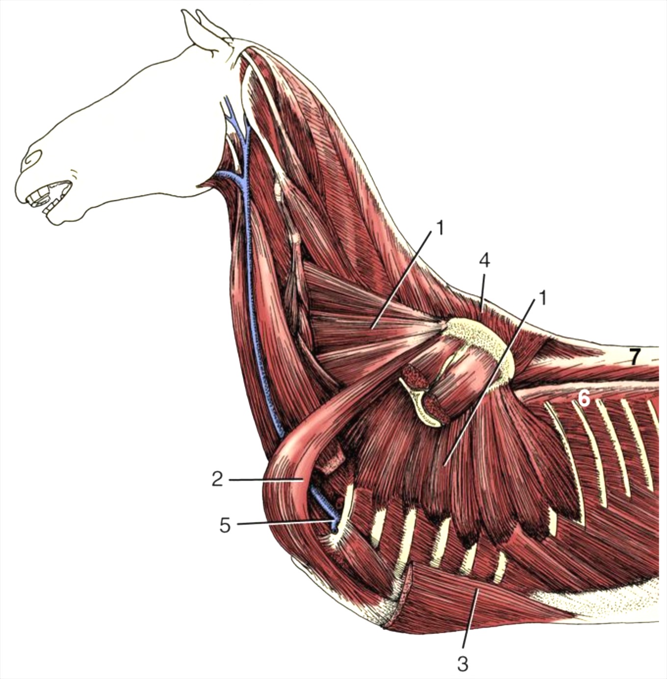
Figure 3-8. Equine, deep muscles attaching the forelimb to the trunk. 1, serratus ventralis m. 2, subclavius m. 3, ascending deep pectoral m. 4, rhomboideus m. 5, axillary vessels turning around first rib into forelimb 6, iliocostalis m. 7, longissimus m. (Modified from TVA Fig. 23-5)
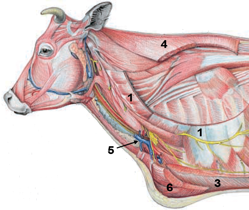

(Duplicate) Figure 1-4. Deep muscles of the ox (upper) and horse (lower) attaching the forelimb to the trunk. 1, serratus ventralis; 2, subclavius (only horse, very underdeveloped/absent in ox); 3, ascending deep pectoral; 4, rhomboideus; 5, axillary vessels turning around first rib into forelimb; 6, superficial pectorals. (Equine: Modified from Dyce, Sack and Wensing, 4th ed.; Bovine: Modified from Budras and Habel, 1st ed.)
Dissection Videos for this Section of Material
External Thorax
- Pony (Watch from 0:00-5:10): https://youtu.be/E5mxf-eIaAc
- Calf (Watch from 0:00-4:51): https://youtu.be/ARg0TsYopKM
