
Part 4: Distal Thoracic Limb
Abby Brown
IMPORTANT NOTE: Two important differences you will note in the distal limb when comparing ruminant to equine: the ruminant has multiple digits (versus the equine single digit) and the ruminant has no distinct check ligaments (versus the equine with two distinct check ligaments).
Skin reflection & Dissection of the antebrachium
- ALL forelimbs: Remove the remaining skin from the distal limb (down to the hoof/claws).
-
- On the medial side of the limb, make an incision through the remaining skin, down to the hoof/claws.
- Extend the incision around the top margin of the hoof/claws (at the coronet) and carefully reflect and remove the skin, then discard it.
- Calf Specimen: Note that this skin reflection will require you to remove the dewclaws with the skin as you reflect/remove it.
- Be cautious of nerves and vessels as you remove the skin! Leave superficial nerves and vessels on the limb as much as possible.
- Pony Specimen: Be particularly careful in the mid-cannon region where there is a communicating branch of the palmar nerves superficial to the digital flexor tendons that you will need to identify.
2. As we move on to our dissection of the antebrachium, note that there is a dense ‘sleeve’ of antebrachial (deep) fascia covering the muscles of the antebrachium.
-
- As needed, make incisions through the deep antebrachial fascia and carefully reflect and then remove it as you work through this dissection.
-
Calf Specimen: Note that this antebrachial fascia is especially tough/thick in the calf specimens.
-
- As needed, make incisions through the deep antebrachial fascia and carefully reflect and then remove it as you work through this dissection.
3. ALL forelimbs: Begin by identifying the extensor muscle group of the antebrachium, which is made up of the muscles on the craniolateral aspect of the antebrachium. This group will include extensors of the carpus and extensors of the digit(s). Similar to the dog/cat dissection, the main difference to note between these muscles is that the tendons of the extensors of the carpus will cross the carpus and then stop, while the tendons of the extensors of the digit(s) will continue past the carpus down to the digit(s).
4. ALL forelimbs: For orientation purposes, return to the biceps brachii m. in the proximal limb and trace the lacertus fibrosus distally onto the extensor carpi radialis m. Identify and isolate the extensor carpi radialis m. and trace it to its insertion on the cannon bone (metacarpal region). (Figs. 1-8 and 1-10)
-
- The extensor carpi radialis m. is found cranially (craniolaterally) in the antebrachium and is part of the extensor muscle group; specifically, it is an extensor of the carpus.
- The tendon of the extensor carpi radialis m. crosses the carpal joint to insert on the cannon bone (metacarpal region), but note that the tendon will run deep to the extensor retinaculum (band of fascia) extending across the dorsal aspect of the carpal region. This retinaculum binds down the extensor tendons that cross the dorsal aspect of the carpus and helps keep them in place.
- Identify the extensor retinaculum and attempt to define its proximal and distal margins.
- Trace the tendon of the extensor carpi radialis m. to where it inserts on the cranial aspect of the proximal cannon region (on the metacarpal tuberosity)
- Dissection Note: Note that the tendon of the extensor carpi obliquus m. will cross over the tendon of the extensor carpi radialis as you near the carpus. You may either work around this tendon or transect it (if needed) to trace the tendon of the extensor carpi radialis to its insertion.
5. ALL forelimbs: Moving lateral to the extensor carpi radialis m., identify the common digital extensor m. and trace its tendon(s) distally toward the digit(s) (as far as the hoof/claws). (Figs. 1-8 and 1-10)
-
- The common digital extensor m. is part of the extensor muscle group and, as its name implies, is an extensor of the digit(s). The tendon(s) of the common digital extensor m. cross the carpal joint and extend to the distal phalanx (P3) of the digit(s).
- Calf Specimen: The common digital extensor m. will have two main heads (medial and lateral) and two corresponding tendons that extend distally. The medial tendon will extend to the distal phalanx (P3) of digit 3, while the lateral tendon will split in the distal cannon region and extend to P3 of both digits 3 and 4.
- Pony Specimen: Note that the tendon of the common digital extensor m. is markedly wider as it nears its insertion on the extensor process of the distal phalanx (P3), which is covered by the hoof wall. In the pony, the common digital extensor m. also has a slight radial head in addition to the main humeral head. The small radial head’s tendon separates and joins the lateral digital extensor m. in the cannon region. (This variation is not significant for our dissection purposes.)
- Note that the tendon(s) of the common digital extensor m. will run deep to the extensor retinaculum (band of fascia) extending across the dorsal aspect of the carpal region. This retinaculum binds down the extensor tendons that cross the dorsal aspect of the carpus and helps keep them in place.
- If needed, cut and reflect the extensor retinaculum to trace these tendons distally toward the digit(s).
6. ALL forelimbs: Moving lateral to the common digital extensor, identify another extensor of the digit(s), the lateral digital extensor m., and trace its tendon distally. (Figs. 1-8 and 1-10)
-
- The lateral digital extensor m. is a fairly small muscle with a tendon that crosses the carpal joint on its lateral side, running deep to/through the lateral collateral ligament of the carpus (Fig. 1-12/6), and extending to the proximal phalanx (P1) of the digit. Trace its tendon of insertion distally to the digit.
- Calf Specimen: Note that in the calf there will be a layer of thick fascia surrounding this muscle; reflect/remove the fascia as need to view the lateral digital extensor m.
- Pony Specimen: Trace the lateral digital extensor m. tendon to its insertion on the proximal end of P1.
- Calf: Trace the lateral digital extensor m. tendon to its insertion on digit 4. (In the calf, the lateral digital extensor inserts on the middle and distal phalanges (P2 and P3) of digit 4.)
- Dissection Note: Note that, in the forelimb, the tendons of the lateral and common digital extensor muscles do not join distally. (In the hind limb, the comparable extensor tendons will join together distally.)
- The lateral digital extensor m. is a fairly small muscle with a tendon that crosses the carpal joint on its lateral side, running deep to/through the lateral collateral ligament of the carpus (Fig. 1-12/6), and extending to the proximal phalanx (P1) of the digit. Trace its tendon of insertion distally to the digit.
7. ALL forelimbs: Locate/palpate the accessory carpal bone on the palmar aspect of the carpus. Identify and isolate the ulnaris lateralis m. (aka extensor carpi ulnaris m.) and trace it to its insertion on the accessory carpal bone. (Figs. 1-8/19 and 1-10/14)
-
- The ulnaris lateralis m. is the most caudolateral muscle of the extensor muscle group and is categorized as an extensor of the carpus.
8. ALL forelimbs: Move to the caudomedial aspect of the limb and identify the flexor muscle group of the antebrachium. This group will include flexors of the carpus and flexors of the digit(s). Again, the main difference to note between these is that the tendons of the flexors of the carpus will cross the carpus and then stop, while the tendons of the flexors of the digits will continue past the carpus down to the digit(s).
-
-
Before you dissect these muscles and trace flexor tendons distally, move to the region of the carpus and note the flexor retinaculum (thick band of fascia) extending across the palmar aspect of the carpal region (covering the carpal canal). This retinaculum binds down the flexor tendons that pass through the carpal canal. Attempt to define its proximal and distal borders. (Note that this retinaculum will be more distinguishable as you complete the dissection of the flexor muscles.)
-
9. ALL forelimbs: Return to the accessory carpal bone on the palmar aspect of the carpus. Identify and isolate flexor carpi ulnaris m. (note there are two heads of this muscle: humeral & ulnar) and trace it to is insertion on the accessory carpal bone. (Fig. 1-9/19, 20)
-
- The flexor carpi ulnaris m. is the most caudomedial muscle of the flexor muscle group and is categorized as a flexor of the carpus.
- Dissection Note: The ulnaris lateralis (aka extensor carpi ulnaris) and the flexor carpi ulnaris mm. are two strong muscles that insert on the accessory carpal bone. These two muscles form a narrow ‘V’ shape at their insertion.
- Note that within this ‘V’ shape lie the ulnar nerve and a long tendon of the ulnar head of the deep digital flexor muscle, which may resemble a nerve; these are especially prominent in the pony.
- Note that the flexor carpi ulnaris has two heads, a larger humeral head (which may be partially covered by the flexor carpi radialis cranially) and a smaller ulnar head; these two heads may not be as prominent in the calf specimen. (You do not need to identify the two heads individually.)
10. ALL forelimbs: Moving cranially from the flexor carpi ulnaris m., while still on the medial side of the limb, identify the flexor carpi radialis m. (Fig. 1-9/17) and trace its tendon to its insertion on the cannon bone (metacarpal region).
-
- The flexor carpi radialis m. is the most craniomedial muscle of the flexor muscle group and is categorized as a flexor of the carpus.
11. ALL forelimbs: Between the two flexors of the carpus that you just dissected, look for the flexors of the digit(s). Identify the superficial digital flexor m. (SDF) and, in the pony, the proximal (radial) check ligament. As you complete this dissection you will also identify and transect the flexor retinaculum to trace the tendon of the SDF distally into the digit(s).
-
-
On the medial aspect of the forelimb, isolate and identify the main belly (the humeral head) of the superficial digital flexor m. (SDF).
- Calf Specimen: Note that in the calf there will be two parts to the main belly of the superficial digital flexor m.
-
Isolate the SDF muscle to the level of the carpus.
- Identify the flexor retinaculum, which is the thick band of fascia that helps hold the flexor tendons in the carpal canal.
- Make a (vertical) cut along the long axis of the flexor retinaculum to completely transect it. (Note that the flexor retinaculum is especially thick in the calf specimen.)
-
Trace the tendon of the SDF m. as it travels through the carpal canal and continues distally into the digit.
-
Pony Specimen: Reflect or elevate the flexor carpi radialis m., and also elevate the muscle belly of the SDF to expose the proximal (radial) check ligament. This is the tendinous radial head of the SDF m., present only in the horse, extending from the radius to join the main part of the SDF m. tendon (Fig. 1-11/4’).
-
Be sure you are able to identify the proximal (radial) check ligament on dried demonstration (museum) specimens as well. (These dried museum specimens are fair game on assessments!)
-
- Calf Dissection Note: In the calf, you may see a small, underdeveloped structure, similar to a check ligament, connecting the SDF to the DDF (which we will dissect next). This variation is not significant for our dissection purposes.
-
12. ALL forelimbs: Just deep to the superficial digital flexor m., identify the other major flexor of the digit(s), the deep digital flexor m. (DDF) and, in the pony, identify the associated distal (carpal) check ligament.
-
- Deep to the SDF muscle, identify the deep digital flexor m. (DDF). This is the largest of the flexor muscles of the forelimb and is made up of several heads that need not be separated out. (i.e., you do not need to identify all of the heads that make up the deep digital flexor m.)
- Trace the tendon of the DDF m. to the carpal canal. Elevate the DDF tendon out of the carpal canal and follow it to the proximal portion of the metacarpus.
- Pony Specimen: Identify the large distal (carpal) check ligament (also known as the accessory ligament of the deep digital flexor tendon) that joins the DDF tendon on the deep surface in the upper third of the cannon region (Fig. 1-11/6).
- Be sure you are able to identify the distal (carpal) check ligament on dried demonstration (museum) specimens as well. (These dried museum specimens are fair game on assessments!)
- Calf Dissection Note: As noted previously, in the calf, you may see a small, underdeveloped structure, similar to a check ligament, connecting the SDF to the DDF . This variation is not significant for our dissection purposes.
13. ALL forelimbs: Identify the carpal canal, which is the groove/space just medial to the accessory carpal bone that is occupied by the digital flexor tendons.
-
- Pony Specimen: In the pony, elevate the deep and superficial digital flexor tendons out of the carpal canal with a probe to help you identify this space.
- Calf Specimen: In the calf, it may be difficult to elevate the deep and superficial digital flexor tendons out of the carpal canal due to the thick connective tissue, but you should still identify the location of the carpal canal.
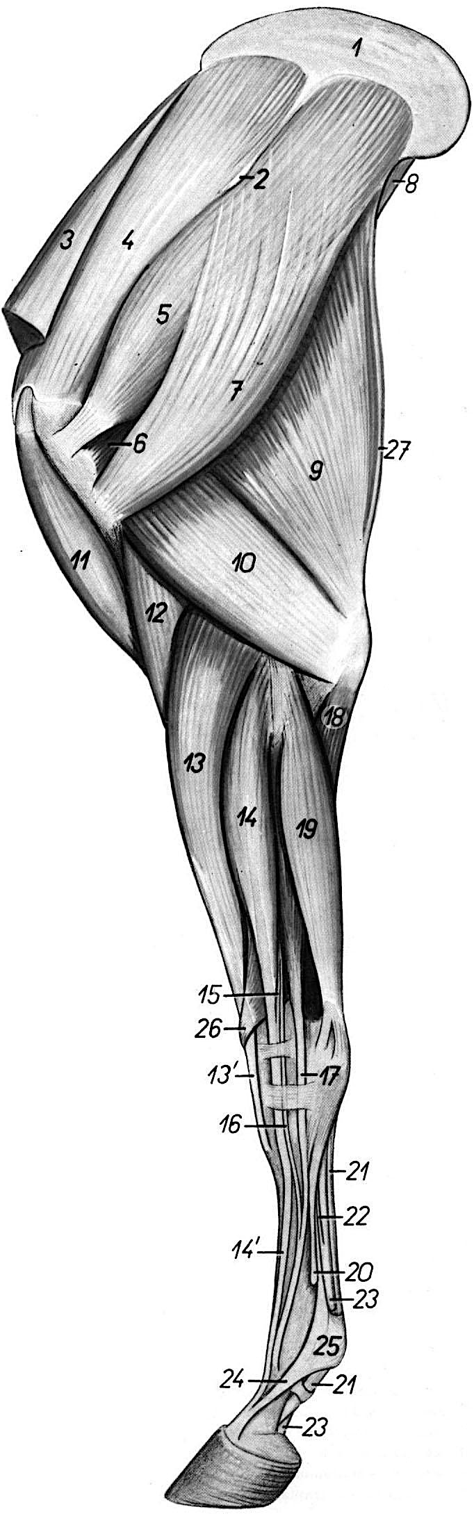 |
(Duplicate) Figure 1-8. Equine left forelimb, lateral view. 1 Scapular cartilage2 Spine of scapula3 Cranial deep pectoral m. (aka subclavius)4 Supraspinatus m.5 Infraspinatus m.6 Teres minor m.7 Deltoideus m.8 Teres minor m.9 Long head of triceps brachii m.10 Lateral head of triceps brachii m.11 Biceps brachii m.12 Brachialis m.13, 13’ Extensor carpi radialis m.14, 14’ Common digital extensor m.15, 16 Rudimentary extensor tendons.17 Lateral digital extensor m.18 Ulnar head of deep digital flexor (DDF) m.19 Ulnaris lateralis m.20 Lateral splint bone = Mc 421 Superficial digital flexor tendon (SDFT)22 Carpal (distal) check ligament23 Deep digital flexor tendon (DDFT)24 Extensor branch of suspensory ligament25 Locus of proximal sesamoid bones26 Extensor carpi obliquus m.27 Tensor fasciae antebrachii m. |
|
|
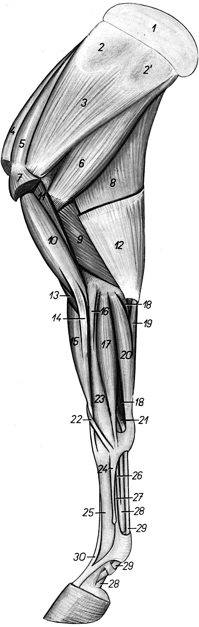
(Duplicate) Figure 1-9. Equine right forelimb, medial view. 1 Scapular cartilage2, 2’ Insertion area of serratus ventralis m.3 Subscapularis m.4 Cranial deep pectoral m. (subclavius)5 Supraspinatus m.6 Teres major m.7 Insertion of caudal deep pectoral m.8 Long head of triceps brachii m.9 Medial head of triceps brachii m.10 Biceps brachii m.11 Coracobrachialis m.12 Tensor fasciae antebrachii m.13 Brachialis m.14 Lacertus fibrosus15 Extensor carpi radialis m.16 Medial collateral ligament of elbow joint17 Flexor carpi radialis m.18 Superficial digital flexor (SDF) m.19 Ulnar head of flexor carpi ulnaris m.20 Humeral head of flexor carpi ulnaris m.21 Accessory head of SDF (proximal check ligament)22 Extensor carpi obliquus m.23 Radius24 Mc 2 = medial splint bone25 Mc 3 = cannon bone26 Carpal (distal) check ligament27 Suspensory ligament = interosseous tendon28 Deep digital flexor tendon (DDFT)29 Superficial digital flexor tendon (SDFT)30 Common digital extensor tendon |
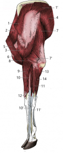 |
(Duplicate) Figure 1-10. Bovine left forelimb, lateral view. 1, 1’ trapezius m.2 supraspinatus m.3 deltoideus m.4 latissimus dorsi m.5 brachiocephalicus m.6 biceps brachii m.7, 7’ long and lateral heads of triceps brachii m.8 brachialis m.9 extensor carpi radialis m.10, 10’ common digital extensor m. and the tendon of its lateral belly11, 11’ lateral digital extensor m. and its tendon12 extensor carpi obliquus m.13 ulnar head of deep digital flexor m.14 ulnaris lateralis m.(Modified from Dyce, Sack and Wensing, 4th ed.) |
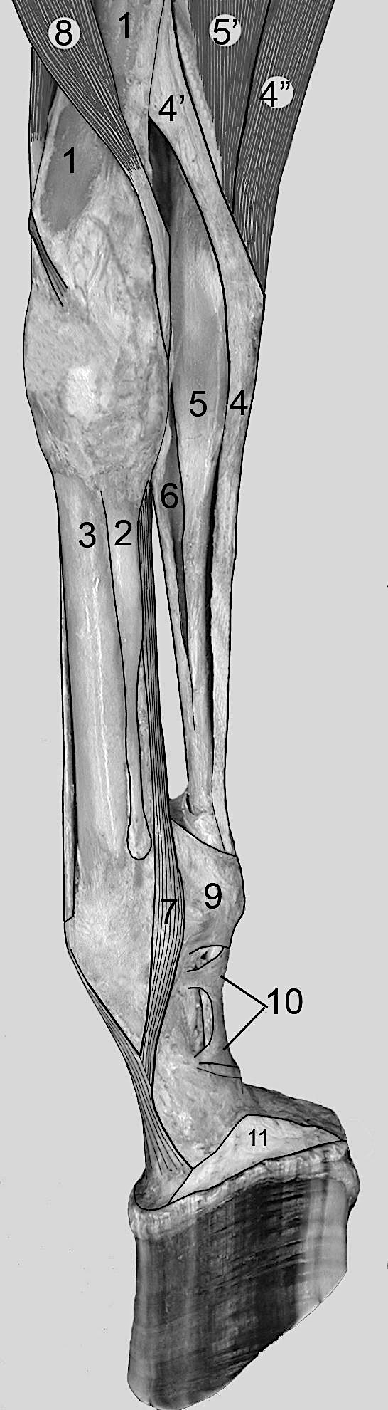 |
Figure 1-11. Equine distal right forelimb, medial view. Check ligaments shown.
1, Radius2, Metacarpal 23, Metacarpal 34, Superficial digital flexor (SDF) tendon4’, Proximal check ligament (SDF radial head)4”, SDF muscle (humeral head)5, Deep digital flexor (DDF) tendon5’, DDF muscle (humeral heads)6, Distal check ligament (accessory ligament of the DDFT)7, Suspensory ligament (interosseous tendon)8, Flexor carpi radialis m.9, Palmar annular ligament10, Proximal digital annular ligament11, Collateral cartilage of P3Image notes:
|
ARTERIES & NERVES of the THORACIC LIMB (Cont.)
14. ALL forelimbs: Identify the median a. and the radial a. (Fig. 1-6)
-
- As mentioned previously in the proximal forelimb dissection, the brachial a. changes names to median a. after the common interosseous branch; identify the median a. in all forelimbs.
- Trace the median a. as it courses distally through the antebrachium to reach the carpal canal.
- The median a. gives rise to another branch, the radial a., just before entering the carpal canal in the pony, and further proximally in the calf.
- The radial a. is fairly small and will be seen branching cranially from the median a.
- You should attempt to locate/identify the radial a. in your specimen, but also be sure to identify it in both species of demonstration specimens.
- The median a. continues distally; the median a. courses through the carpal canal and then changes names as it continues distally. The name change location is dependent upon the species you are dissecting. This will be discussed in the subsequent steps of the dissection.
15. ALL forelimbs: Re-identify and trace the median n. running alongside the median a. through the antebrachium. Trace the median n. to the level of the carpal canal.
16. ALL forelimbs: Trace the median a. into the carpal canal. In the pony, identify and trace the medial palmar a. distally. (Fig. 1-6)
-
- Dissection Note: The flexor carpi radialis m. may somewhat obscure the view as you trace the median a. distally. Do the best you can to work around this muscle to view the path of the median a. as you near the carpal canal, but if necessary, you may transect the flexor carpi radialis m. to trace the median a.
- Pony Specimen: In the pony, the median a. changes names and is continued as the medial palmar a. This name change occurs just after/just distal to the carpal canal (i.e., at the level of the carpus). Distal to the carpus, the main blood supply to the equine digit is the medial palmar a. Identify the medial palmar a. in the pony specimens.
- Calf Specimen: In the calf, the median a. changes names and is continued as the palmar common digital a. III. This name change occurs just before/just proximal to the fetlock (i.e., at the level of the fetlock). Between the carpus and fetlock, the main blood supply to the bovine digits is still called the median a. At the fetlock, the name change occurs – so the main blood supply from the fetlock to the digits comes from the palmar common digital a. III. You do not need to trace/identify the palmar common digital a. III in the calf.
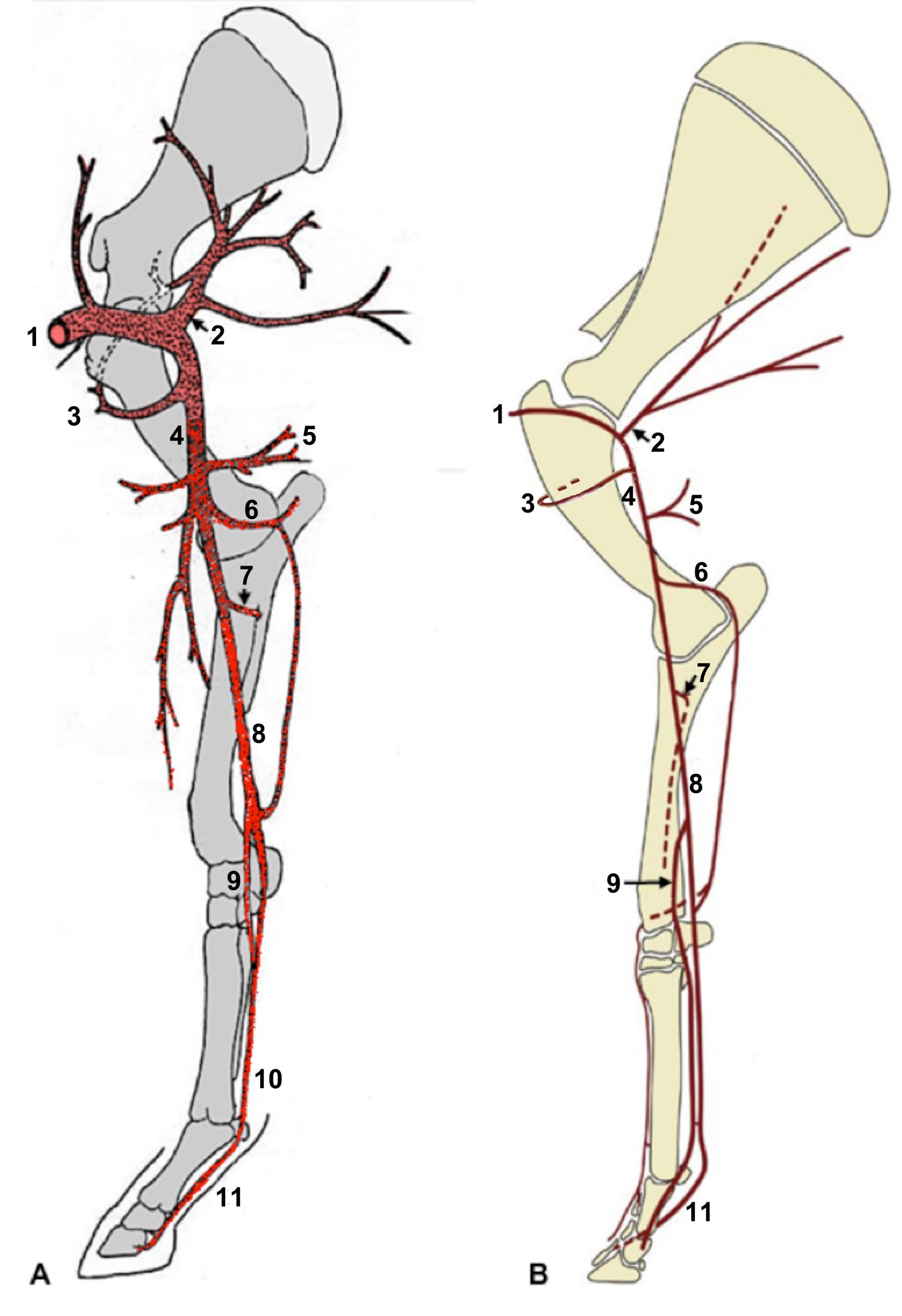
(Duplicate) Figure 1-6. Arteries of the right forelimb of the (A) horse and (B) bovine, medial view. 1, Axillary a.; 2, subscapular a.; 3, cranial circumflex humeral a.; 4, brachial a.; 5, deep brachial a.; 6, collateral ulnar a.; 7, common interosseous a.; 8, median a.; 9, radial a.; 10, medial palmar a.; 11, (A) palmar digital a. (eq), (B) palmar common digital a. (bov) Note: The medial palmar and palmar digital aa. are paired in the bovine, i.e., one per digit. (A: Drawing by A. Weber; B: Modified from Dyce, Sack and Wensing, 4th ed.).
carpus
17. ALL forelimbs: Move to the dorsal aspect of the carpus. Take note of the multiple tendons covering the dorsal aspect of the carpal joint (pony: 2 tendons, calf: 3 tendons).
-
- Looking at the dorsal surface of the carpal joint, moving from medial to lateral, these tendons are the extensor carpi radialis m. tendon (medially) and the common digital extensor m. tendon(s) (laterally) (Fig. 1-8/13,13’,14,14’).
- Re-identify the extensor carpi radialis m. (previously identified) by following its tendon proximally.
- Re-identify the common digital extensor m. (previously identified) by following its tendon proximally, and then trace its tendon(s) distally.
-
- Recall that the extensor carpi radialis tendon inserts on the cranial aspect of the proximal cannon region (metacarpal tuberosity) while the common digital extensor tendon extends much further, extending down through the metacarpus to the extensor process of P3.
-
- Looking at the dorsal surface of the carpal joint, moving from medial to lateral, these tendons are the extensor carpi radialis m. tendon (medially) and the common digital extensor m. tendon(s) (laterally) (Fig. 1-8/13,13’,14,14’).
18. ALL forelimbs: Open the joint capsules of the carpus to identify the individual carpal bones.
-
- On the dorsal side of the carpus, work between the extensor tendons to open the joint capsules and identify the individual carpal bones.
- Notice that the bones of each row are tightly bound together, but the two rows are easily separable on the dorsal surface.
- Note that the distal row of carpal bones is tightly bound to the metacarpal bones and, therefore, the carpal/metacarpal joint is difficult to expose. In contrast, the joint capsule of the radial carpal and middle carpal (intercarpal) joints is more abundant, allowing a greater range of motion and easier removal of the capsule.
19. ALL forelimbs: Note the presence of medial and lateral collateral ligaments of the carpal joint. These ligaments are found on the medial and lateral sides of the limb respectively (as implied by their names) and help provide stability to the carpus. Attempt to identify/visualize the medial and lateral collateral ligaments of the carpal joint. (Fig. 1-12/9)
-
- Dissection Note: These collateral ligaments may be difficult to see, especially in the calf specimens.
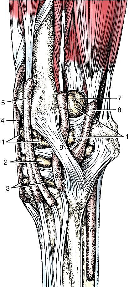
Figure 1-12. (Left) Synovial structures of the left carpus of the horse, lateral view. 1, radiocarpal joint capsule; 2, middle carpal (intercarpal) joint capsule; 3, carpometacarpal joint capsule; 4, tendon sheath of extensor carpi radialis; 5, tendon sheath of common digital extensor; 6, tendon sheath of lateral digital extensor; 7, tendon sheath of superficial and deep digital flexors (carpal sheath); 8, tendon sheath of ulnaris lateralis; 9, lateral collateral ligament. (Modified from TVA Fig. 23-18)
metacarpus & Digit(s)
20. We will now complete our dissection of the forelimb by moving distally to the metacarpus and digit(s). As you dissect you should be palpating the structures to develop your palpation skills (palpation is not limited to the rectum!). Most structures in the equine distal limb are skin deep and therefore easily palpated. Also, most tendons and ligaments lie over a firm bone that stabilizes the palpable structures making them easier to feel.
21. ALL (PONY) forelimbs: On the medial side of the cannon region, re-identify the medial palmar artery medial to the digital flexor tendons; trace it distally to where it branches. Identify the medial and lateral palmar digital aa. (Fig. 1-6/7)
-
- Note the medial palmar nerve adjacent to the medial palmar artery; you will identify this in the next step.
- Trace the medial palmar artery distally where it runs deep to the digital flexor tendons and divides into medial and lateral palmar digital arteries on medial and lateral sides of the limb, respectively; identify the medial and lateral palmar digital arteries.
22. ALL (PONY) forelimbs: Move proximally into the antebrachium and re-identify the median n. running alongside the median a.; trace it to the carpal canal. At the level of the carpal canal, identify the medial palmar nerve; trace it distally. Identify the lateral palmar nerve. Identify the medial and lateral palmar digital nerves. Identify the communicating branch of the palmar nerves. (Fig. 1-13/7)
-
-
On the medial side of the antebrachium, re-identify the median n. and trace it to the carpal canal.
-
As it enters the carpal canal, the median n. changes names to medial palmar n. and continues distally.
-
In the mid-cannon region, adjacent to the medial palmar artery, identify the medial palmar nerve. Trace the medial palmar n. distally to find and identify the lateral palmar n.
-
In the carpal canal, the medial palmar n. gives rise to the lateral palmar n.; attempt to dissect this branching point in the carpal canal and identify the lateral palmar n.
-
-
Note that the ulnar n. also contributes to the lateral palmar n. but need not be dissected.
-
-
-
-
In the mid-cannon region, on the palmar surface of the superficial digital flexor tendon, find the communicating branch of the palmar nerves connecting the medial and lateral palmar nerves.
-
Dissection Note:This communication crosses the superficial surface of the flexor tendons in an oblique fashion, from lateral to medial going “up” the leg.
-
-
-
- Trace the medial & lateral palmar nerves distally and identify the medial and lateral palmar digital nerves where they run alongside the medial and lateral palmar digital arteries over the fetlock joint.
- Continue to trace the digital nerves and vessels as far distally as possible.
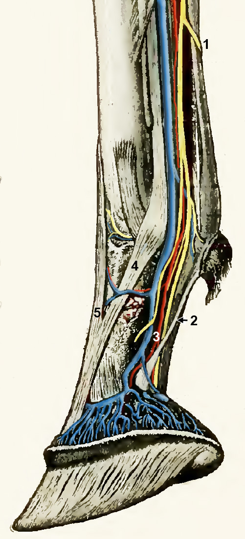
Figure 1-13. Right forelimb digit of the horse, medial view. 1, communicating branch between medial and lateral palmar nn.; 2, ligament of the ergot; 3, neurovascular bundle (palmar digital vein, artery and nerve); 4, extensor branch of suspensory ligament; 5, common digital extensor.
23. ALL (CALF) forelimbs: Note the nerves and arteries of the distal limb, but you do not need to dissect/trace them as in the pony. Identify the dorsal common digital v. in the calf demonstration specimen, as it is of clinical relevance.
-
- You do not need to trace the nerves to the digit of the calf but you should note that there is a similar arrangement of median and ulnar nerves to either side of the digital flexor tendons within the cannon region that resembles the arrangement of the medial and lateral palmar nerves in the pony.
- The calf also has communicating branches between the median and ulnar nerves, but these do not need to be identified.
- You do not need to trace the arteries to the digit of the calf but you should be aware of the difference in arterial supply to the foot of the bovine vs. that of the equine – the median a. continues to the level of the fetlock where it changes names to the palmar common digital a. III. The palmar common digital a. III then splits into axial palmar proper digital arteries III and IV which are the main blood supply to the two digits. (Fig. 1-14)
- You do not need to trace the nerves to the digit of the calf but you should note that there is a similar arrangement of median and ulnar nerves to either side of the digital flexor tendons within the cannon region that resembles the arrangement of the medial and lateral palmar nerves in the pony.
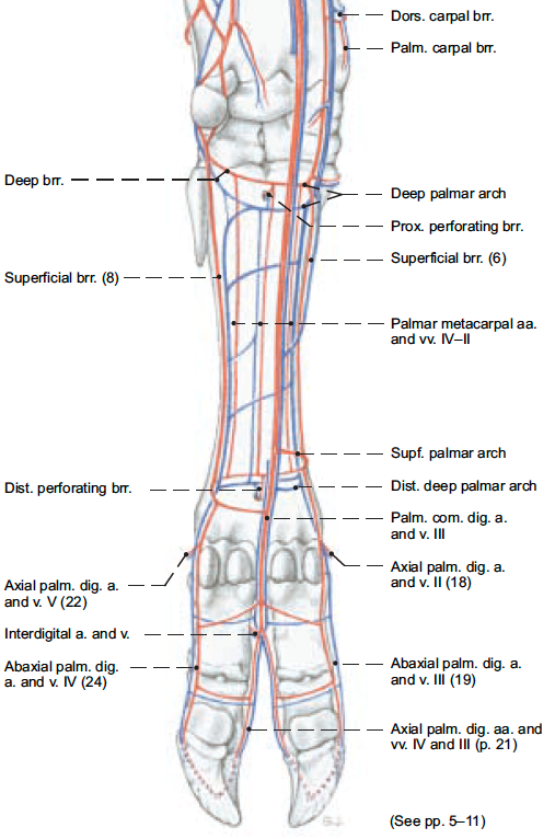
Figure 1-14. Left distal thoracic limb of bovine, palmar view. (Modified from Budras, pg. 8)
-
- One structure of clinical relevance in the bovine is the dorsal common digital v. This vein should be identified on the calf demonstration specimen and is testable information.
- The dorsal common digital v. is found on the dorsal aspect of the distal metacarpus of bovine (and ovine) animals. In the bovine, the dorsal common digital v. may be used during clinical procedures. (Fig. 1-15)
-
- The dorsal common digital v. drains into/joins the accessory cephalic vein in the thoracic limb, which you have already identified. (Think of the accessory cephalic v. as a continuation of the dorsal common digital v. as you move proximally up the limb – more of a name change than separate veins.)
-
- The dorsal common digital v. is found on the dorsal aspect of the distal metacarpus of bovine (and ovine) animals. In the bovine, the dorsal common digital v. may be used during clinical procedures. (Fig. 1-15)
- One structure of clinical relevance in the bovine is the dorsal common digital v. This vein should be identified on the calf demonstration specimen and is testable information.
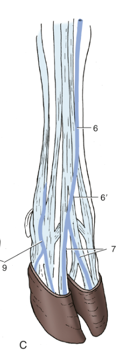 Figure 1-15. C) Right bovine foot, dorsal view. 6, accessory cephalic v.; 6′, dorsal common digital v. III; 7, dorsal digital vv.; 9, abaxial palmar digital vv. (TVA)
24. On the demonstration animals, identify the structures described below that are associated with the distal thoracic limb. (See Figures 1-16 & 1-17) Please note that all terms in this section shown in regular bold font are testable material on quizzes and exams – even though you are not dissecting them on your own specimens! (Be able to identify these structures on demonstration (museum) specimens as well!)
-
- In both the pony and the calf, the digital flexor tendons have been transected in the distal cannon region (in the pony the transection should be distal to the communicating branch of the palmar nerves).
- Reflect the distal portion of the digital flexor tendons distally, off of the sesamoid bones.
- This reflection will require severing the palmar annular ligament (Figs. 1-11/9, 1-17/5) of the fetlock (see next step), which binds the digital flexor tendons in the groove formed by the sesamoid bones.
- Identify the palmar annular ligament, and define its borders by dissection.
- Sever the palmar annular ligament with a longitudinal incision midway between its attachments to the sesamoid bones.
- Identify and define the borders of the proximal and distal digital annular ligaments. (Fig. 1-11/10, 1-17/6,7,8 and TVA 610, 733)
- Note that these ligaments are located distal to the palmar annular ligament.
- Calf Specimen: There will typically be one proximal and one distal digital annular ligament associated with each of the principal digits (often referred to collectively as ‘digital annular ligaments’).
- Sever the proximal digital annular ligament with a longitudinal cut through its attachments on one side. Reflect the ligament off of the digital flexor tendons.
- If needed, sever the distal digital annular ligament with a similar cut.
- Trace/reflect the distal portion of the digital flexor tendons.
- Dissection Note: Just like the arrangement in the carnivore, the deep digital flexor tendon (DDFT) appears to pass through the superficial digital flexor tendon (SDFT) (Fig. 1-14, TVA 609). Actually, the SDFT ends by branching around the DDFT and inserts on the proximal end of P2. This area, where the ‘switch’ occurs, is referred to as the flexor manica.
- Calf Specimen Notes: The ligamentous arrangement within the two principal digits of the calf will be similar to that seen in the single digit of the horse. However, additionally, you should identify the distal interdigital ligament as well.
- This may be difficult in the dissection specimens so be sure to identify the distal interdigital ligament on the plastinated museum specimen of the bovine foot that is available in the demonstration area.
- In both the pony and the calf, the digital flexor tendons have been transected in the distal cannon region (in the pony the transection should be distal to the communicating branch of the palmar nerves).
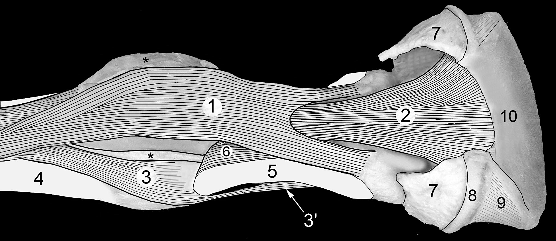
Figure 1-16. Equine fetlock and digit, palmar oblique view. The hoof and much of the digital cushion have been removed to expose the distal portion of the deep digital flexor tendon (DDFT). 1, Superficial digital flexor tendon (SDFT); 2, deep digital flexor tendon (DDFT); 3, suspensory ligament (interosseous tendon); 3’, extensor branch of suspensory lig.; 4, distal end of Mc 3 (cannon bone); 5, P1 (long pastern bone); 6, oblique sesamoidean ligament; 7, collateral cartilage of P3; 8, coronary dermis; 9, laminar dermis; 10, dermis of sole. Both flexor tendons lie in a groove formed by the sesamoid bones on the palmar aspect of the fetlock (Fig. 3-3/6). The flexor tendons are bound by the palmar annular ligament which has been removed. However, the cut edges of the palmar annular ligament are indicated by asterisks (*). Before dissection the digital cushion filled the space between the collateral cartilages.
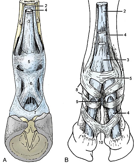
Figure 1-17. A. Equine foot. B. Bovine foot. Palmar view. 1, Equine: splint bones; 2, suspensory ligament (interosseous tendon); 3, superficial digital flexor; 4, deep digital flexor; 5, palmar annular ligament; 6, equine: proximal digital annular ligament; 7, equine: distal digital annular ligament; 8, bovine: digital annular ligament; 9, bovine: proximal interdigital ligament; 10, bovine: distal interdigital ligament. (Modified from TVA Figs. 23-29, 30-7)
25. ALL forelimbs: Identify the suspensory ligament (aka interosseous tendon). Identify the extensor branches of the suspensory ligament. (Be able to identify these structures on demonstration (museum) specimens as well!)
-
- On the palmar surface of the cannon bone, identify the suspensory ligament (aka interosseous tendon). (Note that in the pony it lies between the splint bones.) (Figures 1-9/27, 1-11/7, 1-16/3, and 1-17/2, 1-18/1)
-
The suspensory ligament is the clinical term for the interosseous tendon; in the dog, the interosseous muscle is fleshy, but in the horse it is largely tendinous. In the calf, it may appear somewhat fleshy, but is also largely tendinous.
-
- Note that the suspensory ligament splits to form lateral and medial branches that attach on the “outside” (abaxial side) of the proximal sesamoid bones.
- This distal arrangement of the suspensory ligament is similar for each of the two principal digits of the calf (i.e., ‘splits’ into medial and lateral branches for each toe) and is also similar between front and hind limbs of both species.
- Beyond the sesamoid bones, identify the extensor branches of the suspensory ligament. (Figs. 1-8/24 and 1-18/1’)
- The extensor branches obliquely cross the sides of the long pastern bone to attach dorsally on the common digital extensor tendon (from which they derive their name).
- Again, this distal arrangement of the suspensory ligament is similar for each of the two principal digits of the calf and also similar between front and hind limbs of both species.
- Important Note: The suspensory ligament is part of the suspensory apparatus.
- Suspensory apparatus = suspensory ligament (aka interosseous tendon) + proximal sesamoid bones + distal sesamoidean ligaments
-
- In both the horse and ox these structures act together to support the fetlock against gravity.
-
- Suspensory apparatus = suspensory ligament (aka interosseous tendon) + proximal sesamoid bones + distal sesamoidean ligaments
- On the palmar surface of the cannon bone, identify the suspensory ligament (aka interosseous tendon). (Note that in the pony it lies between the splint bones.) (Figures 1-9/27, 1-11/7, 1-16/3, and 1-17/2, 1-18/1)
26. On the demonstration PONY, identify the structures described below that are associated with the distal thoracic limb. (See Figure 1-18) Please note that all terms in this section shown in regular bold font are testable material on quizzes and exams – even though you are not dissecting them on your own specimens! (Be able to identify these structures on demonstration (museum) specimens as well!)
-
- Reflect the digital flexor tendons distally to expose the distal sesamoidean ligaments.
- Identify the superficial (straight) sesamoidean ligament (Fig. 1-18/4)
- This ligament extends from the proximal sesamoids to the proximal end of P2 where it inserts between the insertions of the superficial digital flexor tendon.
- Flex the digit enough to elevate the superficial sesamoidean ligament in order to identify the underlying middle (oblique) sesamoidean ligament (Fig. 1-18/5).
- Dissection Note: There are also deep (cruciate) sesamoidean ligaments present, but these need not be dissected and can be viewed on plastinated demonstration specimens.
- Be able to identify these ligaments on plastinated demonstration (museum) specimens as well, as they are fair game for assessments!
- Calf Note: You do not need to dissect or visualize the distal sesamoidean ligaments of the calf. However, you should note that the ox does not have the straight sesamoidean ligament, but it does have the oblique and deep sesamoidean ligaments similar to those found in the pony but duplicated for each principal digit.
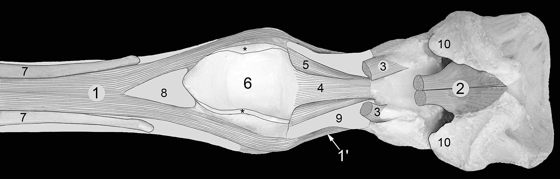
Figure 1-18. Equine distal forelimb, palmar view. The hoof and much of the digital cushion have been removed to expose the distal deep digital flexor tendon (DDFT). 1, Suspensory ligament (interosseous tendon); 1’, extensor branch of suspensory ligament; 2, deep digital flexor tendon (DDFT);3, superficial digital flexor tendon (SDFT); 4, straight sesamoidean ligament; 5, oblique sesamoidean ligament; 6, groove formed by the sesamoid bones; 7, splint bones (Mc 2 and 4); 8, Mc 3 (cannon bone); 9, P1 (long pastern bone); 10, collateral cartilage of P3. Both digital flexor tendons lie in a groove (6) formed by the sesamoid bones. The flexor tendons are bound by the palmar annular ligament which has been removed. However, the cut edges of the palmar annular ligament are indicated by asterisks (*). Notice that the suspensory ligament (interosseous tendon) lies on the palmar surface of the cannon bone and splits at the level of the end of the splint bones. Typically, the medial splint bone, Mc2, is slightly longer than Mc4.
Dissection Videos for this Section of Material
Distal Thoracic Limb
- Pony
- Distal Thoracic Limb – Attached:https://www.youtube.com/watch?v=mx1OZkrQUws
- Distal Thoracic Limb – Detached:https://www.youtube.com/watch?v=IJwUZio9nEE
- Calf
- Distal Thoracic Limb – Attached:https://www.youtube.com/watch?v=uyTGTL16S-g
- Distal Thoracic Limb – Detached: https://www.youtube.com/watch?v=kee3BkLRGGc&t=1s
