
Part 2: Lungs and Bronchi
Abby Brown
Lungs
- ALL specimens: Identify the lobes of the lungs and the principal (primary) bronchi (left and right). Note that the lobes of the lungs are named according to the branching of the principal (primary) bronchi from the trachea.
- ALL (PONY) specimens: In the equine, lungs lack fissures between the lobes, but the lobes are still based on the branching of the left and right principal bronchi.
- The left lung is divided into 2 lobes, these are the cranial and caudal lobes. The cranial and caudal lobes are separated by a cardiac notch.
- The right lung is divided into cranial, caudal, and accessory lobes. The cranial and caudal lobes are separated by a cardiac notch.
-
- Dissection Note: The horse lacks the right middle lobe.
-
- Identify these lung lobes in the pony specimens.
- ALL (CALF) specimens: The bovine (and pig) are similar in lung lobation to the dog and cat and have distinct fissures between the lobes.
- The left lung consists of 2 lobes, a cranial lobe and a caudal lobe.
-
- Note that the cranial lobe is further subdivided into cranial and caudal parts separated by a cardiac notch and fissure.
-
- The right lung consists of 4 lobes: a cranial lobe, a middle lobe, a caudal lobe and an accessory lobe.
-
- Note that the cranial lobe is further subdivided into cranial and caudal parts separated by a cardiac notch and fissure. The cranial lobe also has a tracheal bronchus associated with it that branches off of the trachea (cranial to the bifurcation into left and right primary bronchi).
- Reflect the cranial part of the cranial lobe of the right lung to identify the tracheal bronchus in the calf specimens. (Note that this is considered a lobar bronchus and not a primary bronchus.)
-
- Identify these lung lobes in the calf specimens.
- The left lung consists of 2 lobes, a cranial lobe and a caudal lobe.
- ALL specimens: Attempt to identify where the trachea bifurcates into the left and right principal (primary) bronchi in each specimen by reflecting the lung lobes. (This may be easiest to see from the right side.)
- ALL (PONY) specimens: In the equine, lungs lack fissures between the lobes, but the lobes are still based on the branching of the left and right principal bronchi.
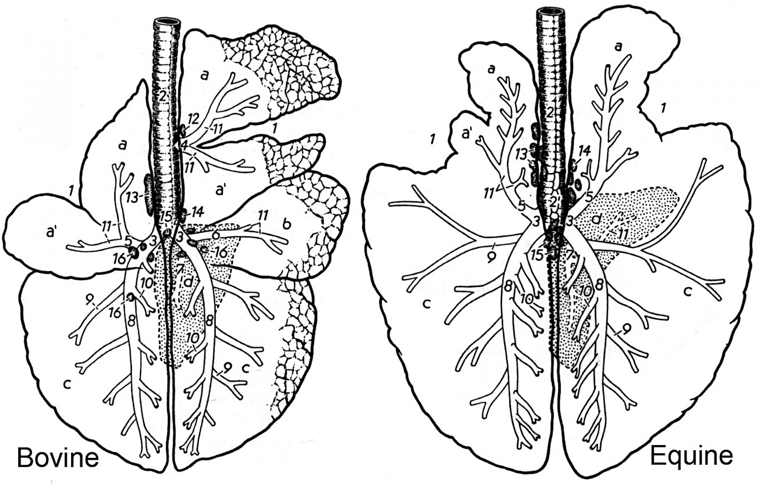
Figure 4-1. Bovine and equine lungs, dorsal schematic view. 1, cardiac notch; 2, trachea; 3, primary bronchus; 4, tracheal bronchus (not in horse); 5, cranial bronchus; 6, middle bronchus (not in horse); 7, accessory bronchus; 8, caudal bronchus; 9-11, segmental bronchi; 12-15, tracheobronchial lymph nodes; 16, pulmonary lymph nodes; a, cranial parts of cranial lobe(s); a’, caudal parts of cranial lobe(s); b, middle lung lobe; c, caudal lung lobes; d, accessory lung lobe.
2. Summary of species differences to note on the lungs:
-
- CALF (and pig): A separate bronchus to the right cranial lobe arises cranial to the primary bifurcation of the trachea in the calf (and pig); this is called the tracheal bronchus (Fig. 4-1/4). This is considered a lobar bronchus.
- CALF: Subdivided cranial lobes are found in the calf (on both left and right lungs). The left and right cranial lobes are each subdivided into cranial and caudal parts in the calf. These cranial and caudal parts are separated by a cardiac notch and fissure (on each side, left and right).
- CALF: Lobulation (distinct lobules separated by fibrous tissue within the lung parenchyma) is obvious in the calf (and pig).
- PONY (and sheep): Lack obvious lobulation (distinct lobules separated by fibrous tissue within the lung parenchyma).
- PONY: Equine lungs lack obvious fissures between the lobes.
- PONY: Equine lungs lack the middle right lobe.
Major Structures Close to the Lungs (in situ)
3. ALL specimens, LEFT side: Reflect (but do not remove) the left cranial lung lobe; identify the left vagus n. (with left recurrent laryngeal branch) and the tracheobronchial lymph nodes. Reflect (but do not remove) the left caudal lung lobe to identify the caudal mediastinal lymph nodes.
-
- CALF Dissection Note: In the calf, there will be abundant thymic tissue that you will need to work around/through to locate the structures within the thorax.
- Reflect the left cranial lung lobe caudally, toward the hilus, and find the left vagus nerve on the wall of the cranial mediastinum (Fig. 4-2/14).
- Trace the left vagus n. caudally to the point where the left recurrent laryngeal n. is given off and wraps around the base of the aorta.
- After wrapping around the aorta, the left recurrent laryngeal n. will travel cranially along the left side of the trachea.
- Locate the tracheobronchial lymph nodes on the cranial edge of the root of the left lung.
- Reflect the left caudal lung lobe cranially and locate the caudal mediastinal lymph nodes lying between the caudal esophagus and the aorta.
- PONY Dissection Note: Note that the pony has a tracheobronchial lymphocenter. This lymphocenter is composed of the tracheobronchial lymph nodes and the caudal mediastinal lymph node, which drains the abdominal structures. Note the closeness of the tracheobronchial lymphocenter to the left recurrent laryngeal n.
- CALF Dissection Note: Note the proximity of the large caudal mediastinal lymph node(s) to the dorsal branches of the vagus nerve(s), dorsal to the esophagus.
4. ALL specimens, RIGHT side: Reflect (but do not remove) the right cranial lung lobe; identify the right vagus n. (with right recurrent laryngeal branch) and attempt to see the tracheobronchial lymph nodes. Reflect the right caudal lung lobe; attempt to identify the caudal mediastinal lymph nodes (visible from both right and left sides).
-
- Reflect the right cranial lung lobe caudally, toward the hilus, and find the right vagus nerve on the wall of the cranial mediastinum.
- Trace the right vagus n. caudally and attempt to identify where the right recurrent laryngeal n. is given off and wraps around the subclavian artery. (Note that the right recurrent laryngeal nerve can be difficult to see.)
- After wrapping around the subclavian a., the right recurrent laryngeal n. will travel cranially along the right side of the trachea.
- Attempt to locate the tracheobronchial lymph nodes on the cranial edge of the root of the right lung.
- Reflect the right caudal lung lobe cranially and attempt to locate the caudal mediastinal lymph nodes lying between the caudal esophagus and the aorta.
- PONY Dissection Note: Note that the pony has a tracheobronchial lymphocenter. This lymphocenter is composed of the tracheobronchial lymph nodes and the caudal mediastinal lymph node, which drains the abdominal structures.
- CALF Dissection Note: In the ruminant (and pig), the tracheobronchial lymph nodes may also be found cranial to the tracheal bronchus. Note the proximity of the large caudal mediastinal lymph node(s) to the dorsal branches of the vagus nerve(s), dorsal to the esophagus.
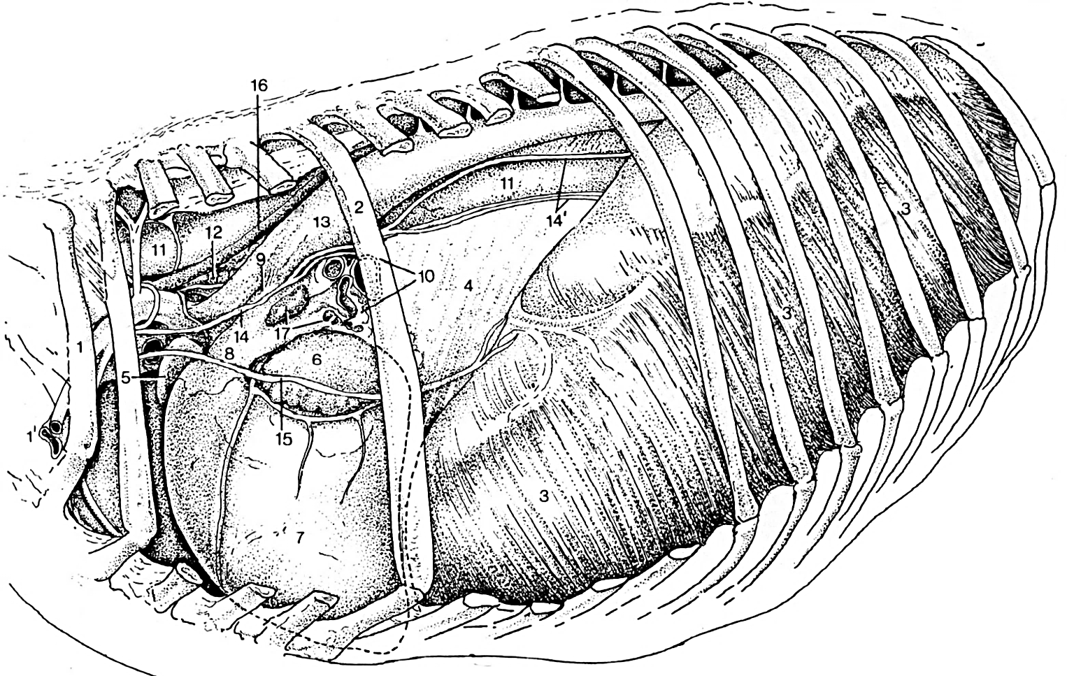
Figure 4-2. Equine thorax, left lateral view. 1, rib 1; 2, rib 6; 3, diaphragm; 4, caudal mediastinum; 5, right auricle; 6, left auricle; 7, left ventricle; 8, pulmonary trunk; 9, ligamentum arteriosum; 10, cut surface of root of the lung; 11, esophagus; 12, trachea; 13, aorta; 14, vagus n.; 14’, dorsal and ventral vagal trunks; 15, phrenic n.; 16, thoracic duct; 12, tracheobronchial lymph node.
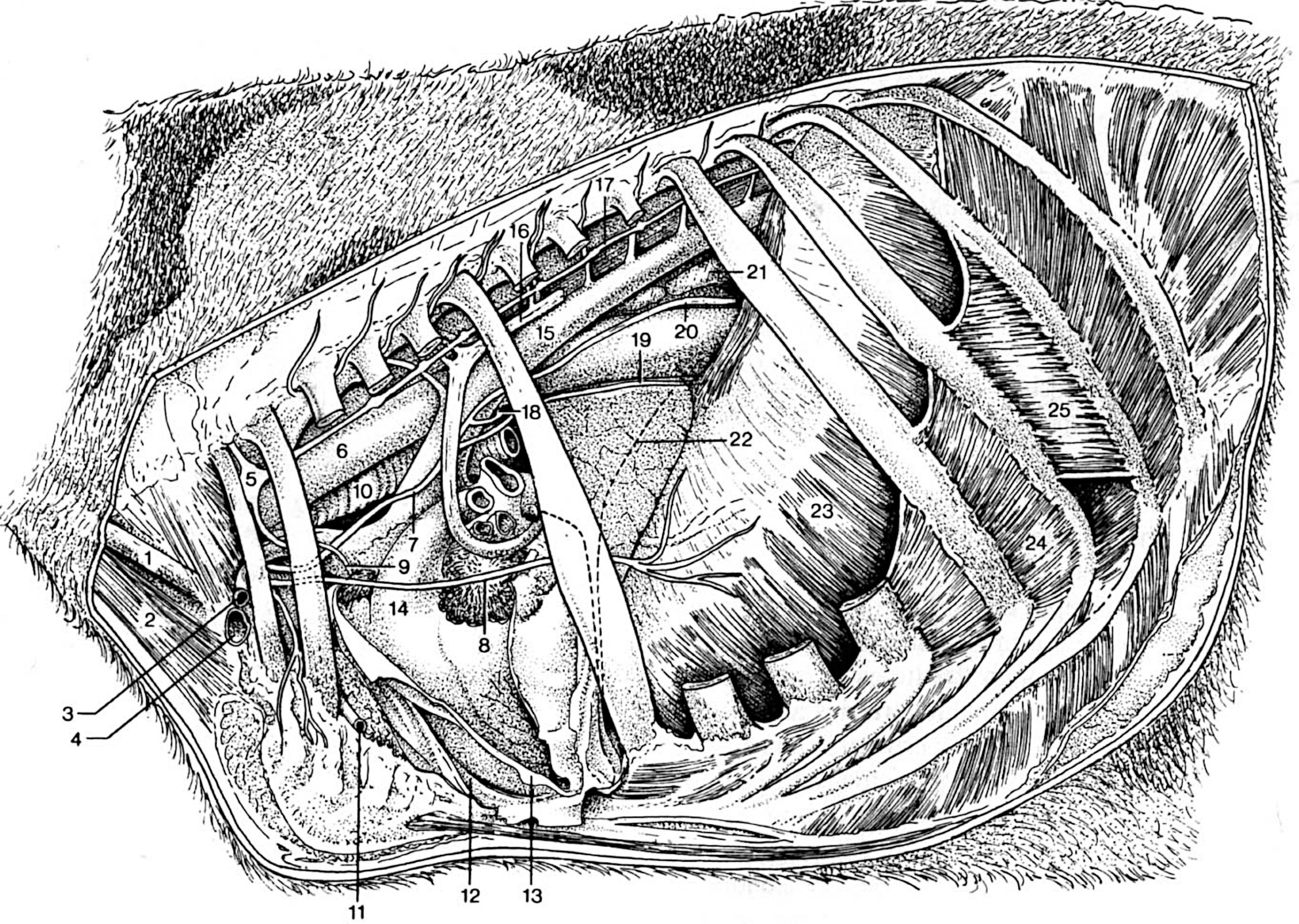
Figure 4-3. Bovine thorax, left lateral view. The left azygous vein (16) passes around the cut surface of the root of the left lung. 1, external jugular vein; 2, sternocephalicus m.; 3, axillary a.; 4, axillary v.; 5, cervicothoracic ganglion; 6, esophagus; 7, left vagus n.; 8, (left) phrenic n.; 9, cardiac n.; 10, trachea; 11, internal thoracic a.; 12, mediastinal pleura; 13, reflected pericardium; 14, pulmonary trunk; 15, aorta; 16, left azygous v.; 17, sympathetic trunk; 18, left recurrent laryngeal n.; 19, ventral vagal trunk.; 20, dorsal vagal trunk.; 21, caudal mediastinal lymph node; 22, cranial extent of the diaphragm; 23, diaphragm; 24, internal intercostal m.; 25, external intercostal m.
Lung removal and isolated lungs
5. ALL specimens: Carefully remove the lungs on both left and right sides by transecting all structures at the root of lung.
-
- Transect the root of the LEFT lung near the hilus and remove the entire lung as a single unit – not separate lobes! Do this by cutting the left principal (primary) bronchus (before the branching of the lobar (secondary) bronchi) and transecting all of the pulmonary vessels of the lung.
- The root of the lung contains a principal bronchus and pulmonary vessels. You will need to transect these structures in order to remove the lung.
-
- Dissection Note: The root of the lung refers to the vessels and nerves entering/leaving the lung, versus the hilus, which refers to the region of the lung where these structures are located.
-
- Be careful to avoid severing the vagal nerves just dorsal and ventral to the root of the lung!
- The root of the lung contains a principal bronchus and pulmonary vessels. You will need to transect these structures in order to remove the lung.
-
On the RIGHT side, slip the accessory lobe of the right lung over the caudal vena cava. (Be careful not to tear the accessory lobe as you do this; it can be tricky to free it from around the caudal vena cava!)
-
Transect the entire root of the RIGHT lung near the hilus (similar to the left lung) and remove the entire lung as a single unit – not separate lobes! Again, be very careful to avoid cutting vagal nerves that course across the heart near the hilus of the lung!
-
The root of the lung contains a principal bronchus and pulmonary vessels. You will need to transect these structures in order to remove the lung.
-
ALL (CALF) specimens: In the calf, you will also need to cut the tracheal bronchus on the right side, cranial to the root of the right lung (Fig. 4-1).
-
Be very careful during removal of the right lung in the calf. It is difficult to remove as one whole piece, so just know that it may come apart and may need to be removed in pieces (and that’s ok!).
-
-
- As you remove the lungs, leave the heart within the thorax to study it ‘in situ’ (in place within the body) in the subsequent parts of this chapter.
- Transect the root of the LEFT lung near the hilus and remove the entire lung as a single unit – not separate lobes! Do this by cutting the left principal (primary) bronchus (before the branching of the lobar (secondary) bronchi) and transecting all of the pulmonary vessels of the lung.
6. ALL specimens: After removal of both lungs, within the thorax, identify the trachea. Trace the trachea caudally to the point where it branches into the principal (primary) bronchi (left and right). (Terminology Note: ‘bronchi’ is plural, ‘bronchus’ is singular.)
7. ALL specimens: Locate and re-identify tracheobronchial lymph nodes in your specimen. These may be attached to the isolated lungs or still within the thorax – usually near the bifurcation of the trachea. (Fig. 4-1)
-
- CALF Dissection Note: The tracheobronchial lymph nodes may be easier to find on the calf specimens due to their larger size.
8. ALL specimens: On the now isolated lungs, trace the very short principal (primary) bronchi as they branch into lobar (secondary) bronchi.
-
-
Note that there is one primary bronchus for each lung and, branching from that primary bronchus, one lobar bronchus for each lung lobe.
- CALF Dissection Note: Though the tracheal bronchus usually branches cranial to the bifurcation of the primary bronchi, it is considered a lobar (secondary) bronchus.
-
Identify the lobar bronchi entering each of the lung lobes.
-
Note that the bronchi will have cartilaginous rings, which will help differentiate them from arteries and veins.
-
- Dissection Note: Beyond these primary and secondary divisions there are 3rd, 4th, 5th degree bronchi, etc.
-
9. ALL specimens: On the isolated lungs, attempt to distinguish between pulmonary veins and pulmonary arteries.
-
-
Observe the (cut surface of the) main pulmonary artery entering each lung (one on the left lung and one on the right lung) and note that the main artery then branches into smaller lobar pulmonary arteries. In latex injected specimens, these arteries may appear blue (if blue media was used and reached the lungs) or not injected if only red media was used.
-
Note that the arteries have thicker walls than the veins.
-
Note the orientation of the pulmonary artery relative to the bronchi. (The pulmonary arteries are dorsal and lateral relative to the bronchi.)
-
-
Observe the (cut surfaces of the) pulmonary veins leaving the lungs. Usually you will see a single pulmonary vein from each lobe (aka lobar pulmonary vv.). In latex injected specimens, these veins may appear red (if the injection reached the lungs).
-
Note the orientation of the pulmonary veins relative to the bronchi. (The pulmonary veins are ventral and medial relative to the bronchi.)
-
-
Note that in latex injected specimens, the coloration seen in the pulmonary arteries and veins is opposite of what you would expect based on the coloration of all other arteries and veins in the injected specimens. Be sure you are able to explain why this would be the case!
-
10. ALL specimens: On the isolated lungs, review the lung lobes (and associated terms) previously described when you viewed the lungs ‘in situ’ (in place within the body).
Dissection Videos for this Section of Material
Lungs, Bronchi and Lymph Nodes
- Pony
- Internal Thorax, Left and Right sides: https://youtu.be/E-4ar4mecXg
- Watch from 0:00-3:48 and 14:16-17:25 (left side)
- Watch from 13:58-16:24, 16:40-17:07 and 19:35-20:15 (right side)
- Internal Thorax, Left and Right sides: https://youtu.be/E-4ar4mecXg
- Calf
- Internal Thorax, Left side (Watch from 0:00-3:33 and 5:38-6:07): https://youtu.be/Lgb_J8z6w8s
- Internal Thorax, Right side (Watch from 0:00-2:24) : https://youtu.be/JlWBLuScrTU
- Dried Lungs (Equine, Bovine, Porcine): https://youtu.be/Sem3DpefZ48
