
Part 3: Distal Pelvic Limb
Abby Brown
IMPORTANT NOTE: The Guide instructions for this section will refer to either “ALL hind limbs” (meaning attached and detached) or to the “DETACHED” or “ATTACHED” hind limbs.” This added instructional note is needed because the dissection of of some regions may be different for attached vs. detached limbs.
Surface Anatomy
1. ALL hind limbs: On the medial aspect of the distal tibia identify the prominent tuberosity, the medial malleolus (noted in the osteology study in the application portion of the course). On the live horse, and possibly some cadaver limbs, you should be able to note the cranial branch of the medial saphenous vein where it can be seen crossing the dorsal (flexor) surface of the hock (this will be dissected later in this part of the chapter). As with the forelimb, you should also note the following superficial structures:
-
- Pony specimen: Note the chestnut on the distal medial aspect of the tarsal (hock) region of the horse and also the ergot on the plantar aspect of the fetlock region.
- Calf specimen: Identify the dewclaws on the plantar surface of the limbs.
- In both species, observe the coronet (skin to hoof transition).
Skin reflection
2. ALL hind limbs: Make a skin incision on the medial side of the hind limb from the level of the stifle down to mid-cannon; reflect the remaining skin from the hind limb and discard it.
-
- On the medial side of each hind limb, make a skin incision from the level of the stifle joint down to the mid-cannon region.
- On the pony, as you proceed and remove the skin distally, try to preserve the cranial branch of the medial saphenous v. on the limb.
- On the calf, as you proceed and remove the skin distally, try to preserve the cranial branch of the lateral saphenous v. on the limb.
- Make a circular incision around the leg in the middle of the cannon bone.
- Reflect the remaining skin off the hind limb and discard it.
Stifle Dissection
** Important Dissection Note: Study of the available ligamentous (dry) preparations in the demonstration area of the lab will be helpful to prepare yourself to perform the following dissection.**
3. ALL hind limbs: Identify the stifle region. Incise, reflect, and remove fat and fascia in order to identify the three patellar ligaments: medial, middle (intermediate), and lateral; these ligaments are attached to the patella. You should also identify and isolate the medial and lateral collateral ligaments of the stifle joint.
-
- Identify the region of the stifle joint. Palpate this region to feel the patella and the three patellar ligaments. Incise, reflect, and remove the sheets of fascia covering the stifle joint.
- Dissection Note: Removal of this fascia in stages, while palpating as you go, will enable you to see the three patellar ligaments before going further into the stifle joint itself.
- Remove some of the abundant adipose tissue deep to and surrounding the patellar ligaments in order to isolate them.
- Identify the three patellar ligaments: medial, middle (intermediate), and lateral. Their name indicates their position on the limb (e.g., medial patellar ligament is on the medial side, the lateral is on the lateral side, and the middle is in the middle/between the other two).(Fig. 2-9/3, 4, 5)
- Note that all three of the patellar ligaments attach to the patella and also the tibial tuberosity (which you should also palpate in your specimen).
- Identify and isolate the medial and lateral collateral ligaments of the stifle joint on the medial and lateral sides of the stifle, respectively (Fig. 2-9/7, 6).
- Identify the region of the stifle joint. Palpate this region to feel the patella and the three patellar ligaments. Incise, reflect, and remove the sheets of fascia covering the stifle joint.
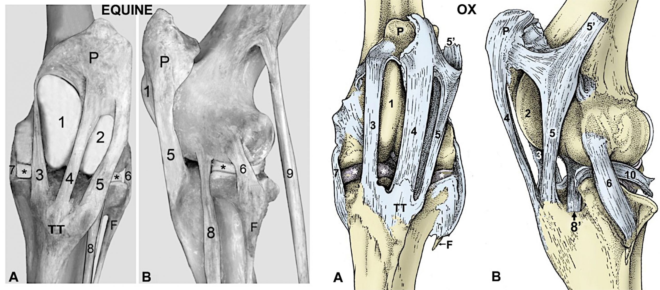
Figure 2-9. Left stifle equine (ligamentous preparation) and ox (drawing modified from TVA figure 31-4) A, cranial view; B, lateral view. 1, Medial ridge of femoral trochlea; 2, lateral ridge of femoral trochlea; 3, medial patellar ligament; 4, middle patellar ligament; 5, lateral patellar ligament; 5’, attachment of biceps femoris m. (shown in ox); 6, lateral collateral ligament; 7, medial collateral ligament; 8, fibularis (peroneus) tertius tendon; 8’ combined tendon of long digital extensor and fibularis (peroneus) tertius tendon (ox only); 9, superficial digital flexor tendon (shown in equine); 10, tendon of popliteus m. (shown in ox); F, fibula; P, patella; TT, tibial tuberosity; *, menisci.
4. ALL hind limbs: Incise the joint (femoropatellar) capsule laterally. Free the patella as directed and reflect it distally. Flex the stifle joint to expose and identify the cranial and caudal cruciate ligament.
-
- Attachments of the joint capsule subdivide the stifle joint into three sacs: the medial femoral tibial, femoropatellar, and lateral femorotibial. You will proceed by opening the joint capsule, but do not cut through the three major patellar ligaments!
- Incise the joint (femoropatellar) capsule on its lateral side and continue the cut around the most proximal edge of the patella.
- Leaving all of the patellar ligaments attached and intact, make a deep incision across the quadriceps femoris m. just proximal to the patella, cutting completely through the muscular attachment to the patella.
- Reflect the patella distally off of the trochlea of the femur.
- Flex the stifle joint as much as possible to expose the cranial and caudal cruciate ligament, which lies between the medial and lateral femoral condyles.
- Identify the cranial cruciate ligament, which inserts cranially on the tibia (between the meniscal cartilages).
- Identify the caudal cruciate ligament, which inserts caudally on the tibia.
Dissection of Craniolateral Muscles of the Crus
5. On the craniolateral aspect of the crus, we will now identify the muscles that are flexors of the tarsus and extensors of the digit(s). The main difference to note between these types of muscles is that the tendons of the flexors of the tarsus will cross the tarsus and then stop, while the tendons of the extensors of the digit(s) will continue past the tarsus down to the digit(s).
6. ALL (PONY) hind limbs: Incise and remove fascia covering the craniolateral muscles of the crus; identify and isolate the long digital extensor m. and the fibularis (peroneus) tertius m.
-
- Incise and remove the fascia covering the cranial aspect of the hind limb below the stifle.
- Identify and isolate the long digital extensor m. (Fig. 2-11/5) on the craniolateral aspect of the limb. (This is the main extensor of the digit in the horse.)
- Deep to the long digital extensor m., identify and isolate the fibularis (peroneus) tertius m. (Figs. 2-11, 2-12/2, 2-13/1, and 2-16/PT).
- Do this by freeing the belly of the long digital extensor, working underneath the caudal edge of it with your fingers or a dissection probe.
- Elevate the long digital extensor to facilitate viewing of the underlying fibularis tertius m.
- Note that the fibularis tertius m. is a flexor of the tarsus, but it is entirely tendinous in the horse.
- The fibularis tertius m. is part of the reciprocal apparatus (Fig. 2-10) which results in flexion of the tarsus when the stifle is flexed.
- Terminology Note: ‘Peroneus’ is the Greek equivalent of the Latin term ‘fibularis’. Use of either term is acceptable.
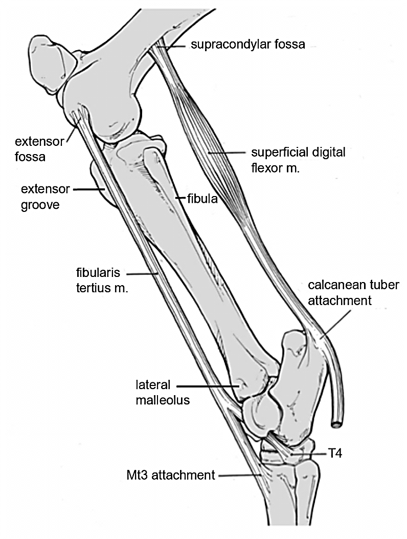
Figure 2-10. Equine left hind limb, lateral view. Reciprocal apparatus: active flexion or extension of the stifle joint results in similar passive actions of the tarsal joint.
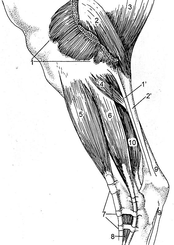
Figure 2-11. Equine left crus, lateral view. 1, biceps femoris m. (cut to expose 2); 1’, tendon of the biceps femoris m.; 2, gastrocnemius m.; 2’, tendon of the gastrocnemius m.; 3, semitendinosus m.; 4, soleus m.; 5, long digital extensor m.; 6, lateral digital extensor m.; 7, extensor retinacula; 8, short digital extensor m.; 9, tendon of the superficial digital flexor m.; 10, deep digital flexor m.
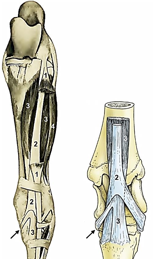
Figure 2-12. Equine left crus, cranial view. Right image is a closer view of relationship of insertions of fibularis (peroneus) tertius and cranial tibial mm. 1, Long digital extensor tendons (much of the muscle has been removed); 2, fibularis (peroneus) tertius m.; 3, cranial tibial m.; arrows, cunean tendon (medial branch of cranial tibial tendon); 4, lateral digital extensor.
7. ALL(CALF) hind limbs: Incise and remove fascia covering the craniolateral muscles of the crus; identify and isolate the fibularis (peroneus) tertius m. and the fibularis (peroneus) longus m. Identify and isolate the long digital extensor m. (with medial and lateral heads).
-
- Incise and remove the fascia covering the cranial aspect of the hind limb below the stifle.
- Superficially, on the craniolateral aspect of the limb, identify and isolate the fibularis (peroneus) tertius m., which is a flexor of the tarsus. (Fig. 2-13/B-1)
- Note that in contrast to the equine, in the ruminant this muscle is fleshy and superficial to the long digital extensor m. rather than tendinous and deep to it.
- Moving slightly lateral to the fibularis tertius m., identify and isolate another flexor of the tarsus, the fibularis (peroneus) longus m. (Fig. 2-13/B-1′)
- Deep to the fibularis tertius m., identify and isolate the main extensor of the digits, the long digital extensor m. (Fig. 2-13/B-3).
- In the calf, note that the long digital extensor muscles is composed of two heads, medial and lateral; identify the medial and lateral heads of the long digital extensor m.
8. ALL hind limbs: Identify and isolate the cranial tibial m. and the cranial tibial artery.
-
- Deep to the long digital extensor m. and fibularis tertius m., lying directly on the craniolateral surface of the tibia, identify the cranial tibial m. (Fig. 2-12/3, 2-13/2)
- Note that the cranial tibial m. is a flexor of the tarsus.
- Elevate the cranial tibial m. and identify the underlying cranial tibial a., which is the continuation of the popliteal artery.
- Calf specimen: In the calf, it may be easiest to identify the cranial tibial a. by working in under the cranial tibial muscle from the lateral side, deep to the long digital extensor m.
- Deep to the long digital extensor m. and fibularis tertius m., lying directly on the craniolateral surface of the tibia, identify the cranial tibial m. (Fig. 2-12/3, 2-13/2)
9. ALL hind limbs: Identify another extensor of the digit(s), the lateral digital extensor m.
-
- On the lateral side of the crus, caudal to the long digital extensor m., identify the lateral digital extensor m. (Figs. 2-11/6 and 2-13/4).
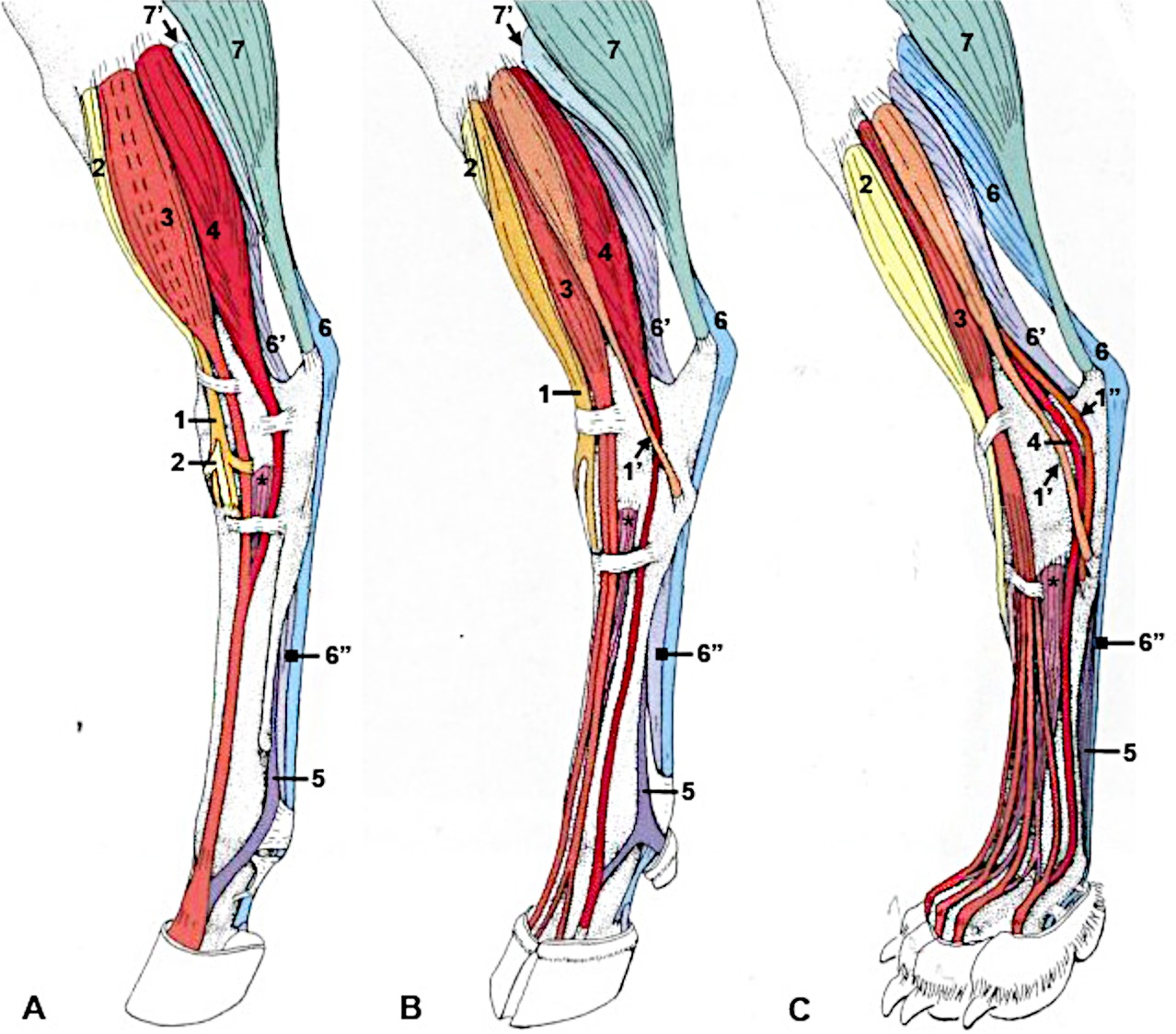
Figure 2-13. Comparative muscle so the crus region, craniolateral view. A, Equine, B, bovine and C, dog. 1, Fibularis (peroneus) tertius (absent in dog); 1′, fibularis longus (absent in equine); 1″, fibularis brevis (absent in equine and ox); 2, cranial tibial m. (note: cunean tendon is the medial branch of insertion); 3, long digital extensor m.; 4, lateral digital extensor m.; 5, interosseus tendon (suspensory ligament in ungulate; ~ entirely tendinous in equine); 6, superficial digital flexor tendon (SDFT).; 6′, deep digital flexor tendon (DDFT), 6″ combined SDFT and DDFT; 7, gastrocnemius m.; 7′, soleus m. (~absent in dog).
Dissection of Caudomedial Crus
10. ALL hind limbs: Re-identify the sciatic nerve and trace it distally to identify the (common) fibular n. and the tibial n.
-
- (Re-) Identify the large sciatic nerve on the lateral side of the limb, under the previously reflected biceps femoris m./gluteobiceps m.
- Trace the sciatic n. distally to the point where it branches into the larger tibial n. and smaller (common) fibular n. and identify these branches. (Fig. 2-14)
- Identify the tibial n. diving in between the medial and lateral heads of the gastrocnemius m.
- Identify the (common) fibular n. that extends slightly cranially as it travels distally in the limb.
Figure 2-14. Equine left hind limb deep dissection, lateral view. 1, superficial gluteal m.; 2, middle gluteal m.; 3, biceps femoris m.; 4, semitendinosus m.; 5, semimembranosus m.; 6, gastrocnemius m.; 7, sciatic n.; 8, tibial n.; 9, common fibular (peroneal) n. 9′, deep branch, 9″, superficial branch; 10, cutaneous n. (fibular); 11, cutaneous n. (tibial) and lateral saphenous v.; 12, dorsal metatarsal a.; 13, popliteal lymphocenter; 14, lateral plantar vessels and n.; the following are only labeled in ox: 15, coccygeus m.; 16, adductor m.; 17, caudal gluteal n. Image modified from Budras.
11. ALL (CALF) hind limbs: Note the (deep) popliteal lymph node.
-
- In the calf specimen, look for the (deep) popliteal lymph node caudal to the stifle, near the branching of the tibial and fibular nerves. It will likely be surrounded by some amount of fat.
- Pony Dissection Note: There may be a small popliteal lymphocenter in this same region in the pony specimen, but the small lymph nodes are often removed during dissection.
12. On the caudal aspect of the crus, we will now identify the muscles that are extensors of the tarsus and flexors of the digit(s). The main difference to note between these types of muscles is that the tendons of the extensors of the tarsus will cross the tarsus and then stop, while the tendons of the flexors of the digit(s) will continue past the tarsus down to the digit(s).
13. ALL hind limbs: Identify the major extensor of the tarsus, the gastrocnemius m.; identify and separate the medial and lateral heads. Identify the (common) calcanean tendon. Transect and reflect the lateral head of the gastrocnemius m.
-
- Identify the gastrocnemius m. (Figs. 2-2, 2-11/2, 2-13/7, 2-14/6) caudal to the stifle, and deep to the previously reflected biceps femoris m./gluteobiceps m.
- Identify the medial and lateral heads of the gastrocnemius m. and carefully separate them.
-
- Use the tibial nerve as a landmark as it dives between the two heads; separate the lateral head from the medial head and also separate both heads from the underlying superficial digital flexor m.
-
- Identify the medial and lateral heads of the gastrocnemius m. and carefully separate them.
- Trace the tendons of the medial and lateral heads of gastrocnemius to the calcanean tuber and identify the (common) calcanean tendon.
- Dissection Note: The (common) calcanean tendon is created by tendinous contributions from several muscles, including the biceps femoris/gluteobiceps, gastrocnemius, and superficial digital flexor mm.
- Carefully transect the lateral head of the gastrocnemius m. near its origin (but don’t cut too deeply!). Reflect the lateral head distally to expose the superficial digital flexor m. in between the two heads of the gastrocnemius m.
- Dissection Note: A small soleus muscle (Fig. 2-13/7′) may be found on the cranial edge of the lateral head of the gastrocnemius, but it is of no significance and need not be dissected. (Note that this muscle is rudimentary in ruminants and may not be seen.)
- Identify the gastrocnemius m. (Figs. 2-2, 2-11/2, 2-13/7, 2-14/6) caudal to the stifle, and deep to the previously reflected biceps femoris m./gluteobiceps m.
14. ALL hind limbs: Identify one of the flexors of the digit(s), the superficial digital flexor m. (SDF), and displace its tendon off of the calcanean tuber as it runs over the hock to identify the calcaneal bursa.
-
- Identify the superficial digital flexor m. (SDF) (Figs. 2-11/9, 2-13/6, 2-15/3, 2-16/S) between (and covered by) the heads of the gastrocnemius m.
- Dissection Note: The SDF muscle is largely tendinous in the pony but fleshy in the calf.
- Trace the SDF tendon distally, over the point of the hock, and isolate it as far as the mid-tarsal region.
- On the medial side of the point of the hock, carefully transect alongside the tendon of SDF and dissect the lateral edge of the tendon off of the calcanean tuber. Then, displace/slide the tendon laterally (off the bone) to expose and identify the subtendinous calcaneal bursa.
- Identify the superficial digital flexor m. (SDF) (Figs. 2-11/9, 2-13/6, 2-15/3, 2-16/S) between (and covered by) the heads of the gastrocnemius m.
 |
(Duplicate) Figure 2-2. Bovine left hind limb, lateral view. 1, tensor fasciae latae m.2, middle gluteal m.3, ischial tuber4, 4’, 4’’ gluteobiceps m. (transected at 4’’)5, semitendinosus m.6, lateral head of gastrocnemius m.7, rudimentary soleus m.8, cranial tibial m.9, 9’, peroneus tertius m.10, 10’, 10’’, long digital extensor m.11, 11’, peroneus longus m.12, lateral digital extensor m.13, lateral digital flexor m.14, tendon of superficial digital flexor m.15, combined tendon of deep digital flexor mm.16, interosseous (suspensory ligament) (Modified from TVA Fig. 31-2) |
15. ALL hind limbs: Identify the flexor retinaculum. Re-identify the tibial n. and trace it distally to identify the medial and lateral plantar nerves. Transect the flexor retinaculum and trace the plantar nerves.
-
- On the medial aspect of the tarsus, identify the thick band of fascia, the flexor retinaculum, binding down the tendons and nerves running through the tarsal canal. (Fig. 2-16, A/R)
- Re-identify the tibial nerve that you used to aid in separating the medial and lateral heads of the gastrocnemius m.
- Trace the tibial nerve distally to the point where it branches. Near the calcaneus, the tibial nerve divides into the medial and lateral plantar nerves which pass through the tarsal canal.
- Identify the medial and lateral plantar nerves.
- Transect the flexor retinaculum with a vertical cut and proceed to trace the medial and lateral plantar nerves as far distally as possible.
- Calf Dissection Notes: The flexor retinaculum will be extremely thick and tough to cut through in the calf specimen. Also, the lateral plantar n. may be difficult to trace through the tarsal canal.
- Distally, note that the plantar nerves lie adjacent to the digital flexor tendons, similar to the palmar nerves of the forelimb.
16. ALL hind limbs: Identify the other major flexor of the digit(s), the deep digital flexor m. (DDF); identify the medial and lateral heads of the DDF. Elevate the DDF tendon out of the tarsal canal; identify the tarsal canal.
-
- On the caudal surface of the tibia, deep to the gastrocnemius m. identify the deep digital flexor m. (DDF) (Figs. 2-11/10, 2-13, 2-15).
- Note that this muscle is mainly composed of a large lateral head and a small medial head (and also includes the caudal tibial m. in both pony and calf).
- Identify the large lateral head of the deep digital flexor m.
- Incise and remove the fascia on the medial aspect of the limb to see the smaller medial head of the deep digital flexor m. Trace the tendon of the medial head distally to see it join the main tendon of the DDF. (Fig. 2-16, A).
- Elevate the DDF tendon of the out of the tarsal canal to identify it.
- Recall that the tarsal canal is the groove/space just medial to the calcaneus that is occupied by the digital flexor tendons.
-
- Dissection Note: The calcaneus serves as the lateral wall of the tarsal canal and the medial wall of the canal is the flexor retinaculum.
-
- Recall that the tarsal canal is the groove/space just medial to the calcaneus that is occupied by the digital flexor tendons.
- On the caudal surface of the tibia, deep to the gastrocnemius m. identify the deep digital flexor m. (DDF) (Figs. 2-11/10, 2-13, 2-15).
Figure 2-15. Equine right crus, caudal view. A, superficial and B, deep muscles. 1, gastrocnemius m., 2, soleus m., 3, superficial digital flexor m., 4, lateral digital extensor m., 5, 5’, 5’’, medial and lateral deep digital flexors and caudal tibial mm., 6, femoral condyles, 7, popliteus m., 8, medial malleolus. (Modified from TVA Fig. 24-14)
Tarsal/Metatarsal Dissection
17. ALL (PONY) hind limbs: Identify the cranial branch of the medial saphenous vein. (Clinically, this vein is significant when performing a cunean tenectomy.) Elevate and trace the tendon of the long digital extensor m. to identify the three extensor retinacula and then find the point where the long and lateral digital extensor tendons join.
-
- Begin on the cranial aspect of the limb at the mid-cannon region to identify the cranial branch of the medial saphenous vein.
- Dissection Note: This vessel crosses the flexor surface of the tarsus.
- Species Note: The horse (and cat) have well-developed medial saphenous veins. In the bovine (and dog) the lateral saphenous vein (which does not accompany a like-named artery) is more obvious.
- Re-identify the long digital extensor m. and elevate it; trace its tendon distally, cutting the three extensor retinacula (Fig. 2-11/7) on the cranial aspect of the tarsus as you proceed so that the long digital extensor tendon can be elevated to expose deeper structures.
- Trace the tendon of the long digital extensor m. to the mid-cannon region where the lateral digital extensor m. tendon joins it.
- Comparative Note (pony): In the equine hind limb, the tendons of the long and lateral digital extensors join, but in the equine forelimb, the lateral and common extensor tendons do not join.
- This is one trick to use when distinguishing a distal ‘fore’ from a distal ‘hind’ limb in the horse.
- Begin on the cranial aspect of the limb at the mid-cannon region to identify the cranial branch of the medial saphenous vein.
18. ALL (CALF) hind limbs: Attempt to identify the cranial branch of the lateral saphenous vein. Elevate and trace the tendon of the long digital extensor m. to identify the two extensor retinacula and expose deeper structures.
-
- In the bovine, the lateral saphenous vein is more obvious than the medial saphenous vein that was identified in the pony. Attempt to identify the cranial branch of the lateral saphenous v. crossing the flexor surface of the tarsus.
- Re-identify the long digital extensor m. and elevate it; if needed, trace its tendons distally, cutting the two extensor retinacula on the cranial aspect of the tarsus as you proceed so that the long digital extensor tendons can be elevated to expose deeper structures.
19. ALL hind limbs: Identify the dorsal pedal artery and the dorsal metatarsal artery.
-
- Lateral to the lateral digital extensor tendon, identify the dorsal metatarsal a.
- Note that the dorsal metatarsal a. is a continuation of the cranial tibial a. after it passes over the dorsal surface of the tarsus as the dorsal pedal a.
- Trace the dorsal metatarsal a. proximally and identify the dorsal pedal artery crossing over the tarsus.
- Trace the dorsal metatarsal a. distally to a groove between the cannon bone (Mt 3) and the lateral splint bone (Mt 4) in the pony and between (dorsal to) the proximally fused Mt 3-Mt4 bones in the calf.
- Lateral to the lateral digital extensor tendon, identify the dorsal metatarsal a.
20. ALL (PONY) hind limbs: Expose the tendon of the cranial tibial m. as it comes through the fibularis tertius m. tendon and then branches into two parts; identify the cunean tendon.
-
- On the dorsal surface of the tarsus, expose the cranial tibial m. tendon as it passes through the fibularis (peroneus) tertius m. tendon and then branches into two parts: a strong medial band, called the cunean tendon, and a dorsal insertion (Fig. 2-12, 2-13, 2-16, TVA 635, left).
- Identify the cunean tendon (Fig. 2-16 B/Cu). (Note the dorsal insertion of the cranial tibial m. tendon as well.) (Fig. 2-16 B/Dc)
- Be sure to look at the dried ligamentous preparations in the demonstration area and be able to identify the cunean tendon on those specimens as well! (Remember, these are fair game for assessments!)
- On the dorsal surface of the tarsus, expose the cranial tibial m. tendon as it passes through the fibularis (peroneus) tertius m. tendon and then branches into two parts: a strong medial band, called the cunean tendon, and a dorsal insertion (Fig. 2-12, 2-13, 2-16, TVA 635, left).
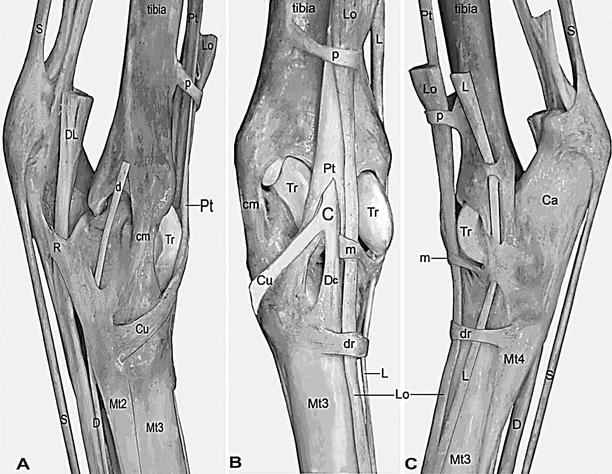 Figure 2-16. Equine left tarsus (ligamentous specimen). Views: A, medial. B, dorsal. C, lateral. Ca, Calcaneus; Tr, trochlea of talus; cm, medial collateral ligament. Digital flexors and retinacula: S, SDFT; D, DDFT; d, DDFT, medial head; DL, DDFT, lateral head; R, flexor retinaculum (mostly removed). Flexor surface of hock and retinacula: Lo, Long digital extensor; L, lateral digital extensor; Pt, peroneus (fibularis) tertius; C, cranial tibial tendon; Dc, dorsal insertion of cranial tibial; Cu, cunean tendon; p, proximal extensor retinaculum; m, middle extensor retinaculum; dr, distal extensor retinaculum.
21. ALL hind limbs: Identify the suspensory ligament (aka interosseous tendon).
-
- On the plantar surface of the cannon bone, identify the suspensory ligament (aka interosseous tendon).
-
The suspensory ligament is the clinical term for the interosseous tendon; in the dog, the interosseous muscle is fleshy, but in the horse it is largely tendinous. In the calf, it may appear somewhat fleshy, but is also largely tendinous.
-
- On the plantar surface of the cannon bone, identify the suspensory ligament (aka interosseous tendon).
Digit(s)
22. We will NOT be dissecting the digit(s) of the hind limb. All structures are essentially the same/equivalent to those found in the forelilmb, and it would be a time consuming and a repetitive exercise to dissect them again in the hind limb. Be sure you can identify all of the listed terms on the museum (dried) specimens available in the demonstration area. (Remember that those are fair game on assessments!) Below are the structures associated with the distal hind limb if you would like to review them – BUT YOU NEED NOT DISSECT ANY FURTHER. (Refer to Figures 1-11 and Figures 1-16 through 1-18 in Chapter 1 for visuals.)
23. ALL hind limbs: Identify the plantar annular ligament, proximal digital annular ligament, and distal digital annular ligament. (Be able to identify these structures on demonstration (museum) specimens as well!)
24. Calf Specimens: The ligamentous arrangement within the two principal digits of the calf will be similar to that seen in the single digit of the horse. However, additionally, you should identify the distal interdigital ligament on the plastinated museum specimen of the bovine foot that is available in the demonstration area.
25. ALL hind limbs: Identify the suspensory ligament (aka interosseous tendon) and the extensor branches of the suspensory ligament. (Be able to identify these structures on demonstration (museum) specimens as well!)
-
- On the plantar surface of the cannon bone, identify the suspensory ligament (aka interosseous tendon).
-
The suspensory ligament is the clinical term for the interosseous tendon; in the dog, the interosseous muscle is fleshy, but in the horse it is largely tendinous. In the calf, it may appear somewhat fleshy, but is also largely tendinous.
-
- Note that the suspensory ligament splits to form lateral and medial branches that attach on the “outside” (abaxial side) of the proximal sesamoid bones.
- This distal arrangement of the suspensory ligament is similar for each of the two principal digits of the calf (i.e., ‘splits’ into medial and lateral branches for each toe) and is also similar between front and hind limbs of both species.
- Beyond the sesamoid bones, identify the extensor branches of the suspensory ligament.
- The extensor branches obliquely cross the sides of the long pastern bone to attach dorsally on the digital extensor tendons (from which they derive their name).
- Again, this distal arrangement of the suspensory ligament is similar for each of the two principal digits of the calf and also similar between front and hind limbs of both species.
- Important Note: The suspensory ligament is part of the suspensory apparatus.
- Suspensory apparatus = suspensory ligament (aka interosseous tendon) + proximal sesamoid bones + distal sesamoidean ligaments
-
- In both the horse and ox these structures act together to support the fetlock against gravity.
-
- Suspensory apparatus = suspensory ligament (aka interosseous tendon) + proximal sesamoid bones + distal sesamoidean ligaments
- On the plantar surface of the cannon bone, identify the suspensory ligament (aka interosseous tendon).
26. ALL (PONY) hind limbs: Identify the distal sesamoidean ligaments: the superficial (straight) sesamoidean ligament and the middle (oblique) sesamoidean ligament.
-
- Dissection Note: There are also deep (cruciate) sesamoidean ligaments present, but these need not be dissected and can be viewed on plastinated demonstration specimens.
- Be able to identify these ligaments on plastinated demonstration (museum) specimens as well, as they are fair game for assessments!
- Calf Note: You do not need to dissect the distal sesamoidean ligaments of the calf. However, you should note that the ox does not have the straight sesamoidean ligament, but it does have the oblique and deep sesamoidean ligaments similar to those found in the pony but duplicated for each principal digit.
27. Additional note for ALL (CALF) hind limbs: Similar to the forelimb, one structure of clinical relevance in the bovine is the dorsal common digital v. This vein is found on the dorsal aspect of the distal metatarsus of bovine (and ovine) animals. In the bovine, the dorsal common digital v. may be used during clinical procedures. (Refer to Fig. 1-15 for similar placement of this vein in the forelimb)
-
-
-
- The dorsal common digital v. drains into/joins the cranial branch of the lateral saphenous vein in the pelvic limb, which you have already identified. (Think of the cranial branch of the lateral saphenous v. as a continuation of the dorsal common digital v. as you move proximally up the limb – more of a name change than separate veins.)
-
-
Dissection Videos for this Section of Material
Distal Pelvic Limb
- Pony
- Proximal Pelvic Limb – Attached, watch from 6:38-end: https://youtu.be/B26J-aCqhiU
- Proximal Pelvic Limb – Detached, watch from 5:30-end: https://youtu.be/4z1ezvuzkzk
- Distal Pelvic Limb – Attached: https://youtu.be/QGb7GzOWrs8
- Distal Pelvic Limb – Detached: https://youtu.be/pfpEu6d0OuM
- Calf
- Proximal Pelvic Limb – Attached, watch from 6:19-end: https://youtu.be/rWwfvDhVWmI
- Proximal Pelvic Limb – Detached, watch from 5:08-end: https://youtu.be/lnKg–Y6dxM
- Distal Pelvic Limb – Attached: https://youtu.be/zJo_HknQnRw
- Distal Pelvic Limb – Detached: https://youtu.be/Ls_auz8zu0Q
