
Part 3: Proximal Thoracic Limb
Abby Brown
IMPORTANT NOTE: The Guide instructions will refer to either “ALL forelimbs” (meaning attached and detached) or to the “DETACHED” or “ATTACHED” forelimbs.” This added instructional note is needed because the dissection of the region medial to the scapula/brachium is only possible (accessible) on the “detached” specimens, versus other parts of the dissection which are possible on all specimens.
Skin & Muscle Reflections
- ALL forelimbs: Make a vertical skin incision on the medial side (on all specimens) from the elbow to the mid-cannon bone region. Then, make an incision horizontally encircling the forelimb at mid-cannon.
- Reflect and discard the skin, being careful to preserve the cephalic vein (Fig. 1-5 ) on the medial surface of the antebrachium.
- ALL forelimbs: Make a vertical incision through the superficial pectoral muscles to bisect the transverse superficial pectoral m. and reflect its parts. (This will enable you to see the underlying vessels and nerves.)
- DETACHED forelimbs: Reflect the deep pectoral muscles cranially on the limb, but do not remove them.
Vessels
VEINS of the THORACIC LIMB (Fig. 1-5)
4. ALL forelimbs: Identify the cephalic v. (Fig.1-5) and trace it proximally, as far as the median cubital vein; identify the median cubital v. (Fig. 1-5/11) and note the brachial vv.
-
-
The cephalic vein will be visible on the medial aspect of the forelimb. Identify the cephalic vein and trace as much of its course as possible. (Sometimes the cephalic vein is accidentally removed with the skin.)
-
Note that there is an accessory cephalic vein branch seen more cranially on the limb, which then joins the cephalic, which you should attempt to identify. (Sometimes the accessory cephalic vein is accidentally removed with the skin.)
-
In the pony, the accessory cephalic v. joins the cephalic significantly above the carpus, nearing the elbow region.
-
In the calf, the accessory cephalic v. joins the cephalic further distally on the limb, just proximal to the carpus.
-
-
Trace the cephalic v. proximally to locate and identify the median cubital vein coursing medially.
-
Medially, note the brachial vein(s). Often there are multiple brachial veins present.
-
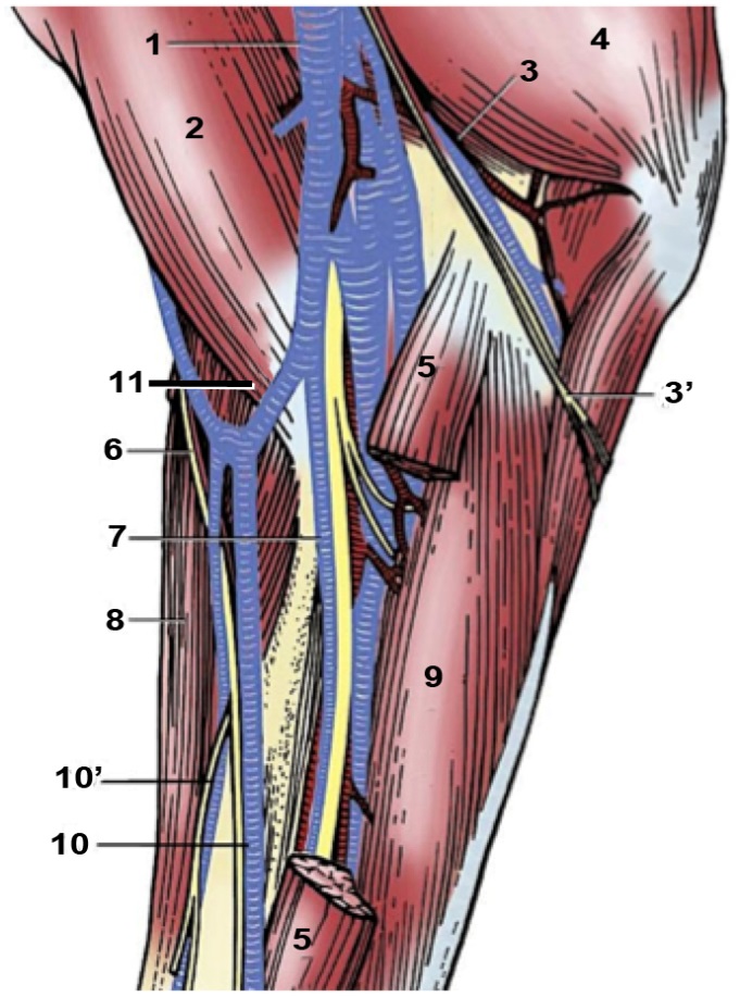
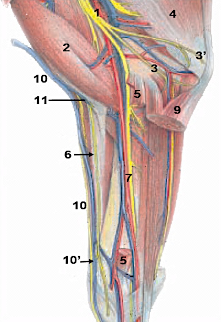
Figure 1-5. Dissection of the medial surface of the right forelimb of the horse (upper) and bovine (lower) 1, multiple brachial veins; 2, biceps brachii m.; 3, ulnar nerve and collateral ulnar vessels; 3’, caudal cutaneous antebrachial nerve; 4, triceps brachii m.; 5, flexor carpi radialis m., resected; 6, medial cutaneous antebrachial nerve; 7, median nerve and vessels; 8, extensor carpi radialis m.; 9, flexor carpi ulnaris m.; 10, 10’, cephalic and accessory cephalic veins; 11, median cubital vein. (left image: Modified from Dyce, Sack and Wensing, 4th ed.; right image: Modified from Budras and Habel, 1st ed.)
ARTERIES of the THORACIC LIMB (Fig. 1-6)
5. DETACHED forelimbs: Identify the axillary a. and v. Trace the axillary artery to identify the subscapular and cranial circumflex humeral aa. (Fig. 1-6).
-
- On the medial side of the detached limb, identify the axillary a. and axillary v.(Remember that the axillary vessels were cut in order to remove the limb from the body.)
- Trace the axillary a. to find and identify the subscapular a.
- The subscapular a. is a large branch off of the axillary artery that will in turn give off several branches into the musculature on the medial aspect of the scapula.
- After the subscapular branch, there will be a smaller artery, the cranial circumflex humeral a. that branches from the axillary. After this branch is given off, the axillary a. changes names to brachial a.
- Identify the cranial circumflex humeral a. and note where the brachial a. begins.
6. DETACHED forelimbs: Identify and trace the brachial a. to identify the common interosseous aa. (Fig. 1-6).
-
- As mentioned previously, the axillary a. changes names to brachial a. after the cranial circumflex humeral a. is given off; identify the brachial a. on the detached forelimbs.
- Continue to trace the brachial a. distally through the brachial region and into the antebrachium.
- As you trace the brachial artery you will need to cut through the thick antebrachial fascia and reflect it.
- Note that you should see three branches given off the caudal aspect of the brachial a., the last of which (the common interosseous a.) you should identify:
- The first branch is the deep brachial a. The deep brachial will dive into the musculature on the caudal aspect of the forelimb. (You do not need to identify this.)
- The second branch is the collateral ulnar a. which will course caudally toward the elbow region. (The collateral ulnar usually gives off a varying number of additional branches as well.) (You do not need to identify this.)
- The third branch is the common interosseous a. which will be given off further distally in the limb, just distal to the elbow joint; identify the common interosseous a.
- The common interosseous a. will dive down into the interosseous space between the radius and ulna. Use your dissection probe to verify the location of this space.
- After the common interosseous branch, the brachial a. changes names and is continued as the median a.
7. ATTACHED forelimbs: Identify the brachial a. and common interosseous a. (Fig. 1-6).
-
- As mentioned for the detached forelimbs, the axillary a. is the main artery supplying the forelimb proximally. The axillary a. will give off a large subscapular a. and a smaller cranial circumflex humeral a. and then the axillary a. changes names to brachial a. (after the cranial circumflex humeral a. is given off).
- Identify the brachial a. on the medial aspect of the attached forelimbs, just deep to the bisected transverse superficial pectoral m.
- Trace the brachial a. distally through the brachial region and into the antebrachium.
- As you trace the artery you will need to cut through the thick antebrachial fascia and reflect it.
- Note that there are three different branches given off the caudal aspect of the brachial a., the third of which (the common interosseous a.) you should identify. (Dissection Note: In the attached forelimb it may be quite difficult to see the deep brachial a. and the collateral ulnar a., but you should be able to dissect and identify the third branch, the common interosseous a.)
- The first branch is the deep brachial a. The deep brachial will dive into the musculature on the caudal aspect of the forelimb. (You do not need to dissect/identify this.)
- The second branch is the collateral ulnar a. which will course caudally toward the elbow region. (The collateral ulnar usually gives off a varying number of additional branches as well.) You may see this artery on the attached forelimb, but it also may not be visible. (You do not need to dissect/identify this.)
- The third branch is the common interosseous a. which will be given off further distally in the limb, just distal to the elbow joint; identify the common interosseous a.
- The common interosseous a. will dive down into the interosseous space between the radius and ulna. Use your dissection probe to verify the location of this space.
- After the common interosseous branch, the brachial a. changes names and is continued as the median a.
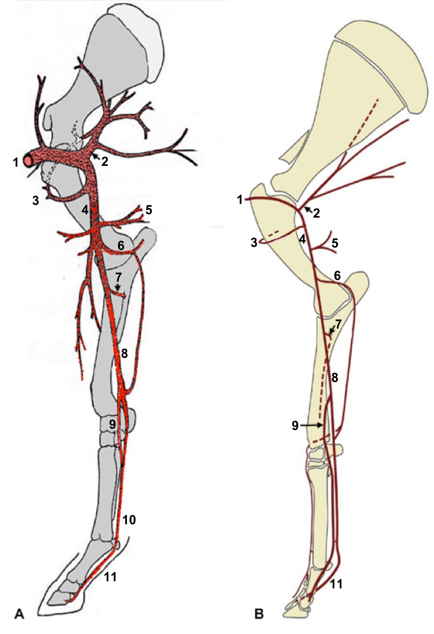
Figure 1-6. Arteries of the right forelimb of the (A) horse and (B) bovine, medial view. 1, Axillary a.; 2, subscapular a.; 3, cranial circumflex humeral a.; 4, brachial a.; 5, deep brachial a.; 6, collateral ulnar a.; 7, common interosseous a.; 8, median a.; 9, radial a.; 10, medial palmar a.; 11, palmar digital aa. Note: The medial palmar and palmar digital aa. are paired in the bovine, i.e., one per digit. (A: Drawing by A. Weber; B: Modified from Dyce, Sack and Wensing, 4th ed.).
nerve and muscle dissections
8. DETACHED forelimbs: Identify the radial, median, ulnar, and musculocutaneous nerves. (Fig. 1-7)
-
- On the medial side of the detached forelimb, identify the following nerves of the brachial plexus: radial, median, musculocutaneous, and ulnar nerves.
- The radial n. is large and will dive caudally into the triceps brachii muscle mass.
- The median n. will course distally on the medial side of the forelimb, down into the antebrachium, where it can be found alongside the median a. Trace the median n. through the brachium, just distal to the elbow. (We will continue to trace this nerve through the antebrachium with the dissection of the distal limb.)
- The musculocutaneous n. shares a common trunk with the median n. but will branch off cranially to supply the musculature. (Dissection Note: There may be more than one musculocutaneous n.)
- The ulnar n. will course caudally toward the elbow region. (Dissection Note: In the calf, the ulnar nerve may share a common trunk with the median n. and then branch off caudally toward the elbow.)
- On the medial side of the detached forelimb, identify the following nerves of the brachial plexus: radial, median, musculocutaneous, and ulnar nerves.
9. ATTACHED forelimbs: Identify the median n.
-
- On the medial side of the attached forelimb, identify the median n. where it courses distally on the medial side of the limb, down into the antebrachium, where it can be found alongside the median a. Trace the median n. through the brachium, just distal to the elbow. (We will continue to trace this nerve through the antebrachium with the dissection of the distal limb.)
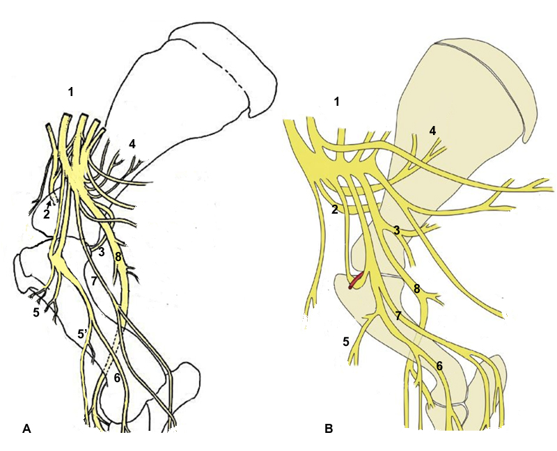
Figure 1-7. Nerves in the right forelimb of the (A) equine and (B) bovine, medial view. 1, Brachial plexus; 2, suprascapular n.; 3, axillary n.; 4, subscapular n.; 5, 5′ musculocutaneous n.; 6, median n.; 7, ulnar n. 8, radial n. (Modified from Dyce, Sack and Wensing, 4th ed.).
10. DETACHED forelimbs: Identify the subscapularis m. and supraspinatus m.
-
- Identify the subscapularis m. on the medial aspect of the limb, covering the medial face of the scapula (Fig. 1-9).
- Identify the supraspinatus m. on the lateral aspect of the limb, filling the space above the spine of the scapula (Figs. 1-8, 1-10).
- Dissection Note: The supraspinatus m. can also be seen from the medial side of the limb (adjacent to the subscapularis m.), overlapping from the lateral side onto the cranial edge of the scapula.
11. ATTACHED forelimbs: Identify the supraspinatus m. on the lateral aspect of the limb, filling the space above the spine of the scapula (Figs. 1-8, 1-10).
12. DETACHED forelimbs: On the medial side of the limb, identify the suprascapular n. and trace it to the fissure between the subscapularis and supraspinatus muscles.
-
- Surrounding the suprascapular n., remove a small rectangular section of the subscapularis m. (and, if needed, a small section of the supraspinatus m. as well); remove enough muscle to expose a small portion of the cranial edge of the scapula.
- Note the close proximity of the suprascapular n. to the cranial edge of the scapula (i.e., where this nerve is vulnerable to damage).
- Surrounding the suprascapular n., remove a small rectangular section of the subscapularis m. (and, if needed, a small section of the supraspinatus m. as well); remove enough muscle to expose a small portion of the cranial edge of the scapula.
13. DETACHED forelimbs: Identify the triceps brachii m. (3 heads: long, lateral and medial).
-
- On the medial side of the limb, trace the radial nerve into the triceps brachii muscle mass.
- Identify the three main heads of the triceps brachii m.: long, lateral and medial heads.
- Just as it is in the dog, the long head is the largest of the three heads of the triceps brachii m.
- . The long head can be seen from both medial and lateral sides.
- The lateral head is smaller and is located at the distal border of the long head on the lateral side of the forelimb.
-
- Isolate the lateral head of the triceps brachii m. by defining its borders.
-
- The medial head is the smallest of the three, and the most difficult to see. The medial head can be seen from the medial side, just cranial to the long head.
- Note that an accessory head of the triceps brachii m. is present in the ruminant but absent in the horse (but you do not need to identify it).
14. DETACHED (pony) forelimbs: In the pony, identify the broad, thin tensor fasciae antebrachii m. on the medial aspect of the limb; the tensor fasciae antebrachii m. is small (or absent) in the calf (and you need not identify it).
-
- If needed, transect the tensor fasciae antebrachii m. through its aponeurotic origin and reflect it distally to facilitate a better view of the long and medial heads of the triceps brachii m.
15. ATTACHED forelimbs: Identify the heads of the triceps brachii m. that are visible from the lateral side, specifically the long and lateral heads.
-
- Just as it is in the dog, the long head is the largest of the three heads of the triceps brachii m. The long head can be seen from both medial and lateral sides, but you will only identify it from the lateral side on your attached limb.
- The lateral head is smaller and is located at the distal border of the long head on the lateral side of the forelimb.
- Isolate the lateral head of the triceps brachii m. by defining its borders.
- Note that an accessory head of the triceps brachii m. is present in the ruminant but absent in the horse (but you do not need to identify it).
16. ALL forelimbs: On the lateral side, re-identify the supraspinatus m., then, identify the infraspinatus m., and deltoideus m. (Fig 1-8)
-
- On the lateral side of the forelimb, begin by re-identifying the supraspinatus m. filling the space above the spine of the scapula.
- Then, identify the infraspinatus m. filling the space below the spine of the scapula.
- Note that in the pony, the infraspinatus m. may somewhat overlap onto the supraspinatus m.
- Identify the deltoideus m., which somewhat overlaps the infraspinatus m. (In the calf, note that the deltoideus m. overlaps the infraspinatus m. extensively.) Note that similar to the dog and cat, the deltoideus m. has two parts.
- Make a horizontal transection (cut) through the deltoideus m. in the distal third of the muscle (near the distal end of the spine of the scapula) and reflect both parts.
17. ALL forelimbs: Transect and reflect the lateral head of the triceps brachii m. to identify the brachialis m.
-
- Transect the lateral head of the triceps brachii m. near its origin on the humerus.
- Reflect the lateral head of the triceps caudally/distally to expose the brachialis m., which lies in the deep brachialis groove of the humerus; identify the brachialis m.
18. ALL forelimbs: Identify, isolate, and transect the biceps brachii m. on the cranial aspect of the humerus and identify the bicipital tendon and the lacertus fibrosus (Figs 1-8 to 1-10).
-
- On the cranial aspect of the humerus, identify and isolate the biceps brachii m.
- Dissection Note: Reflection of the cranial edge of the brachiocephalicus m. will aide in exposing the biceps brachii m.
- Make a transection through the middle of the biceps brachii m. and note the central tendon on the cut surface. (This central tendon is actually on the surface of one of the two biceps heads that are fused in most quadrupeds.)
- Trace the central tendon of the biceps brachii proximally and distally:
- Proximally, trace it to the bicipital tendon, which runs over the intertubercular (bicipital) groove of the proximal humerus. This tendon takes origin on the scapula, from the supraglenoid tubercle.
- Distally, trace it to the cubital (elbow) region where it gives off the lacertus fibrosus (aka long tendon of the biceps brachii m.) (Fig. 1-9/14).
-
- The lacertus fibrosus links the biceps brachii m. and extensor carpi radialis m. tendons to form a passive cable connection from the scapula to the metacarpal tuberosity of the cannon bone (i.e., part of the forelimb stay apparatus).
- Note that the lacertus fibrosus is a very well-developed structure in the pony and underdeveloped in the calf.
-
- Trace the lacertus fibrosus distally onto the extensor carpi radialis m. (which will be dissected with the distal thoracic limb in the next part of this chapter).
- On the cranial aspect of the humerus, identify and isolate the biceps brachii m.
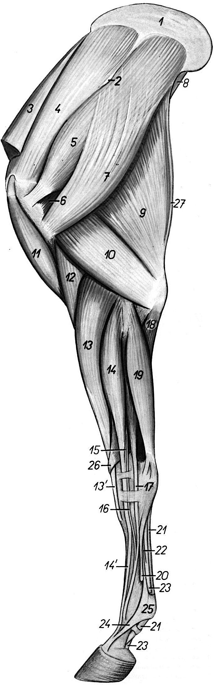 |
Figure 1-8. Equine left forelimb, lateral view. 1 Scapular cartilage2 Spine of scapula3 Cranial deep pectoral m. (aka subclavius)4 Supraspinatus m.5 Infraspinatus m.6 Teres minor m.7 Deltoideus m.8 Teres minor m.9 Long head of triceps brachii m.10 Lateral head of triceps brachii m.11 Biceps brachii m.12 Brachialis m.13, 13’ Extensor carpi radialis m.14, 14’ Common digital extensor m.15, 16 Rudimentary extensor tendons.17 Lateral digital extensor m.18 Ulnar head of deep digital flexor (DDF) m.19 Ulnaris lateralis m.20 Lateral splint bone = Mc 421 Superficial digital flexor tendon (SDFT)22 Carpal (distal) check ligament23 Deep digital flexor tendon (DDFT)24 Extensor branch of suspensory ligament25 Locus of proximal sesamoid bones26 Extensor carpi obliquus m.27 Tensor fasciae antebrachii m. |
|
|
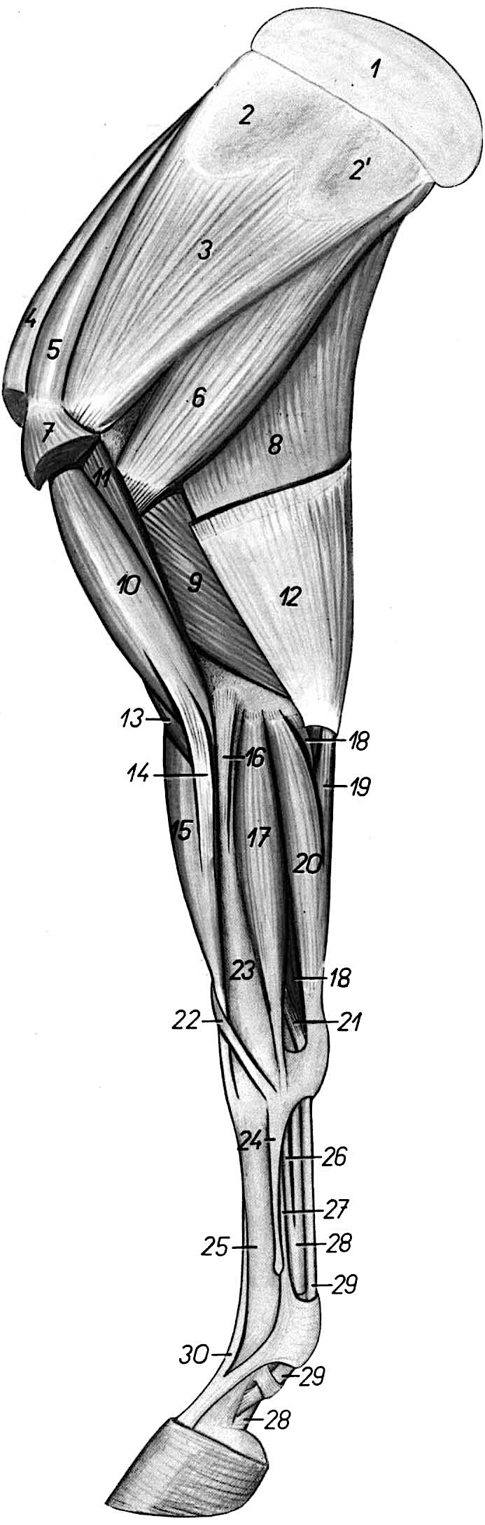
Figure 1-9. Equine right forelimb, medial view. 1 Scapular cartilage2, 2’ Insertion area of serratus ventralis m.3 Subscapularis m.4 Cranial deep pectoral m. (subclavius)5 Supraspinatus m.6 Teres major m.7 Insertion of caudal deep pectoral m.8 Long head of triceps brachii m.9 Medial head of triceps brachii m.10 Biceps brachii m.11 Coracobrachialis m.12 Tensor fasciae antebrachii m.13 Brachialis m.14 Lacertus fibrosus15 Extensor carpi radialis m.16 Medial collateral ligament of elbow joint17 Flexor carpi radialis m.18 Superficial digital flexor (SDF) m.19 Ulnar head of flexor carpi ulnaris m.20 Humeral head of flexor carpi ulnaris m.21 Accessory head of SDF (proximal check ligament)22 Extensor carpi obliquus m.23 Radius24 Mc 2 = medial splint bone25 Mc 3 = cannon bone26 Carpal (distal) check ligament27 Suspensory ligament = interosseous tendon28 Deep digital flexor tendon (DDFT)29 Superficial digital flexor tendon (SDFT)30 Common digital extensor tendon |
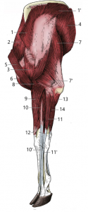 |
Figure 1-10. Bovine left forelimb, lateral view. 1, 1’ trapezius m.2 supraspinatus m.3 deltoideus m.4 latissimus dorsi m.5 brachiocephalicus m.6 biceps brachii m.7, 7’ long and lateral heads of triceps brachii m.8 brachialis m.9 extensor carpi radialis m.10, 10’ common digital extensor m. and the tendon of its lateral belly11, 11’ lateral digital extensor m. and its tendon12 extensor carpi obliquus m.13 ulnar head of deep digital flexor m.14 ulnaris lateralis m.(Modified from Dyce, Sack and Wensing, 4th ed.) |
Dissection Videos for this Section of Material
Proximal Thoracic Limb
- Pony:
- Proximal Thoracic Limb – Attached: https://www.youtube.com/watch?v=u98lsMy1fTU&t=1s
- Proximal Thoracic Limb – Detached: https://www.youtube.com/watch?v=WLdA1NVKEeQ&t=11s
- Calf:
- Proximal Thoracic Limb – Attached: https://www.youtube.com/watch?v=_87o7_8cUJY&t=6s
- Proximal Thoracic Limb – Detached: https://www.youtube.com/watch?v=eQUS0IeuGog
