
Part 5: Heart and Major Vessels
Abby Brown
VESSEL DISSECTION
- ALL specimens: Re-identify the descending (thoracic) aorta within the thorax and attempt to identify the thoracic duct.
- The thoracic duct may be identifiable on the left or right side of the thorax, just dorsal to the descending aorta. Attempt to locate the thoracic duct.
- Dissection Note: If you cannot locate a thoracic duct on your specimen, be sure to check the demonstration specimens.
- The thoracic duct may be identifiable on the left or right side of the thorax, just dorsal to the descending aorta. Attempt to locate the thoracic duct.
- ALL specimens: Identify the azygous vein as described for each species.
- The azygous vein drains the walls of the chest by receiving blood from the intercostal veins, and develops as a paired structure; however, most commonly, only a single vein persists in the adult animal of most species.
- ALL (PONY) specimens: Identify the right azygous v.
- In the equine (and camelid), there is only a right azygous vein present.
-
- Observe/identify the right azygous vein draining into the right atrium of the heart on your pony specimen.
-
- In the equine (and camelid), there is only a right azygous vein present.
- ALL (CALF) specimens: Identify the left azygous v. and note that you may also see a right azygous v. in the calf.
- Ruminants and swine have a predominant left azygous vein which drains into the great cardiac vein and then into the coronary sinus (within the right atrium).
-
- Observe/identify the left azygous vein in your calf specimen as it passes cranial to the left root of the lung (Fig. 4-3/16).
-
- In addition, the ruminant occasionally has a right azygous v. present as either a complete vessel or as a branch of the left azygous v. Look for the presence of a right azygous v. in your calf specimen.
- Ruminants and swine have a predominant left azygous vein which drains into the great cardiac vein and then into the coronary sinus (within the right atrium).
- ALL specimens: Identify the cranial vena cava and caudal vena cava.
- ALL specimens: (Re-) Identify the left and right external jugular vv. (previously identified in Chapter 3)
- ALL (CALF) specimens: Re-identify the right and left internal jugular veins within the carotid sheath (recall that both calf and pig have internal jugular vv.) (previously identified in Chapter 3)
-
Comparative Note: The bovine has a relatively small internal jugular vein, while in the pig, the internal jugular vein is relatively large. The internal jugular vein is usually absent in the equine.
-
- ALL specimens: Re-identify the pulmonary veins (previously identified with the dissection of the lungs in this chapter) and observe them draining into the left atrium of the heart.
heart and major vessels
IMPORTANT NOTE: As you proceed to study the heart, be sure to know/understand the blood flow pattern and general valve locations in addition to identifying the structures. (i.e., right atrium -> right AV valve -> right ventricle -> pulmonic/pulmonary valve -> pulmonary trunk -> pulmonary aa. -> lungs -> pulmonary vv. -> left atrium -> left AV valve -> left ventricle -> aortic valve -> aorta -> out to the body -> right atrium; then repeat the cycle).
Dissection Note: Leave the heart in place inside the chest (in situ) as you perform this dissection.
7. ALL specimens: Leaving the heart in place, dissect around it, removing the pericardium/pericardial sac and fat to expose and identify the structures indicated throughout this dissection.
8. ALL specimens: Incise the pericardium/pericardial sac and reflect it off of the heart. Carefully remove fat from around the heart to expose the coronary groove and coronary vessels.
-
- Recall from Anatomy I that the pericardial sac is thin, but strong, and consists of three inseparable components: serous pericardium (which includes parietal and visceral pericardium), fibrous pericardium, and pericardial mediastinal pleura.
- Identify the visceral pericardium (aka epicardium) that is tightly adhered to the surface of the heart itself.
- Recall that the heart has a base dorsally/cranially (where the great vessels are attached) and an apex ventrally/caudally (usually inclines to the left side).
- Identify the coronary groove (aka coronary sulcus) encircling the heart. This a groove between the atria and ventricles. The coronary groove contains coronary vessels and some amount of fat.
- Dissection Note: There are two other ‘grooves’ but you will not need to identify them specifically on your specimen. These are the subsinuosal interventricular groove (sulcus) and the paraconal interventricular groove (sulcus). These grooves are shallow indentations separating the right and left ventricles of the heart.
9. ALL specimens: Re-identify the cranial vena cava and caudal vena cava to help orient yourself to the positioning of the heart within the thorax. (The cranial vena cava is cranial to the heart, while the caudal vena cava is caudal to it.) These vena cavae drain into the right atrium of the heart.
10. ALL specimens: Identify the right atrium and right auricle.
-
- The right atrium receives the blood from systemic veins and most of the blood from the heart itself.
- The right auricle is a flap-like (or ‘ear-shaped’), blind pouch that is part of the right atrium.
11. ALL specimens: Open the right atrium with a longitudinal incision, extending from the cranial vena cava to the caudal vena cava. Extend another small incision from this cut to the tip of the right auricle (this makes a ‘T’ or ‘J’ shaped incision into the right atrium and auricle). Open the right atrium and clean out any debris found inside.
-
- Note that the right atrium is divided into two parts, a main part and a blind pouch; the main part is the sinus venarum, and the blind pouch is the auricle.
-
- Dissection Notes: Note that there is an interatrial septum between left and right atria; this is within the dorsomedial wall of the sinus venarum and the walls of the auricle are strengthened by interlacing muscular bands called the pectinate muscles. (Pectinate muscles are also found on the lateral wall of the atrium proper.)
-
- If your specimen is injected with latex, attempt to remove the latex from within the atrial chamber.
- Dissection Note: Recall that the internal surfaces of the heart are lined with a thin, glistening membrane called the endocardium.
- Note the opening where the caudal vena cava enters the right atrium caudally. Then, note the opening where the cranial vena cava enters the right atrium cranially. Between these two openings there is a transverse ridge of tissue called the intervenous tubercle. This tubercle diverts incoming blood from the caval veins toward the right atrioventricular orifice. Identify this tubercle in your specimen.
-
- Dissection Note: Note that caudal to the intervenous tubercle you may see a slit-like depression called the fossa ovalis. (This is a remnant of an opening present in the fetus, the foramen ovale, which allowed blood to pass from the right atrium to the left atrium.)
-
- Ventral to the opening of the caudal vena cava into the right atrium, identify the coronary sinus. This is an opening that allows for venous return of most of the blood from the heart itself, via the great cardiac vein.
- Note that the right atrium is divided into two parts, a main part and a blind pouch; the main part is the sinus venarum, and the blind pouch is the auricle.
12. ALL specimens: While still looking at the right atrium, identify the large opening that allows the blood to pass from the right atrium into the right ventricle. This is the right atrioventricular (AV) orifice.
13. ALL specimens: Moving outside of the right atrium, identify where the right ventricle is located in relation to the right atrium. Identify the pulmonary trunk, which is the large vessel arising from the right ventricle cranially and angling to the left. (The pulmonary trunk then gives rise to the left and right pulmonary arteries.) (Fig. 4-2/8)
14. ALL specimens: To the right side of the pulmonary trunk, find the aorta. Note the three parts of the aorta: ascending aorta, aortic arch, descending aorta.
-
-
Attempt to identify the ascending aorta emerging from the heart and coursing cranially for a short distance before it curves to the left.
-
-
Dissection Note: Be aware that the ascending aorta is quite short and is difficult to see.
-
-
- Trace the aortic arch to the descending aorta.
-
- Dissection Note: The descending aorta has two parts: thoracic and abdominal. The portion of the descending aorta cranial to the diaphragm is the thoracic part and the portion of the descending aorta caudal to the diaphragm is the abdominal part. (Note: The abdominal part will not be seen until we enter the abdomen.)
-
- Identify the fibrous connection between the pulmonary trunk and the aortic arch that is the ligamentum arteriosum, which is the remnant of the fetal ductus arteriosus.
- Dissection Note: In the fetus, this was the patent ductus arteriosus that shunts blood directly to the aorta to bypass the fetal lungs.
- Trace the pulmonary trunk to the place where it branches into left and right pulmonary arteries; identify the cut ends of the left and right pulmonary arteries.
-
15. ALL specimens: Make an incision through the wall of the pulmonary trunk at its base and continue this cut distally, through the wall of the right ventricle. (The cut should be made in the middle of the wall of the right ventricle,)
-
-
-
Reflect the right ventricular wall and remove any debris/latex found inside.
- Dissection Note: Note that you will see muscular irregularities of the interior walls of the ventricle; these are called trabeculae carneae.
- At the junction of the right ventricle and the pulmonary trunk, identify the pulmonary (aka pulmonic) valve. The pulmonary valve consists of three semilunar cusps.
-
- Dissection Note: The right ventricle terminates as the funnel-shaped conus arteriosus which then gives rise to the pulmonary trunk.
-
-
-
16. ALL specimens: Re-identify the right atrioventricular (AV) orifice connecting the right atrium and right ventricle and then identify the right atrioventricular (AV) valve (aka tricuspid valve) that allows the orifice to open and close.
-
- Note that the right atrioventricular (AV) valve (aka tricuspid valve) has parietal and septal cusps.
- Identify the chordae tendineae which are the stringy, fibrous bands that attach the cusps to the septal wall of the ventricle by attaching to the papillary muscles.
- The papillary muscles are conical muscular projections in the septal wall of the ventricle that serve as attachment points for the chordae tendineae. Identify the papillary muscles seen in the right ventricle.
-
Dissection Note: There is a possibility that you will see a muscular strand extending across the lumen of the ventricle from the septal wall to the parietal wall. This structure is called the trabecula septomarginalis (aka moderator band). (These are not often seen in the cadavers because they get ripped/broken during the dissection process.)
-
17. ALL specimens: On the left side of the heart, identify the left atrium and left auricle.
-
- The left atrium will have multiple pulmonary veins entering it at the base of the heart. Recall that the left atrium receives the oxygenated blood returning from the lungs.
- The left auricle is a flap-like (or ‘ear-shaped’), blind pouch that is part of the left atrium.
- Dissection Note: The atrium proper will be quite small; most of what is seen of the left atrium is the blind pouch of the left auricle.
18. ALL specimens: Identify the left ventricle and open the left side of the heart with one longitudinal incision through the middle of the left ventricle and another small (separate) incision through the left atrium and left auricle. Remove any debris found inside these chambers.
-
- Note the significant difference in thickness of the left ventricular wall as compared with that of the right ventricular wall.
- As previously noted, the left atrium proper will be quite small; most of what is seen of the left atrium is the blind pouch of the left auricle.
- Dissection Note: Note that the left ventricle will also have trabeculae carneae as the right ventricle did, but they are not as numerous in the left ventricle.
19. ALL specimens: While looking at the left atrium, identify the large opening that allows the blood to pass from the left atrium into the left ventricle. This is the left atrioventricular (AV) orifice. Identify the left atrioventricular (AV) orifice connecting the left atrium and left ventricle.
20. ALL specimens: While looking at the left ventricle, identify the left atrioventricular (AV) valve (aka bicuspid or mitral valve) that allows the orifice to open and close.
-
- Note that the left atrioventricular (AV) valve (aka bicuspid or mitral valve) has parietal and septal cusps.
- Note the large papillary muscles within the ventricle and the chordae tendineae attached to the cusps of the valve (similar to what was seen in the right ventricle).
21. ALL specimens: At the junction of the left ventricle and the aorta, attempt to find/see the aortic valve (aortic root region) and its three semilunar cusps. Since this region is difficult to see, you should also look at the available museum specimens to see this area.
22. ALL specimens: On the outer surface of the heart, re-identify the coronary groove between the atria and ventricles. Identify the right and left coronary aa.
-
- As noted previously, you will need to carefully remove the fat in this groove to expose the vessels indicated.
- Begin at the base of the aorta (aka ascending aorta) as it leaves the heart and attempt to locate the origins of the coronary arteries.
- Identify the right coronary artery and trace it under the right auricle, into the coronary groove.
- ALL (PONY) specimens: In the pony (and pig), the right coronary artery terminates as the subsinuosal interventricular branch (Fig. 4-5/2’); identify the subsinuosal interventricular branch as the termination of the right coronary artery in the pony specimens.
- Identify the left coronary artery and trace it under the pulmonary trunk.
- Note that the left coronary a. is short and divides into the circumflex branch, which lays in the coronary groove, and the paraconal interventricular branch, which projects ventrally between the right and left ventricles. Identify these branches in all specimens.
- ALL (CALF) specimens: In ruminants (and canines), the left (circumflex) coronary artery terminates as the subsinuosal interventricular branch (Fig. 4-5/1′); identify the subsinuosal interventricular branch as the termination of the circumflex branch of the left coronary artery in the calf specimens.
23. ALL specimens: Locate and identify the great cardiac vein which begins in the paraconal interventricular groove and then follows the coronary groove to return blood supplied to the heart by the left coronary artery. The great cardiac vein empties this returned blood into the coronary sinus.
-
-
Re-identify the coronary sinus where the great cardiac vein empties into the right atrium (previously identified within the right atrium).
-
24. ALL specimens: Branching from the aortic arch, identify the brachiocephalic trunk.
-
- (Re-) Identify the aortic arch after the ascending aorta comes out of the heart.
- Identify the large brachiocephalic trunk arising from the aortic arch. (Fig. 4-4/1)
- Comparative Note: Recall that in the dog and cat, both the left subclavian a. and the brachiocephalic trunk arise from the aortic arch. In contrast, the horse and ruminant have only the brachiocephalic trunk that arises from the aortic arch.
25. ALL specimens: Identify the left and right subclavian arteries given off by the brachiocephalic trunk.
26. ALL specimens: The left and right subclavian arteries give off several named branches, but, in the interest of time, we will not dissect these – with the exception of the internal thoracic a.
-
-
Dissection Notes: The major branches given off of the subclavian arteries (both left and right) are the vertebral a., costocervical trunk, superficial cervical a., and internal thoracic a. These branches all arise from the subclavian medial to the first rib (before leaving the thoracic cavity). If you wish to identify these arteries, please view the demonstration specimens.
-
-
The costocervical trunk usually arises as the first branch of the subclavian and courses dorsally. This trunk splits into multiple branches supplying deep regions of the neck and first few intercostal spaces.
-
-
Pony specimen note: In the pony, note that there is an additional branch off of the left subclavian a. called the deep cervical artery. On the right side, this artery arises from the costocervical trunk. (Fig. 4-4/3)
-
-
-
The vertebral a. varies significantly in its branching pattern.
-
-
The vertebral a. may arise from either the costocervical trunk (ruminants) or directly from the subclavian a. (equine).
-
-
Pony specimen note: In the pony, the vertebral a. typically branches from the subclavian a. and dives deep, on the dorsal aspect, to pass through the transverse foramina of the cervical vertebrae.
-
Calf specimen note: Do not spend time looking for the vertebral a. in the calf specimens; you would need to remove quite a bit of muscle to see it branching from the costocervical trunk.
-
-
-
-
-
The superficial cervical a. supplies structures in the caudal neck/craniolateral shoulder region and can be found near the superficial cervical lymph nodes.
-
The internal thoracic a. is the last branch arising from the subclavian a.; it courses ventrally and caudally in the thorax, running along the deep face of the sternum on both left and right sides.; identify the internal thoracic a. on both sides of the thorax.
-
Beyond the first rib (where it rounds the rib cage to exit the thorax) the subclavian a. is continued as the axillary a. which was previously identified in Chapter 1; identify the cut end of the axillary a.
-
-
-
27. ALL specimens: Identify the bicarotid trunk and the right and left common carotid arteries.
-
- After giving off the subclavian arteries, the brachiocephalic trunk is continued cranially for a short distance as the bicarotid trunk; identify the bicarotid trunk.
- To view the bicarotid trunk, approach it from the cranial aspect, ventrally in the neck. (i.e., split the ‘sterno-‘ muscles on the midline ventrally in the neck to find the bicarotid trunk)
- Calf specimen note: You will need to work around/through the abundant cervical thymus in order to view the bicarotid trunk.
- The bicarotid trunk terminates as the right and left common carotid arteries; identify the right and left common carotid arteries in both species.
- Dissection Note: Note that the continuations of the carotid aa. were previously identified in Chapter 3, along with other structures within the carotid sheath of the neck.
- After giving off the subclavian arteries, the brachiocephalic trunk is continued cranially for a short distance as the bicarotid trunk; identify the bicarotid trunk.
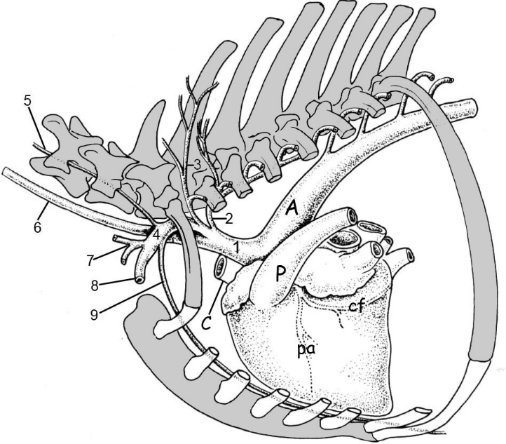
Figure 4-4. Equine great vessels, left lateral view. 1, brachiocephalic trunk; 2, left costocervical trunk; 3, left deep cervical a.; 4, left subclavian a., 5, left vertebral a.; 6, left common carotid a., 7, left superficial cervical a.; 8, left axillary a; 9, left internal thoracic a.; A, aorta; C, cranial vena cava; P, pulmonary trunk; cf, circumflex branch of left coronary a.; pa, paraconal intereventricular branch of left coronary a.
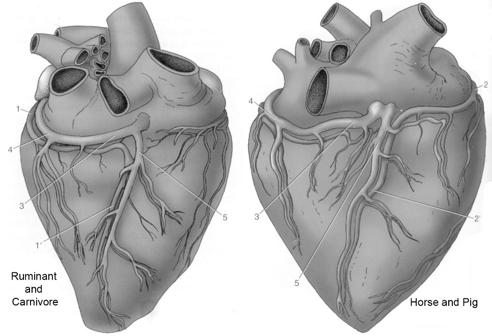
Figure 4-5. Cardiac vessels, right-sided view. 1, circumflex branch of the left coronary a.; 1’, subsinuosal interventricular branch; 2, right coronary a.; 2’, subsinuosal interventricular branch; 3, 4, great cardiac v.; 5, middle cardiac v.
Dissection Videos for this Section of Material
Heart and Major Vessels
- Pony
- Internal Thorax, Left and Right sides: https://youtu.be/E-4ar4mecXg
- Watch from 6:35-12:55 (left side)
- Watch from 16:25-16:36 and 20:15-end (right side)
- Internal Thorax, Left and Right sides: https://youtu.be/E-4ar4mecXg
- Calf
- Internal Thorax, Left side (Watch from 7:10-12:02): https://youtu.be/Lgb_J8z6w8s
- Internal Thorax, Right side (Watch from 4:17-end) : https://youtu.be/JlWBLuScrTU
- Isolated Heart Specimens
- Plastinated Hearts: https://youtu.be/zpHWXT-wXzI
- Fresh Heart 1 (Bovine): https://youtu.be/HO6P43KH6VU
- Fresh Heart 2 (Porcine): https://youtu.be/q1HNMgIAQww
