
Part 4: Female Genitalia
Abby Brown
FEMALE GENITALIA – Equine
-
ALL FEMALE PONY specimens: Identify all parts of the uterus; identify the uterine horns, uterine body, and the cervix.
-
- In the pony specimens, identify the T-shaped uterus with two uterine horns (left and right), a uterine body (where the two uterine horns unite) and a cervix.
-
- Note that of all the common domestic animals, the mare has the greatest degree of fusion of uterine horns to form a uterine body.
- The uterus is T-shaped with no distinct intercornual ligament and the cervix lacks transverse folds, therefore passage of an artificial insemination (A.I.) pipet is easier than in the cow.
-
- In the pony specimens, identify the T-shaped uterus with two uterine horns (left and right), a uterine body (where the two uterine horns unite) and a cervix.
-
-
ALL FEMALE PONY specimens: Identify the ovaries (right and left). Identify the ovulation fossa, ovarian bursa, uterine tube, and infundibulum.
-
- Identify the ovaries (right and left).
-
- Take note of the large size of the equine ovaries (TVA, 569-574) and note that no nerves enter the ovary except those associated with blood vessels.
-
- Identify the ovulation fossa.
-
- The equine ovary is indented at the ovulation fossa. This is the only site of ovulation in the mare.
-
- Identify the ovarian bursa.
-
- The ovarian bursa is formed by the mesosalpinx and mesovarium lateral and cranial to the ovary; this bursa communicates with the peritoneal cavity.
-
- Identify the uterine tube (aka oviduct).
-
- The uterine tube is easily identified running in the mesosalpinx from its opening, the infundibulum, around the cranial aspect of the ovary, to the lateral surface, and then to the uterine horn. The uterine tube is thrown into a tight wave-like path by its muscular walls.
- Note how the uterine tube begins as a wide structure and tapers to a uniform diameter about midway to the uterus. This establishes two indistinct segments, the ampulla and isthmus. The ampullary-isthmic junction is the midway point where a uniform diameter is established.
-
- Identify the ovaries (right and left).
3. ALL FEMALE PONY specimens: Identify the broad ligament of the uterus and its three parts: mesovarium, mesosalpinx, and mesometrium.
-
- The broad ligament of the uterus is the double fold of peritoneum that suspends the female reproductive organs from the body wall. Find this structure on both left and right sides.
- The broad ligament is divided into three parts which you should identify: mesovarium, mesosalpinx, and mesometrium.
-
- The mesometrium is the largest part and attaches to the uterus.
- The mesovarium attaches to the ovary.
- The mesosalpinx attaches the uterine tube to the mesovarium.
-
4. ALL FEMALE PONY specimens: Identify the ovarian proper ligament, suspensory ligament of the ovary, and round ligament of the uterus.
-
- Identify the ovarian proper ligament.
-
- This is the strong fibrous band attaching the caudal pole of the ovary to the cranial end of the uterus.
-
- Identify the suspensory ligament of the ovary.
-
- This is the thickened cranial border of the mesovarium extending to the body wall
-
- Identify the round ligament of the uterus.
-
- This is the free edge of the mesometrium coursing toward the vaginal ring (previously discussed in this chapter).
-
- Recall that the vaginal ring is sealed in all female ungulates.
-
- This is the free edge of the mesometrium coursing toward the vaginal ring (previously discussed in this chapter).
-
- Identify the ovarian proper ligament.
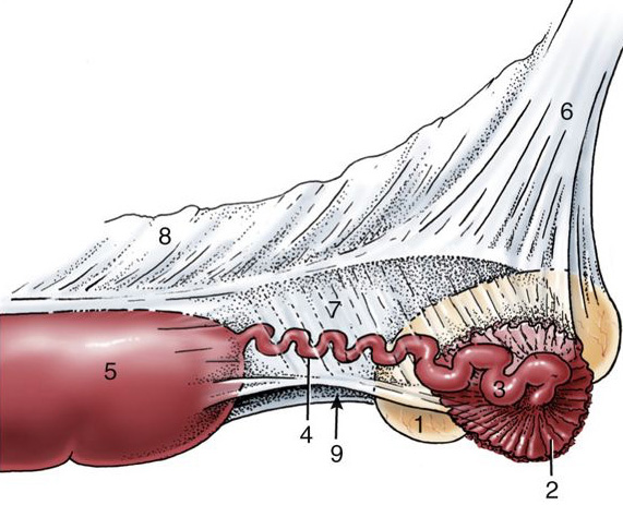
Figure 6-5. Mare; right ovary, uterine tube and part of uterus (lateral view). 1, ovary; 2, infundibulum of tube; 3, ampulla of tube; 4, isthmus of tube; 5, uterine horn; 6, mesovarium; 7, mesosalpinx; 8, mesometrium; 9, arrow indicates entrance to ovarian bursa. (TVA 5-58)
5. ALL FEMALE PONY specimens: Identify the vulva; identify the dorsal and ventral commissures of the vulva.
-
- The vulva contributes to the external opening of the female urogenital tract. Identify the vulva which is made up of two labia, often referred to as vulvar ‘lips’.
-
-
Where the vulvar lips come together and fuse dorsally, identify the dorsal commissure of the vulva.
-
Where the vulvar lips come together and fuse ventrally, identify the ventral commissure of the vulva.
-
-
- The vulva contributes to the external opening of the female urogenital tract. Identify the vulva which is made up of two labia, often referred to as vulvar ‘lips’.
6. ALL FEMALE PONY specimens: Identify the vestibule and vagina. Identify the urethral orifice, urethra, and the external os of the cervix.
-
- Identify the vestibule, extending from the region of the vulva to the vagina.
-
-
Open the vestibule with a dorsal midline incision to view the interior.
-
Within the floor of the cranial part of the vestibule, identify the urethral orifice. The urethral orifice leads to the urethra. Pass a probe through the urethral orifice to help you identify the urethra.
-
-
- Identify the vagina. The vagina is the cavity located between the cervix and the vestibule.
- Re-identify the location of the cervix (of the uterus). Identify the external opening of the cervix into the vagina – this is the external os of the cervix.
- Identify the vestibule, extending from the region of the vulva to the vagina.
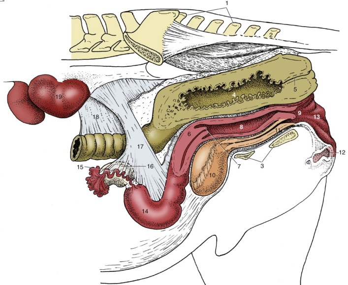
Figure 6-6. Mare, caudal abdominal and pelvic organs; the organs have been sectioned in a paramedian plane with the pelvis. Because of the absence of the intestines, the ovaries hang much lower than they would in the intact animal. 1, sacrum; 2, Cd2; 3, floor of pelvis; 4, rectum; 5, anal canal; 6, cervix; 7, vaginal part of cervix (with external os of cervix); 8, vagina; 9, vestibule; 10, bladder; 11, urethra; 12, clitoris; 13, vulva; 14, left uterine horn; 15, uterine tube; 16, ovary; 17, broad ligament (largely cut awary); 18, descending mesocolon; 19, left kidney. (TVA)
7. ALL FEMALE PONY specimens: Identify the (glans) clitoris, clitoral fossa and the clitoral sinuses.
-
-
Near the ventral commissure of the vulva, in the floor of the vestibule, identify the large mare (glans) clitoris which resembles the glans penis of the male horse.
-
-
Palpate the (glans) clitoris and note the firm rod like crura of the clitoris which are deep to the mucosa of the vestibule.
-
-
-
Identify the clitoral fossa, which is the space surrounding the clitoris ventrally. (This fossa is a homologue of the preputial fossa of the male horse.)
-
-
Note that of all the common domestic species, the clitoris is larger and more developed in the mare than any other domestic animal.
-
-
- Identify the clitoral sinuses.
-
-
- Clitoral sinuses are depressions on the dorsal surface of the equine clitoris. There are usually at least three sinuses: one central, one towards the right, and one towards the left. (See Fig. 6-7)
- These sinuses are only found in the equine, we will not see them in our other large animal species.
-
-
-
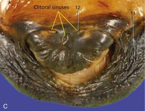
Figure 6-7. Mare (C); An enlargement of the [equine] vulva, showing the glans of the clitoris within the ventral commissure. 11, Right labium; 12, glans of clitoris. Clitoral sinuses are labeled with yellow lines. TVA
FEMALE GENITALIA – BOVINE
8. ALL FEMALE CALF specimens: Identify all parts of the uterus; identify the uterine horns, uterine body, and the cervix. Also identify the intercornual ligament(s) between the uterine horns and the transverse folds of the cervix.
-
- In the calf/bovine specimens, identify all parts of the uterus.
-
- Identify the two long uterine horns (left and right), and note that each horn is coiled into a loose spiral.
- Identify the short uterine body where the two uterine horns unite.
-
- Incise and open the body of the uterus and one of the uterine horns; attempt to identify the uterine caruncles within. (These would be more prominent in a pregnant animal.) Caruncles are the sites of placental attachment. Caruncles, together with cotyledons, from placentomes. (The placentomes and cotyledons would only be present in a pregnant cow.)
-
- Identify the cervix.
-
- Make an incision into the cervix to open it and identify the transverse folds of the cervix. These folds make passage of an artificial insemination (A.I.) pipet more difficult in the cow than in the mare.
-
- Return to the uterine body and horns; look for where the uterine horns diverge from the uterine body and identify the distinct intercornual ligament(s).
-
- Note that the cow has dorsal and ventral intercornual ligaments, but small female ruminants (goat, sheep) only have one intercornual ligament. (In the cow, the dorsal intercornual ligament is the one felt upon rectal palpation.)
-
-
- In the calf/bovine specimens, identify all parts of the uterus.
9. ALL FEMALE CALF specimens: Identify the ovaries (right and left). Identify the ovarian bursa, uterine tube, and infundibulum.
-
- Identify the ovaries (right and left). The ovaries of the cow are rounded ovals (sometimes referred to as ‘almond shaped’).
- Identify the ovarian bursa.
-
- The ovarian bursa is formed by the mesosalpinx and mesovarium lateral and cranial to the ovary; this bursa communicates with the peritoneal cavity.
-
- Identify the uterine tube (aka oviduct).
-
- The uterine tube is easily identified running in the mesosalpinx from its opening, the infundibulum, around the cranial aspect of the ovary, to the lateral surface, and then to the uterine horn. The uterine tube is thrown into a tight wave-like path by its muscular walls.
- Note how the uterine tube begins as a wide structure (at the infundibulum) and tapers to a uniform diameter about midway to the uterus. This establishes two indistinct segments, the ampulla and isthmus. The ampullary-isthmic junction is the midway point where a uniform diameter is established.
-
10. ALL FEMALE CALF specimens: Identify the broad ligament of the uterus and its three parts: mesovarium, mesosalpinx, and mesometrium.
-
- The broad ligament of the uterus is the double fold of peritoneum that suspends the female reproductive organs from the body wall. Find this structure on both left and right sides.
- The broad ligament is divided into three parts which you should identify: mesovarium, mesosalpinx, and mesometrium.
-
-
- The mesometrium is the largest part and attaches to the uterus.
- The mesovarium attaches to the ovary.
- The mesosalpinx attaches the uterine tube to the mesovarium.
-
-
11. ALL FEMALE CALF specimens: Identify the ovarian proper ligament, the suspensory ligament of the ovary, and the round ligament of the uterus.
-
- Identify the ovarian proper ligament.
-
-
- This is the strong fibrous band attaching the caudal pole of the ovary to the cranial end of the uterus.
-
-
- Identify the suspensory ligament of the ovary.
-
-
- This is the thickened cranial border of the mesovarium extending to the body wall
-
-
- Identify the round ligament of the uterus.
-
-
- This is the free edge of the mesometrium coursing toward the vaginal ring (previously discussed in this chapter).
-
- Recall that the vaginal ring is sealed in all female ungulates.
-
- This is the free edge of the mesometrium coursing toward the vaginal ring (previously discussed in this chapter).
-
-
- Identify the ovarian proper ligament.
12. ALL FEMALE CALF specimens: Identify the vulva; identify the dorsal and ventral commissures of the vulva.
-
- The vulva contributes to the external opening of the female urogenital tract. Identify the vulva which is made up of two labia, often referred to as vulvar ‘lips’.
-
-
Where the vulvar lips come together and fuse dorsally, identify the dorsal commissure of the vulva.
-
Where the vulvar lips come together and fuse ventrally, identify the ventral commissure of the vulva.
-
-
- The vulva contributes to the external opening of the female urogenital tract. Identify the vulva which is made up of two labia, often referred to as vulvar ‘lips’.
13. ALL FEMALE CALF specimens: Identify the vestibule and vagina. Identify the urethral orifice, urethra, suburethral diverticulum and the external os of the cervix.
-
- Identify the vestibule, extending from the region of the vulva to the vagina.
-
-
Open the vestibule with a dorsal midline incision to view the interior.
-
Within the floor of the cranial part of the vestibule, identify the urethral orifice. The urethral orifice leads to the urethra. Pass a probe through the urethral orifice to help you identify the urethra.
-
-
Identify the suburethral diverticulum on the floor of the vestibule. This is a small blind ‘pouch’ just caudal/ventral to the urethral orifice.
-
-
-
-
- Identify the vagina. The vagina is the cavity located between the cervix and the vestibule.
- Re-identify the location of the cervix (of the uterus). Identify the external opening of the cervix into the vagina – this is the external os of the cervix.
- Identify the vestibule, extending from the region of the vulva to the vagina.
14. ALL FEMALE CALF specimens: Identify the (glans) clitoris and clitoral fossa.
-
-
Near the ventral commissure of the vulva, in the floor of the vestibule, identify the (glans) clitoris which is much smaller in the cow than in the mare.
-
Palpate the (glans) clitoris and note the firm rod like crura of the clitoris which are deep to the mucosa of the vestibule.
-
-
Identify the clitoral fossa, which is the space surrounding the clitoris ventrally.
-
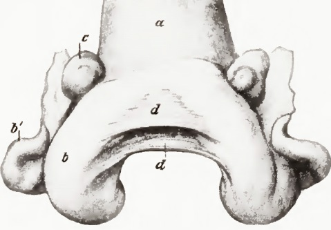
Figure 6-8. Cow uterus (contracted); dorsal view. a, Body of uterus; b,b’, horn of uterus; c, ovary; d, d, intercornual ligaments. (Sisson Grossman Fig. 535)
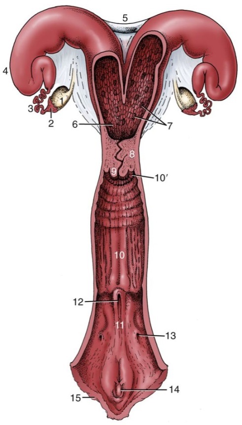
Figure 6-9. Cow reproductive tract, opened dorsally. 1, ovary; 2, infundibulum; 3, uterine tube; 4, horn of uterus; 5, intercornual ligaments; 6, body of uterus; 7, caruncles; 8, cervix; 9, vaginal part of cervix; 10, vagina; 10’, fornix; 11, vestibule; 12, external urethral opening; 13, opening of major vestibular gland; 14, clitoris; 15, vulva. (TVA 5-59)
FEMALE GENITALIA – PORCINE
15. ALL FEMALE PIG specimens: Identify all parts of the uterus; identify the uterine horns, uterine body, and the cervix. Also identify the mucosal prominences of the cervix.
-
-
In the pig specimens, identify the uterus which has very long uterine horns (that have multiple curves and coils) and a very short uterine body.
-
Identify the cervix which is unusually long.
-
-
Open the cervix and identify the mucosal prominences (TVA, 773, right (11)) that are present, rather than transverse folds as in the cervix of the cow.
-
-
-
16. ALL FEMALE PIG specimens: Identify the ovaries (right and left).
-
- Identify the ovaries (right and left) and notice the irregular surface of the sow ovary due to the numerous follicles.
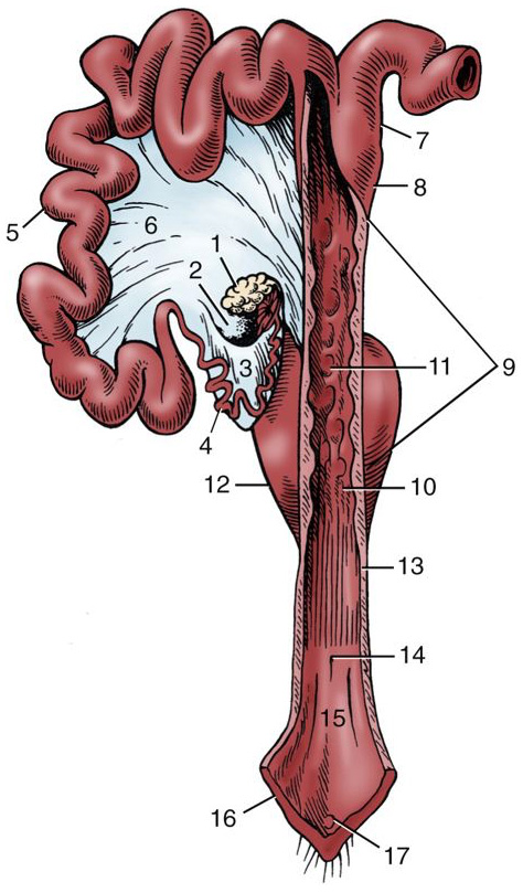
Figure 6-10. Sow reproductive tract opened dorsally in part; the right uterine horn and ovary are not shown. 1, left ovary; 2, ovarian bursa; 3, mesosalpinx; 4, uterine tube; 5, uterine horn; 6, broad ligament; 7, parallel segments of uterine horns; 8, body of uterus; 9, cervix; 10, external uterine orifice (aka external os of the cervix); 11, mucosal prominences; 12, urinary bladder, 13, vagina; 14, external urethral orifice; 15,vestibule; 16, vulva; 17, glans of clitoris. (TVA Fig
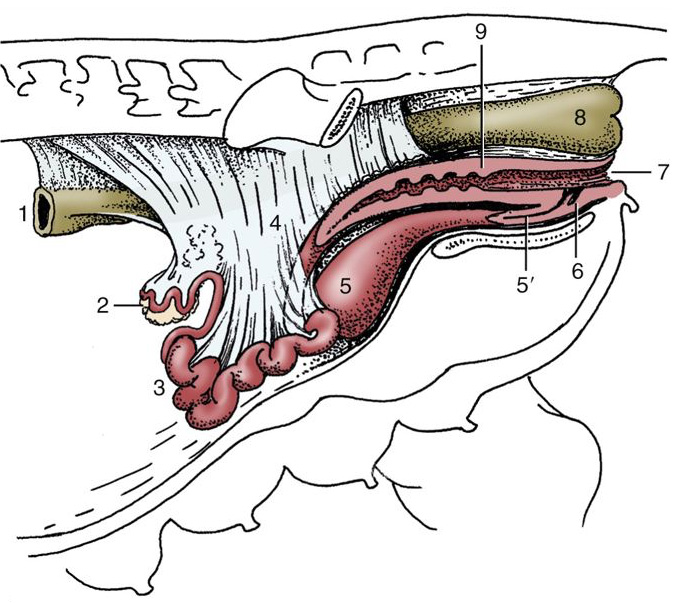
Figure 6-11. Sow reproductive tract in situ. (The presence of the intestines in the intact animal causes the ovaries and uterine horns to lie more dorsally than shown here.) 1, Descending colon; 2, ovary; 3, uterine horns; 4, broad ligaments; 5, urinary bladder, 5’ urethra; 6, suburethral diverticulum; 7, vulva; 8, rectum. Image source: TVA Fig. 35-3, 35-4)
UDDER (AKA MAMMARY GLANDS)
Important Note: Be sure to identify the structures of the udder and teat on dried/plastinated museum specimens available in lab – these are fair game on assessments!
17. ALL FEMALE specimens: Identify the udder (aka mammary gland(s)) and open one of the teats with an incision to view the internal structures. Identify any supernumerary teats that may be present.
-
- PONY specimens: The udder is small and easily removed from the mare (TVA, 584). It consists of a pair of mammary glands, with one teat on each.
-
- Note that any teats present in excess of the normal number of teats would be called supernumerary teats. (See dried museum specimen of cow udder.) Identify any supernumerary teats that may be present on your specimen.
-
- CALF specimens: The udder of the cow consists of four ‘quarters’ (mammary glands). Each quarter has one teat. (Note that small ruminants only have two mammary glands, hence two teats.)
-
- Note that the udder will be small on immature calves and the structures may be difficult to see (so be sure to look at the dried museum specimens of cow udders).
- Note that any teats present in excess of the normal number of teats would be called supernumerary teats. (See dried museum specimen.) Identify any supernumerary teats that may be present on your specimen.
-
- PONY specimens: The udder is small and easily removed from the mare (TVA, 584). It consists of a pair of mammary glands, with one teat on each.
18. ALL FEMALE specimens: Identify the teat, the teat orifice(s), papillary duct(s), lactiferous sinus (made up of the teat sinus and gland sinus), and lactiferous ducts.
-
- PONY specimens:
-
- Each teat has two teat orifices. These are the external openings of the teat where milk is expressed.
- Each teat orifice leads into a papillary duct (aka ‘teat canal’ or ‘streak canal’). In the mare, since there are two teat orifices, there are two papillary ducts on each teat (one corresponding with each teat orifice). These papillary ducts drain separate lactiferous sinuses.
- Note that the papillary ducts connect the lactiferous sinus to the teat orifice.
- Each lactiferous sinus is made up of a teat sinus and a gland sinus.
-
- The portion within the teat is the teat sinus.
- The portion within the gland is the gland sinus.
-
- Each gland sinus receives milk from lactiferous ducts.
-
-
- Open one of the teats and sinuses on your female pony specimen and attempt to see these structures.
- Simplified summary of milk flow: lactiferous ducts -> gland sinus -> teat sinus -> papillary duct -> teat orifice
-
- CALF specimens:
-
- Each teat has one teat orifice. This is the external opening of the teat where milk is expressed.
- The teat orifice leads into the papillary duct (aka ‘teat canal’ or ‘streak’ canal). The papillary duct drains the lactiferous sinus.
- Note that the papillary ducts connect the lactiferous sinus to the teat orifice.
- The lactiferous sinus is made up of a teat sinus and a gland sinus.
-
- The portion within the teat is the teat sinus.
- The portion within the gland is the gland sinus.
-
- The gland sinus receives milk from lactiferous ducts.
-
-
- Open one of the teats and sinuses on your female calf/bovine specimen and attempt to see these structures.
- Simplified summary of milk flow: lactiferous ducts -> gland sinus -> teat sinus -> papillary duct -> teat orifice
-
- PONY specimens:
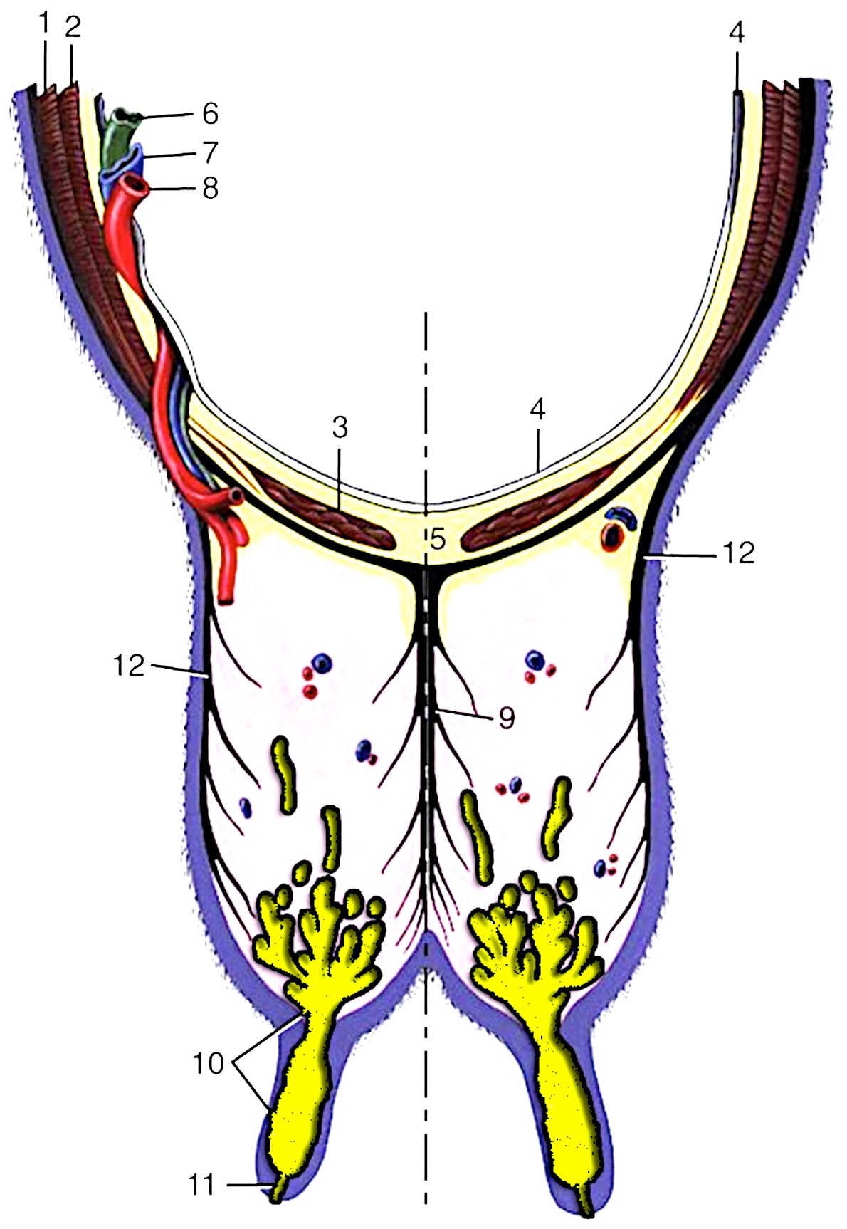
Figure 6-12. Transverse section of the abdominal floor and cranial quarters of the bovine udder. 1, external abdominal oblique; 2, internal abdominal oblique; 3, rectus abdominis; 4, peritoneum; 5, linea alba; 6, lymph vessel; 7, external pudendal vein; 8, external pudendal (mammary) artery; 9, medial suspensory ligament; 10, lactiferous sinus; 11, papillary duct; 12, lateral laminae of suspensory apparatus. (Modified from TVA Figure 29-39).
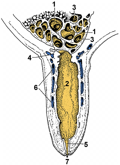
Figure 6-13. Section of a cow’s teat and lactiferous sinus. 1, 2, Lactiferous sinus = gland sinus (1) and teat sinus (2); 3, openings of lactiferous ducts into the lactiferous sinus; 4, submucosal venous ring; 5, papillary duct; 6, venous plexus in teat wall; 7, teat orifice. (Modified from TVA 3rd ed., Fig. 31-5)
Dissection Videos for this Section of Material
Genitalia
- Pony
- Female Pony:
- Internal Pelvis & Genitalia: https://youtu.be/KFFRO7oT1-k
- Female Pony:
- Isolated specimens
- Female:
- Mare & Cow Genitalia: https://youtu.be/vTCY7CEWCbE
- Bovine Mammary Gland: https://youtu.be/SzUG6ScaHU8
- Female:
