
Part 1: Abdominal Wall
Abby Brown
IMPORTANT NOTE: The following dissection should be completed on both sides of each specimen (unless otherwise specified).
abdominal wall
- ALL specimens: If needed, on both left and right sides of your specimen, incise any remaining skin of the thorax and abdominal regions along the dorsal midline. Reflect the skin ventrally and remove/discard it.
- In male specimens, avoid damaging the prepuce when reflecting the skin (if possible).
- ALL specimens: On the RIGHT side, cranial to the thigh, attempt to identify the subiliac lymph node (calf)/subiliac lymphocenter (pony).
- ALL (CALF) specimens: On the right side (with the attached hind limb), identify the large, oval subiliac lymph node on the cranial edge of the thigh muscles, about midway between the tuber coxae and the fold of the flank.
- ALL (PONY) specimens: On the right side (with the attached hind limb), attempt to find the subiliac lymphocenter on the cranial edge of the thigh muscles, about midway between the tuber coxae and the fold of the flank.
- ALL specimens: On left and right sides, on the lateral aspect of the abdominal wall, identify the external abdominal oblique (EAO) m. and its elastic covering, the tunica flava abdominis.
- The external abdominal oblique (EAO) m. is the most powerful of all expiratory muscles. This muscle is covered by a pale yellow elastic sheet of deep fascia known as the tunica flava abdominis (Terminology Note: flava = yellow). This elastic sheet is difficult to observe in the dog but thick in large ungulates.
- Dissection Note: The EAO has a costal part arising from the last several ribs as well as a lumbar part arising from the lumbar region of the thoracolumbar fascia. It inserts on the tuber coxae, the body of the ilium, and on the ventral midline, via an extensive aponeurosis, along the linea alba and the prepubic tendon.
- Identify the external abdominal oblique m. and the tunica flava abdominis that covers it.
- The external abdominal oblique (EAO) m. is the most powerful of all expiratory muscles. This muscle is covered by a pale yellow elastic sheet of deep fascia known as the tunica flava abdominis (Terminology Note: flava = yellow). This elastic sheet is difficult to observe in the dog but thick in large ungulates.
- ALL specimens: Transect the external abdominal oblique m. and reflect it ventrally, to the level of the rectus abdominis m; identify the rectus abdominis m.
- On both left and right sides, transect the external abdominal oblique m. (EAO) along the last rib and also along its attachment to the costal arch.
- Detach the EAO from the lumbar epaxial mm. and carefully transect it along the cranial edge of the thigh muscles.
- Reflect the EAO ventrally until the lateral edge of the rectus abdominis m. is reached.
- Dissection Note: As you reflect the EAO, avoid damaging the underlying internal abdominal oblique m.!
- Identify the rectus abdominis m. running along the ventral midline of the belly.
- Dissection Note: This muscle inserts on the pubis via the prepubic tendon.
- LEFT side only: Transect the EAO along the edge of the rectus abdominis m. and discard it; this will allow you to more clearly identify and isolate the rectus abdominis m. on the left side.
- Comparative Note: The rectus abdominis m. is proportionately wider in ungulates than it is in the dog. This enables ungulates to bear the weight of herbivorous guts.
- ALL specimens: Identify the internal abdominal oblique (IAO) m.; transect and reflect it to the level of the end of the last rib. In the calf specimens, take note of the paralumbar fossa.
- After reflection of the EAO, identify the internal abdominal oblique (IAO) m. that is now visible.
- Dissection Note: The IAO muscle runs cranioventrally from the tuber coxae toward the costal arch and also inserts on the linea alba and prepubic tendon via an aponeurosis.
- On both sides, transect the IAO just caudal to the last rib and begin reflecting it caudally.
- Dissection Note: As you reflect the IAO avoid damaging the underlying transversus abdominis m.!
- Use your hand to work underneath the IAO and separate it from the transversus abdominis m. as far back as the deep surface of the tuber coxae.
- Transect the IAO m. about 2 cm from where it originates from the tuber coxae and reflect the muscle ventrally, but only as far as the level of the end of the short last rib.
- Note that the IAO muscle is thickest at its origin from the tuber coxae and becomes thinner as it projects cranially and ventrally.
- ALL (CALF) specimens: As you reflect the IAO take note of the paralumbar fossa caudal to the last rib.
- The thinness of the internal abdominal oblique m. behind the last rib in the bovine is the reason why the muscle “sinks in” to form the paralumbar fossa.
-
- Comparative Note: The pony (equine) also has a paralumbar fossa, but it is not as significant/sizable as in the bovine, so you need not identify it.
-
- The paralumbar fossa is the most common surgical site for entry into the ruminant abdomen. This triangular depression is bounded by the following landmarks:
-
- Dorsally: the lumbar transverse processes and the epaxial muscle dorsal to the processes.
- Cranially: the last rib
- Ventrally: an oblique muscular thickening of the IAO m. extending from the tuber coxae to the costal arch.
-
- The thinness of the internal abdominal oblique m. behind the last rib in the bovine is the reason why the muscle “sinks in” to form the paralumbar fossa.
- After reflection of the EAO, identify the internal abdominal oblique (IAO) m. that is now visible.
- ALL specimens: Identify the transversus abdominis (TA) m. and note the ventral branches of lumbar spinal nn.
- After reflection of the IAE, identify the transversus abdominis m. that has been uncovered.
- On the surface of the transversus abdominis m. note the large ventral branches of lumber nerves (L1 & L2). You should also look for branches of the deep circumflex iliac vessels emerging deep to the tuber coxae.
- ALL specimens: On both sides, transect the transversus abdominis m. (and underlying peritoneum) and reflect it ventrally as far as the level of the end of the last rib, but no farther.
-
- This opening will provide access for palpation and some visualization of the caudally positioned viscera.
8. ALL specimens: To visualize some of the more cranially placed abdominal organs transect, and then reflect, the lateral part of the diaphragm (on both sides) away from the ribcage (i.e., a “diaphragmatic window”). Leave about 5-10 cm of the diaphragm remaining connected on the midline.
-
-
-
- This opening will provide some visualization but will still retain the visceral contents within the abdominal cavity.
-
-
-
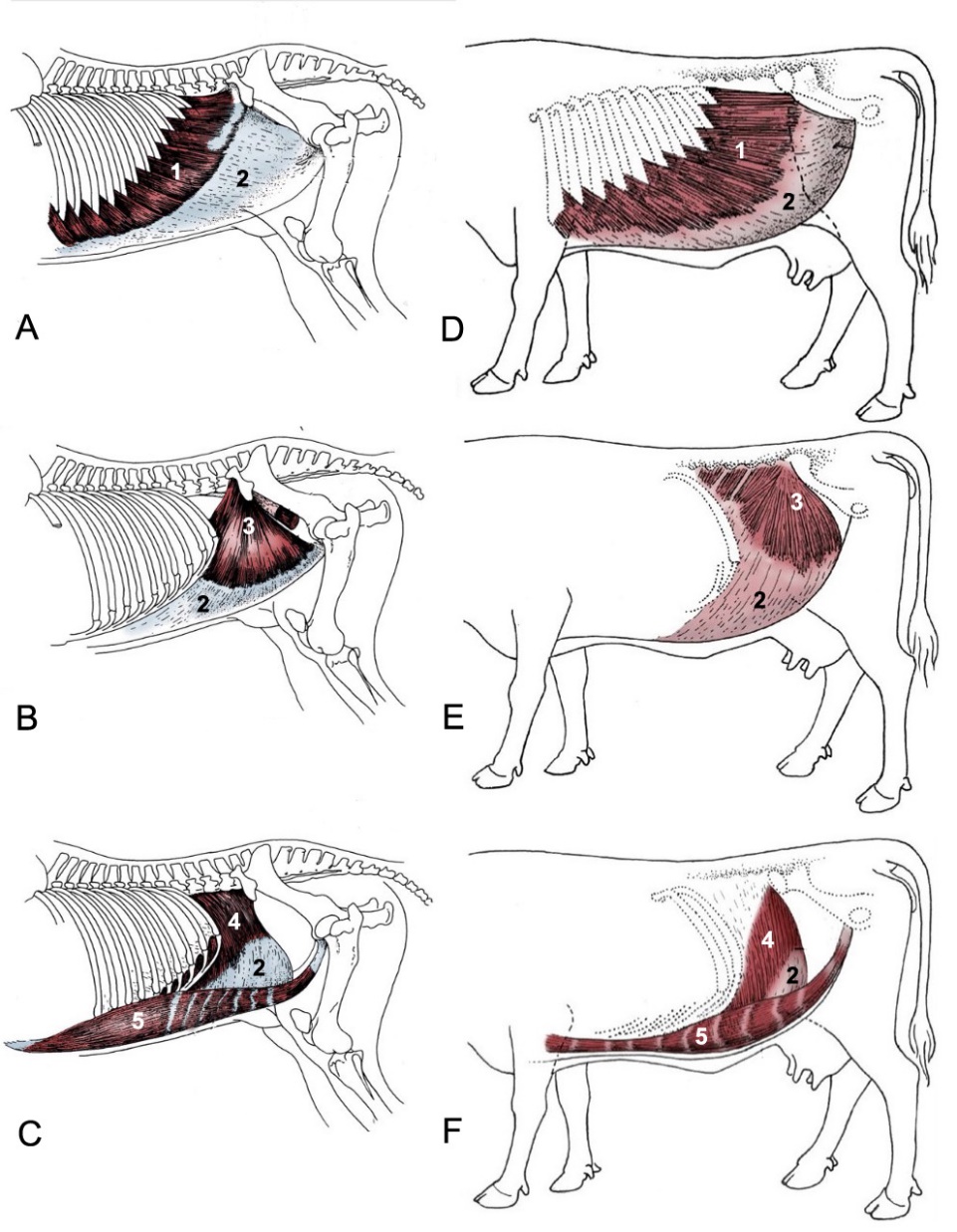
Figure 5–1. Abdominal Wall of the Horse (A-C) and Ox (D-F). 1, external abdominal oblique m. 2, aponeurosis of abdominal wall muscles 3, internal abdominal oblique m. 4, transversus abdominis m. 5, rectus abdominis m. (Modified from TVA Figs. 21-4 and 28-1)
Dissection Videos for this Section of Material
Abdominal Wall
- Pony, In situ Abdomen, Left and Right sides: https://youtu.be/oHUvRk-urG8
- Watch from 0:00-4:24 (Left side)
- Watch from 4:44-7:29 (Right side)
- Calf, In situ Abdomen, Left and Right sides: https://youtu.be/IvQkIFzW-Rc
- Watch from 0:00-4:15 (Left side)
- Watch from 4:31-6:36 (Right side)
