
Part 2: Extrinsic Muscles and Associated Structures
Abby Brown
Extrinsic Forelimb Muscles and Associated Structures
- Identify the brachiocephalicus m. and dissect its borders.
- Remove cervical fascia to expose the parallel edges of the brachiocephalicus m. and the cervical trapezius m. Note that the brachiocephalicus m. covers the shoulder region and is attached to the humerus.
- In the horse, the brachiocephalicus muscle is fused with the omotransversarius muscle; this will be discussed later during the dissection of the neck.
- As you dissect, note that in the ox (calf), the cleidocephalicus portion of the brachiocephalicus m. has two parts (cleido-occipitalis and cleidomastoideus) while the horse cleidocephalicus only has one part (cleidomastoideus). Note that these will be seen more clearly during the dissection of the neck.
- Remove cervical fascia to expose the parallel edges of the brachiocephalicus m. and the cervical trapezius m. Note that the brachiocephalicus m. covers the shoulder region and is attached to the humerus.
- Calf Specimen: Identify the omotransversarius m. (calf only).
- In the ox, the omotransversarius m. is separate from the brachiocephalicus m., similar to the carnivore. Identify the omotransversarius m. extending from the shoulder region toward the head in the calf specimens.
- Pony specimen: Identify the cutaneous colli m. (horse only)
- In the horse, observe that there is another superficially located muscle in the ventral neck region, the cutaneous colli m. (with a thinner ‘sphincter colli’ subdivision as you move up the neck toward the head). The cutaneous colli will cover/overlap the ventral edge of the brachiocephalicus muscle and will be further dissected/identified in Chapter 3 (Fig. 1-3).
- Note that the cutaneous colli m. is virtually non-existent in the calf and other ruminants.
- Identify visible parts of the serratus ventralis m. Recall that this muscle has two parts: serratus ventralis cervicis (attaching on the neck) and serratus ventralis thoracis (attaching on the thorax). As we proceed through the dissection, more of this muscle will become visible/identifiable. (Figure 1-2, SVC and SVT)
- **Note**: In Figure 1-2 the serratus ventralis m. (SVC, SVT) lies deep to the other extrinsic forelimb muscles, and to the forelimb itself, attaching deep to the scapula. As noted, this muscle will be observed more fully as you proceed with this dissection.
- Identify the superficial pectoral muscles (2 parts: descending and transverse) and the superficial cervical lymph nodes.
-
- The pectoral muscles (Fig 1-2, SP and DP) originate from the sternum and attach to the medial side of the forelimb.
- In the horse, the superficial pectoral m. (Fig. 1-3) is well-developed and is sometimes referred to as the ‘breast muscle’.
- The superficial pectoral muscle is made up of descending and transverse parts in both horse and ox. Separate the edges of the superficial pectoral muscles from surrounding tissues, and also differentiate the descending part from the transverse part.
- While dissecting the pectorals, take care to preserve the nearby branch of the cephalic vein (Fig. 1-3).
- In the pony, also preserve the fatty tissue along the proximal/dorsal border of the descending superficial pectoral and the distal/ventral border of the cutaneous colli m.; this fatty tissue area contains the superficial cervical lymph nodes.
- In the horse, lymph nodes appear as clusters of small lymph nodes (a.k.a. lymphocenter) in contrast to the larger, fewer, regional lymph nodes found in other animals. Within the fatty tissue just dorsal to the pectoral muscles, identify the superficial cervical lymph nodes in your pony specimen.
- In the calf, there will be a single, large, oval-shaped superficial cervical lymph node on each side (left and right). Lift the caudal edge of the omotransversarius m. and dissect deep to (underneath) it to observe/identify these lymph nodes.
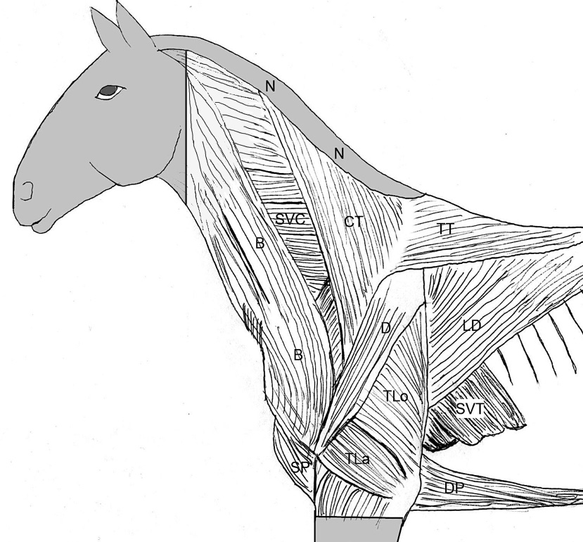
(Duplicate) Figure 1-2. Extrinsic and intrinsic muscles of the horse (upper) and ox (lower). (N) Indicates the location of the nuchal fatty crest present only in the horse. Extrinsic muscles: CT, Cervical trapezius; TT, thoracic trapezius; B, brachiocephalicus;SP, superficial pectorals; DP, deep pectorals; LD, latissimus dorsi;SVC, serratus ventralis cervicis; SVT, serratus ventralis thoracis. Labeled intrinsic forelimb muscles: D, Deltoideus; TLo, long head of the triceps brachii; TLa, lateral head of the triceps brachii. (Not shown is the cutaneous trunci m. which may come off when the skin is reflected. If present, it will cover the upper forelimb and structures caudal to it.) (Modified from Budras and Habel, 1st ed.)
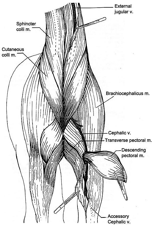
Figure 1-3. Equine, cranial view of the superficial pectoral muscles and the base of the neck.
6. Transect both parts of the superficial pectoral muscles adjacent to the sternum, approximately 1-2 cm from the ventral midline, and reflect them to see the underlying deep pectoral muscles. Do this on both right and left sides.
7. Identify the deep pectoral mm. (Pony: 2 parts: ascending deep pectoral and subclavius; Calf: ascending deep pectoral only)
-
-
In the pony, there are two parts to the deep pectoral m., the ascending deep pectoral and subclavius mm.
-
In the calf only the ascending deep pectoral m. is well developed.
- The ascending deep pectoral (also called the caudal deep pectoral in the horse) is the largest pectoral muscle and is found ventral to the serratus ventralis m. It runs adjacent to the sternum on ventral midline. Identify the ascending deep pectoral m. and define its borders in both pony and calf specimens.
- In the pony, identify the subclavius m. (also called the cranial deep pectoral in the horse).
- The subclavius is a large fusiform muscle extending from the sternum to the cranial aspect of the forelimb in the horse, but in the ox this muscle is very underdeveloped (i.e., you do not need to find it in the calf specimens).
-
8. Identify the trapezius m. (2 parts: cervical and thoracic) and define its borders. Transect both parts with an arching cut from cranial to caudal and reflect them to reveal the rhomboideus m. underneath.
-
- Moving dorsally on the lateral aspect of the limb, identify the flat, triangular trapezius m., which has a cervical part and a thoracic part. The trapezius m. extends from the dorsal midline to the spine of the scapula. Define the borders of the trapezius by dissection.
- Make an arching cut (similar to the dog/cat dissection) from the middle of the cranial aspect of the cervical trapezius across the entirety of the muscle to the caudal border of the thoracic trapezius. Reflect the proximal portion upward (toward the dorsal midline) and the distal portion downward (toward the spine of the scapula). This will reveal the underlying rhomboideus m. and expose a greater portion of the cervical part of the serratus ventralis muscle.
9. Identify the latissimus dorsi m.; transect it caudal to the limb and reflect it dorsally.
-
- Caudal to the forelimb, identify and define the edges of the latissimus dorsi muscle by dissection.
- Transect the latissimus dorsi m. about 2-3 cm caudal to its insertion on the deep face of the limb. (This will leave enough of the muscle attached to the forelimb for later identification.)
- Carefully reflect the latissimus dorsi m. dorsally, toward the midline of the back, keeping the muscle attached dorsally via its aponeurotic attachment to the thoracolumbar fascia.
10. Identify the two parts of the rhomboideus m. (cervical and thoracic); transect and reflect both parts. In the pony, identify the well-developed dorsoscapular ligament found deep to the rhomboideus m.
-
- At the most proximal edge of the scapula, identify the rhomboideus m. and note that it has cervical and thoracic parts, both of which insert on the deep face of the scapula.
- The horse and ox do not have the rhomboideus capitis subdivision of this muscle as is seen in the dog/cat.
- Transect both portions of the rhomboideus m. approximately 1 cm from the scapular insertion.
- In the horse, once the rhomboideus m. is transected, reflect it and take notice of the underlying structure, which is a modification of the thoracolumbar fascia that forms a well-developed ligamentous sheet called the dorsoscapular ligament. This ligament extends from near-midline dorsally to the deep face of the scapula.
- Dissection Note: Adducting the distal portion of the limb, i.e., pushing it toward the midline/other side of the animal, will help you visualize the dorsoscapular ligament.
- The dorsoscapular ligament is less developed in the ox, hence need not be identified. Due to this difference, in the calf, you do not need to transect the rhomboideus m. of the right forelimb.
- **NOTE**: The Ungulate Dissection Web Site Lab 1 – Image 17 provides you with a glimpse of the nuchal ligament of the horse, but you do not need to uncover this structure now; it will be covered in detail when we cover the neck region.
- At the most proximal edge of the scapula, identify the rhomboideus m. and note that it has cervical and thoracic parts, both of which insert on the deep face of the scapula.
Removal of LEFT Forelimb
You will remove the LEFT forelimb of the hanging specimens by completing transections of the extrinsic muscles. (NOTE: Remove only the LEFT forelimb of the hanging specimens by completing transections of extrinsic muscles as directed.) Please proceed with the LEFT forelimb removal instructions as outlined below:
- In both species, transect the brachiocephalicus m. at least one full hand-width cranial to the shoulder and reflect the distal portion.
- **NOTE**: In the pony, be cautious! Try not to cut into the omohyoideus muscle that lies directly underneath the brachiocephalicus m.!
- In the pony, as you reflect the brachiocephalicus m. distally (toward the leg), identify the underlying omohyoideus muscle (well-developed only in the horse) and define its borders.
- In the pony, after identification of the omohyoideus m., transect it about 5 cm cranial to its origin from the shoulder.
- The omohyoideus m. is poorly developed in ruminants, so you do not need to identify it on the calf specimens.
- **NOTE**: In the pony, be cautious! Try not to cut into the omohyoideus muscle that lies directly underneath the brachiocephalicus m.!
- In the calf, transect the omotransversarius m. about 4-5cm cranial to the shoulder.
- Distally, cross the left forefoot over the right forefoot, pulling each foot toward the opposing side (resembling an ‘X’). This will displace the left scapula laterally. (In the horse, you can now fully visualize, and then transect, the well-developed dorsoscapular ligament.)
- If present, transect the dorsoscapular ligament (of the left limb) and any remaining portions of muscle keeping the dorsal border of the scapula attached to the body.
- In the pony, make your transection horizontally through the dorsoscapular ligament, mid-way between its origin and insertion.
- In the calf, if there is a (underdeveloped) dorsoscapular ligament present, transect it with a horizontal incision mid-way between its origin and insertion.
- Uncross the limbs and abduct (pull) the left foot laterally (up and away from the body) to aid in transection of the left deep pectoral muscles in the next step.
- Transect the subclavius m. (only well-developed in the horse) approximately 10 cm from its insertion on the scapular region. (This should be done just above the point of the cephalic vein and the superficial cervical lymph nodes.)
- Transect the cephalic v., leaving an identifiable portion on both the body and the limb for later orientation.
- Transect the ascending deep pectoral m. adjacent (parallel) to the sternum, approximately 3 cm from the ventral midline.
- Further abduct (lift) the left forelimb laterally and transect the vessels and nerves entering the deep face of the limb.
- As you continue to abduct (lift) the left forelimb laterally, transect the scapular attachment of the serratus ventralis m. close to where it meets the deep surface of the scapula (i.e., leave the main portion of this muscle attached to the thorax).
- This will complete the removal of the left forelimb. (Fig. 1-4 shows the muscles that will be left on the trunk.)
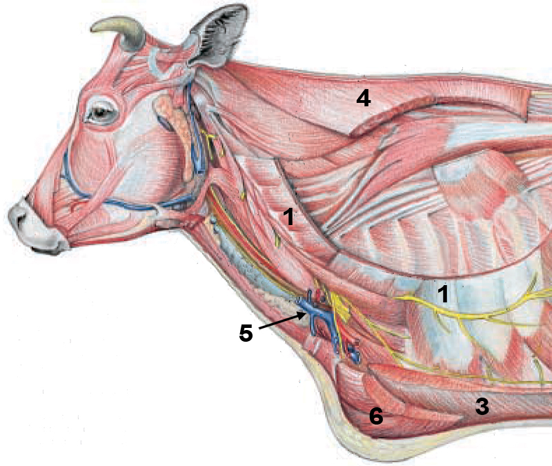
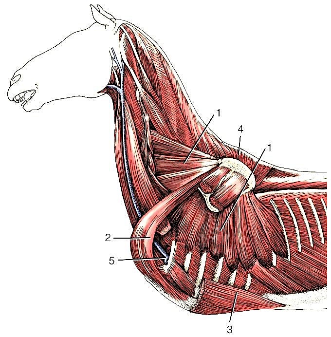
Figure 1-4. Deep muscles of the ox (upper) and horse (lower) attaching the forelimb to the trunk. 1, serratus ventralis; 2, subclavius (only horse, very underdeveloped/absent in ox); 3, ascending deep pectoral; 4, rhomboideus; 5, axillary vessels turning around first rib into forelimb; 6, superficial pectorals. (Equine: Modified from Dyce, Sack and Wensing, 4th ed.; Bovine: Modified from Budras and Habel, 1st ed.)
Dissection Videos for this Section of Material
Extrinsic Forelimb Muscles and Associated Structures
- Pony: Watch from 3:25-end
- Proximal Thoracic Limb:https://www.youtube.com/watch?v=oIi4i8ScZEM&t=824s
- Calf: Watch from 2:45-end
- Proximal Thoracic Limb: https://www.youtube.com/watch?v=hN10FaowIos
