
Part 1: Skinning Your Cadaver
Abby Brown
IMPORTANT NOTE: For this part of the chapter, the Guide directions will refer to either “ALL specimens” (meaning dissect pony and calf specimens in the same way) or to one species in particular, i.e., “ALL (PONY) specimens” or “ALL (CALF) specimens.” This added instructional note is needed because the dissection may differ slightly according to species.
REVIEW of Superficial Structures
- Before you begin to skin out the hind limb of your cadaver, review/identify the superficial structures found on the distal limbs, as described for each species. (See duplicate Figure 1-1)
- Pony Specimen: Identify the chestnut, ergot and coronet.
- In the horse, identify the chestnut on the medial aspect of the hind limb, at the level of the tarsus.
- On the plantar surface of the fetlock, identify the ergot. (Normally, in a non-clipped animal, there is an area of long hair, referred to as the feather that usually obscures the ergot. However, clipping the hair off the fetlock during preparation of the specimen should have exposed this area sufficiently to see the ergot.) Note that the ergot correlates to the metacarpal or metatarsal pad of the dog.
- Observe the skin to hoof transition; this is called the coronet, referring to the ‘crown’ of the hoof. Deep, and distal, to the coronet is a band of dermal tissue (coronary band) which will be discussed later.
- Calf Specimen: Identify the dewclaws and the coronet.
- Identify the dewclaws on the plantar surface of your calf limbs.
- Observe the skin to claw transition; this is called the coronet.
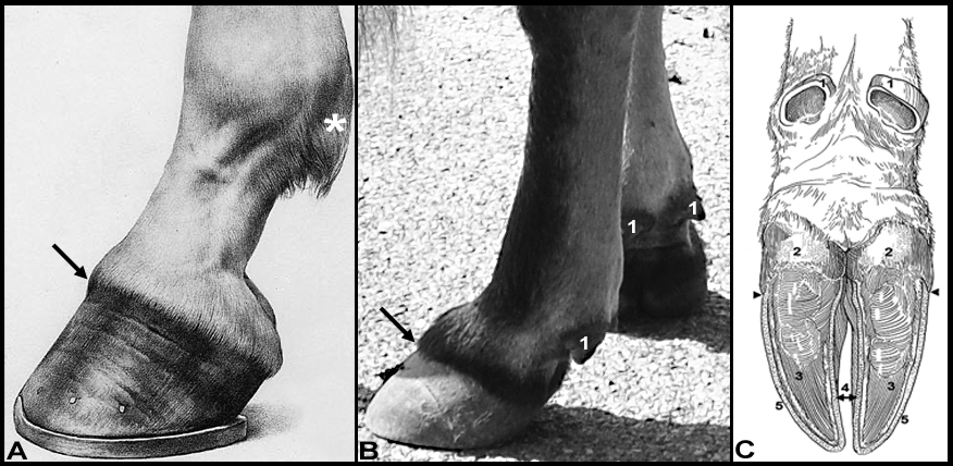 (Duplicate) Figure 1-1. A. Horse front foot, lateral view; B. Bovine front foot, lateral view; C. Bovine foot, palmar/plantar view. *Region of ergot (horse); arrows, coronet (horse and bovine); bovine: 1, Dewclaws (digits 2 and 5); Claw (digits 3 and 4): 2, bulb of claws; 3, sole of claws; 4, wall (axial surfaces); 5, wall (abaxial surfaces); arrowhead, junction of wall and bulb. (Modified from Anatomy and Physiology of Farm Animals; Frandson, Wilke, Fails)
- Pony Specimen: Identify the chestnut, ergot and coronet.
- As you begin the proximal pelvic limb dissection, you should recall that the hind limb is bound to the body by very few extrinsic muscles (in contrast to the forelimb, which has many). The main group of extrinsic muscles is the iliopsoas group, which will be identified later in this dissection. (Recall from Anatomy I that extrinsic muscles have one attachment on the body/trunk and one attachment on the limb itself.) The following dissections will be done on both sides of the pony and calf hanging specimens.
Skinning Your Cadaver
3. ALL specimens: Incise and reflect the skin off of the proximal hind limbs as directed; begin by making a mid-dorsal (dorsal midline) incision from the level of the tuber coxae to the base of the tail.
4. ALL (PONY) specimens: Make a vertical incision, just cranial to the attachment of the hind limb, from the dorsal midline down to the level of the stifle.
5. ALL (CALF) specimens: Make a vertical incision, just cranial to the tuber coxae, from the dorsal midline down to the level of the stifle. (This incision should be made further cranially than the incision made in the pony.)
6. ALL specimens: Make another vertical incision along the caudal edges of the proximal pelvic limb as far as the level of the stifle joint.
-
- For the caudal incision you will carefully encircle the base of the tail and also the anus (as well as the vulva in mare/cow).
- Important Dissection Note: In the areas surrounding the anus and genitalia, the skin is not as thick, therefore deep incisions can be destructive. Use more superficial incisions in these areas and carefully reflect the skin.
- For the caudal incision you will carefully encircle the base of the tail and also the anus (as well as the vulva in mare/cow).
7. ALL specimens: Reflect the large flap of skin you have created; reflect the skin laterally and distally, off of the upper (proximal) pelvic limb and distally as far as the stifle joint, being careful to avoid damage to the anus and the vulva of the mare/cow as previously noted.
Dissection Videos for this Section of Material
Skinning Your Cadaver
- Pony: watch 0:00-1:08
- Proximal Pelvic Limb: https://youtu.be/65hVe2ekzqI
- Calf: watch 0:00-1:17
- Proximal Pelvic Limb: https://youtu.be/e44X-gYwyx4
