
Part 1: Pleural Cavity/Space
Abby Brown
thoracic pleura
IMPORTANT NOTE: Be sure the thoracic cavity has been opened on your specimen before you begin this dissection! (This step was completed at the end of Chapter 3; please reference Chapter 3, Part 3: Opening the Thorax, as needed.)
-
Recall from Anatomy I, that within the thorax you will find pleurae, which are the serous membranes that cover the lungs and line the walls of the thoracic cavity. These pleurae form right and left ‘sacs’ that enclose the pleural cavities/spaces (R & L). Each ‘sac’ consists of visceral and parietal layers; the pleural cavity/space is found between the two layers of pleura (visceral and parietal) within the thoracic cavity. Identify the pleural cavities/spaces in your specimen. Within the pleural cavity of available specimens/species, identify the layers of pleura described.
-
Identify the pulmonary (visceral) pleura that is closely adhered to the surfaces of the lungs.
-
Identify the parietal pleura lining the thoracic wall. Parietal pleura consists of costal, diaphragmatic, and mediastinal pleurae.
-
-
Costal pleura covers the inner surfaces of the ribs (and associated muscles).
-
Diaphragmatic pleura covers the cranial surface of the diaphragm that bulges into the thoracic cavity.
-
Mediastinal pleurae are the layers of pleura that cover the partition between the two pleural cavities (the mediastinum).
-
-
Dissection Notes: The mediastinum includes the two layers of mediastinal pleurae and the space between them. There are two named parts of the mediastinal pleura, pericardial mediastinal pleura and plica venae cavae, but you do not need to identify them in your specimen. The pericardial mediastinal pleura is the pleura covering the heart and the plica vena cavae is a loose fold of pleura that surrounds the caudal vena cava.
-
-
-
-
-
-
Identify the region of the root of the lung which is composed of the pleura and the bronchi, vessels, and nerves entering each lung. At the root of the lung, the mediastinal parietal pleura is continuous with the pulmonary (visceral) pleura via a very small connecting pleural layer.
-
-
Dissection Note: Caudal to the hilus, this connection forms a free border, known as the pulmonary ligament, between the caudal lobe of the lung (both left and right) and the mediastinum.
-
-
- Recall from Chapter 3, that as you opened the thoracic cavity, you identified the line of pleural reflection and the costodiaphragmatic recess through removal of intercostal mm. and soft tissues.
- That dissection demonstrated the line of pleural reflection (the line along which diaphragmatic pleura reflects off the diaphragm and onto the ribs as costal pleura). This clinically important landmark is the caudoventral extent of the pleural cavities.
- The pleura reflect in a narrow angled space between the convex surface of the diaphragm and the thoracic body wall. This space is the costodiaphragmatic recess.
- That dissection demonstrated the line of pleural reflection (the line along which diaphragmatic pleura reflects off the diaphragm and onto the ribs as costal pleura). This clinically important landmark is the caudoventral extent of the pleural cavities.

(Duplicate) Figure 3-9. Equine thorax, lateral view. The dotted line (1) is an outline of the heart; shown in black is where the heart is exposed by the cardiac notch; 2, basal border of lung; 3, line of pleural reflection.
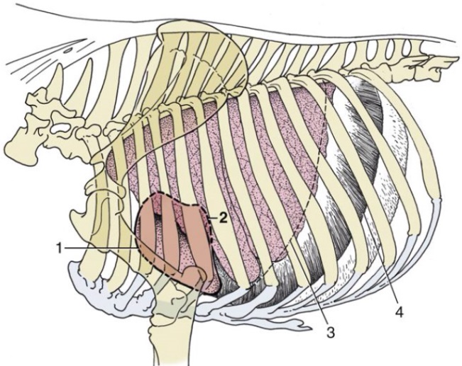
(Duplicate) Figure 3-10. Bovine thorax, lateral view. The dotted line (1) is an outline of the heart; 2, caudal extent of heart; 3, basal border of lung; 4, line of pleural reflection.
