
Part 3: Mediastinum and Diaphragm
Abby Brown
Mediastinal dissection
1. ALL specimens: Identify the mediastinum and note that it can be divided into a cranial part (cranial to the heart), a middle part (containing the heart), a dorsal part (dorsal to the heart), a ventral part (ventral to the heart), and a caudal part (caudal to the heart).
-
- Recall that the mediastinum is the region between the pleural cavities, including the right and left mediastinal parietal pleura. It contains all of the thoracic viscera except the lungs.
- Enclosed in the mediastinum you will find the following: thymus, thoracic lymph nodes, heart, aorta, trachea, esophagus, vagus nerves, as well as other nerves and vessels. You should know the contents of the mediastinum and be able to describe their general placement within the mediastinum.
2. ALL specimens: Within the mediastinum, identify the thymus (if visible) and the esophagus.
-
- In a young animal, a large portion of the cranial mediastinum is occupied by the thymus. Identify any visible thymus in your specimen.
- Dissection Note: The thymus is a bilobed structure that atrophies as the animal ages (i.e., young animals will have a large thymus, old animals will have little to none present). The thymus is located in the cranial mediastinum (cranial to the heart).
- Identify the esophagus within the mediastinum and note that it is found in the dorsal mediastinum (dorsal to the heart). (Figs. 4-2/11 and 4-3/6)
- Dissection Note: Dissect carefully (blunt dissection with scissors) when looking for the esophagus in the dorsal mediastinum so that you do not cut vessels and nerves you still need to identify.
- In a young animal, a large portion of the cranial mediastinum is occupied by the thymus. Identify any visible thymus in your specimen.
3. ALL specimens: In the caudal part of the thoracic cavity, identify the diaphragm which is the muscular partition between the thoracic and abdominal cavities. Note its curvature and slant. After identification of the diaphragm, identify and name its various parts: tendinous center (aka central tendon), lumbar part (diaphragmatic crura: left crus & right crus), costal part, and sternal part.
-
-
The tendinous center (aka central tendon) is a V-shaped tendinous sheet in the middle portion of the diaphragm.
-
The muscular parts of the diaphragm (attached to the tendinous center) are the lumbar, costal, and sternal parts; these are named according to their regions of attachment.
-
-
The sternal part is narrow and is attached to the sternum just cranial to the xiphoid cartilage.
-
The costal part is broader than the sternal part and attaches along the deep face of the ribs.
-
The lumbar part attaches to lumbar vertebrae and forms the diaphragmatic crura; a left crus and a right crus. Note that the right crus is typically larger than the left crus. The crura (plural) are somewhat triangular slips of muscle that surround structures passing through the diaphragm. Separate/dissect the crura by manual dissection, noting their thickness as you do so.
-
-
-
4. ALL specimens: Identify the three ‘passageways’ through the diaphragm, the aortic hiatus, esophageal hiatus, and caval foramen.
-
-
The aortic hiatus is located dorsally in the central diaphragm, between the muscular diaphragmatic crura. It allows for the passage of the aorta, azygos vein(s), and thoracic duct through the diaphragm.
-
The esophageal hiatus is centrally located in the diaphragm, between the muscular diaphragmatic crura. It allows for the passage of the esophagus, esophageal vessels, and vagal nerve trunks through the diaphragm.
-
The caval foramen is usually located slightly to the right side, passing through the central tendon of the diaphragm. It allows for the passage of the caudal vena cava through the diaphragm.
-
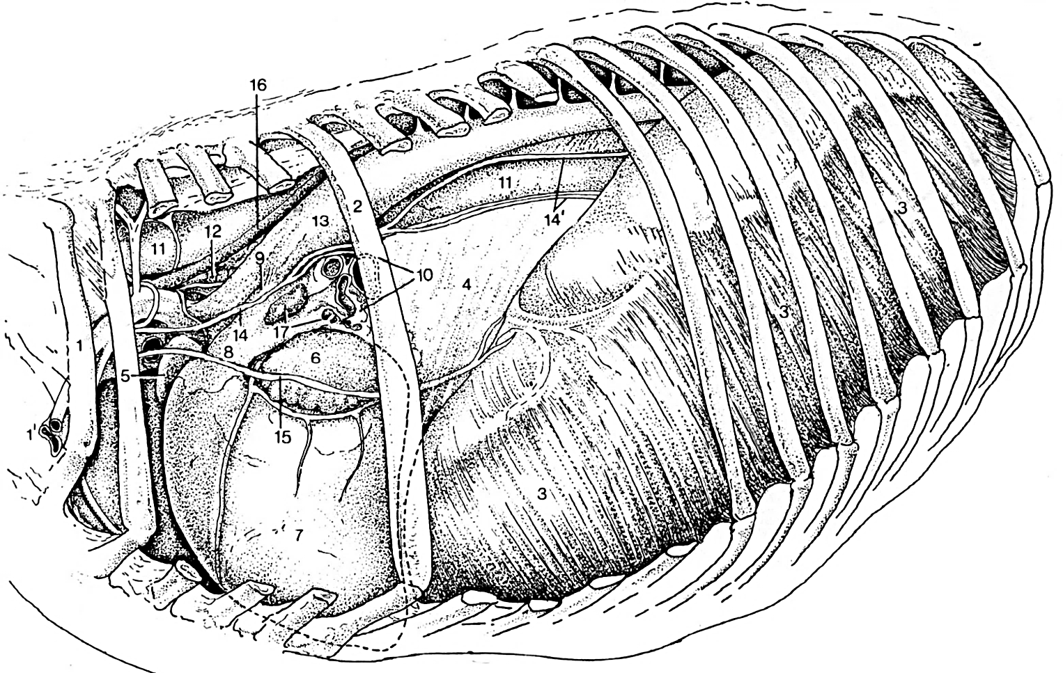
(Duplicate) Figure 4-2. Equine thorax, left lateral view. 1, rib 1; 2, rib 6; 3, diaphragm; 4, caudal mediastinum; 5, right auricle; 6, left auricle; 7, left ventricle; 8, pulmonary trunk; 9, ligamentum arteriosum; 10, cut surface of root of the lung; 11, esophagus; 12, trachea; 13, aorta; 14, vagus n.; 14’, dorsal and ventral vagal trunks; 15, phrenic n.; 16, thoracic duct; 12, tracheobronchial lymph node.
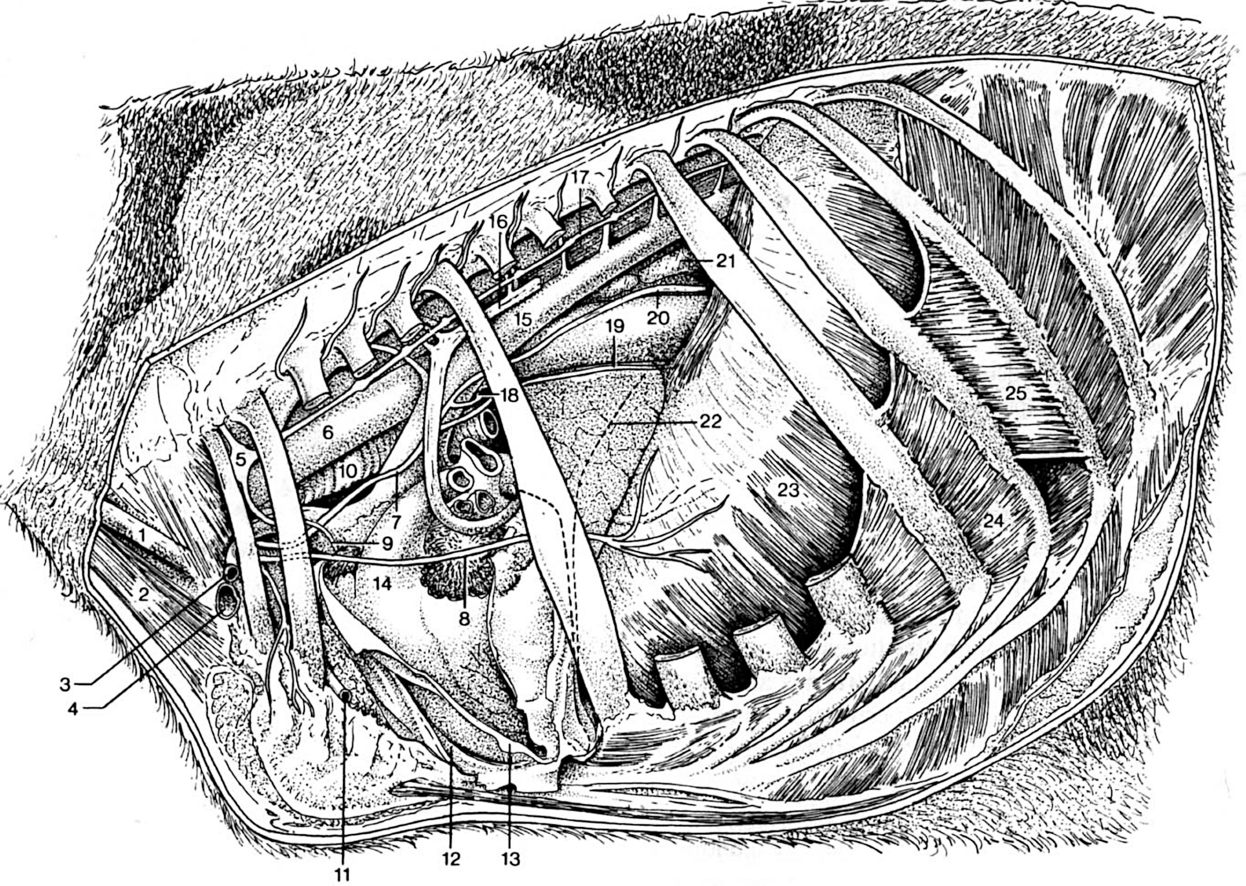
(Duplicate) Figure 4-3. Bovine thorax, left lateral view. The left azygous vein (16) passes around the cut surface of the root of the left lung. 1, external jugular vein; 2, sternocephalicus m.; 3, axillary a.; 4, axillary v.; 5, cervicothoracic ganglion; 6, esophagus; 7, left vagus n.; 8, (left) phrenic n.; 9, cardiac n.; 10, trachea; 11, internal thoracic a.; 12, mediastinal pleura; 13, reflected pericardium; 14, pulmonary trunk; 15, aorta; 16, left azygous v.; 17, sympathetic trunk; 18, left recurrent laryngeal n.; 19, ventral vagal trunk.; 20, dorsal vagal trunk.; 21, caudal mediastinal lymph node; 22, cranial extent of the diaphragm; 23, diaphragm; 24, internal intercostal m.; 25, external intercostal m.
Dissection Videos for this Section of Material
Mediastinum and Diaphragm
- Pony
- Internal Thorax, Left and Right sides: https://youtu.be/E-4ar4mecXg
- Watch from 3:48-4:06, 5:18-5:48 and 12:56-13:58 (left side)
- Watch from 18:20-18:56 (right side)
- Internal Thorax, Left and Right sides: https://youtu.be/E-4ar4mecXg
- Calf
- Internal Thorax, Left side (Watch from 2:25-2:48, 6:08- 7:08 and 12:02-end): https://youtu.be/Lgb_J8z6w8s
- Internal Thorax, Right side (Watch from 3:30-4:00) : https://youtu.be/JlWBLuScrTU
