
Part 5: Male Genitalia
Abby Brown
MALE GENITALIA – ALL
- ALL MALE specimens: Identify the testes/testicles (if they are intact) within the scrotum. Identify the all parts of the epididymis; identify the head, body and tail of the epididymis. Also, identify the testicular bursa.
-
- Identify the scrotum. Open the scrotum with a midline incision and reflect the skin covering each teste/testis (if they are present/intact).
- On the testicles, identify the associated epididymis. The epididymis has 3 parts which you should identify: head, body, and tail of the epididymis. Note that the epididymis is found mostly on the lateral side of each testis.
-
-
The head of the epididymis is the cranial part where the epididymis communicates with the testis.
-
The body of the epididymis is the middle part.
-
The tail of the epididymis is the caudal part which is then continuous with the ductus deferens on the medial side of the testis.
-
-
Dissection Note: The tail of the epididymis is attached to the vaginal tunic and spermatic fascia by the ligament of the tail of the epididymis and the tail of the epididymis is attached to the testis itself by the proper ligament of the testis.
-
-
-
On the lateral side of each testis, identify the testicular bursa (a space between the epididymis and testis).
-
-
Dissection Note: Using the testicular bursa and the position of the head or tail of the epididymis, you should be able to differentiate right versus left testicles.
-
-
-
-
2. ALL MALE specimens: Identify the spermatic cord, ductus deferens, testicular a. & v. and the cremaster m. Identify the vaginal tunics; identify the parietal and visceral vaginal tunics, the mesorchium and mesoductus deferens.
-
- Recall that the spermatic cord is composed of the ductus deferens and the testicular a. & v. and is found attached to the cranial end of each testis.
- Identify the cremaster m., the caudal slip of internal abdominal oblique m. running alongside the spermatic cord.
- Identify the vaginal tunics covering the spermatic cord and extending onto each testicle.
-
- Identify the parietal vaginal tunic, which is the outer layer of the vaginal tunics.
-
- Incise the parietal vaginal tunic over the spermatic cord and testis to see the vaginal cavity and the visceral vaginal tunic.
-
- Identify the visceral vaginal tunic, which is the inner layer of the vaginal tunics. The visceral vaginal tunic is closely applied to the testes and epididymis.
- After incising the parietal layer of the vaginal tunic, identify the mesorchium and mesoductus deferens. The mesorchium and mesoductus deferens are connecting mesenteries of the testis and ductus deferens. (These connecting mesenteries are continuous with the parietal and visceral vaginal tunics.)
-
- The mesorchium is attached to the testicular vessels.
- The mesoductus deferens is attached to the ductus deferens.
-
- Identify the parietal vaginal tunic, which is the outer layer of the vaginal tunics.
-
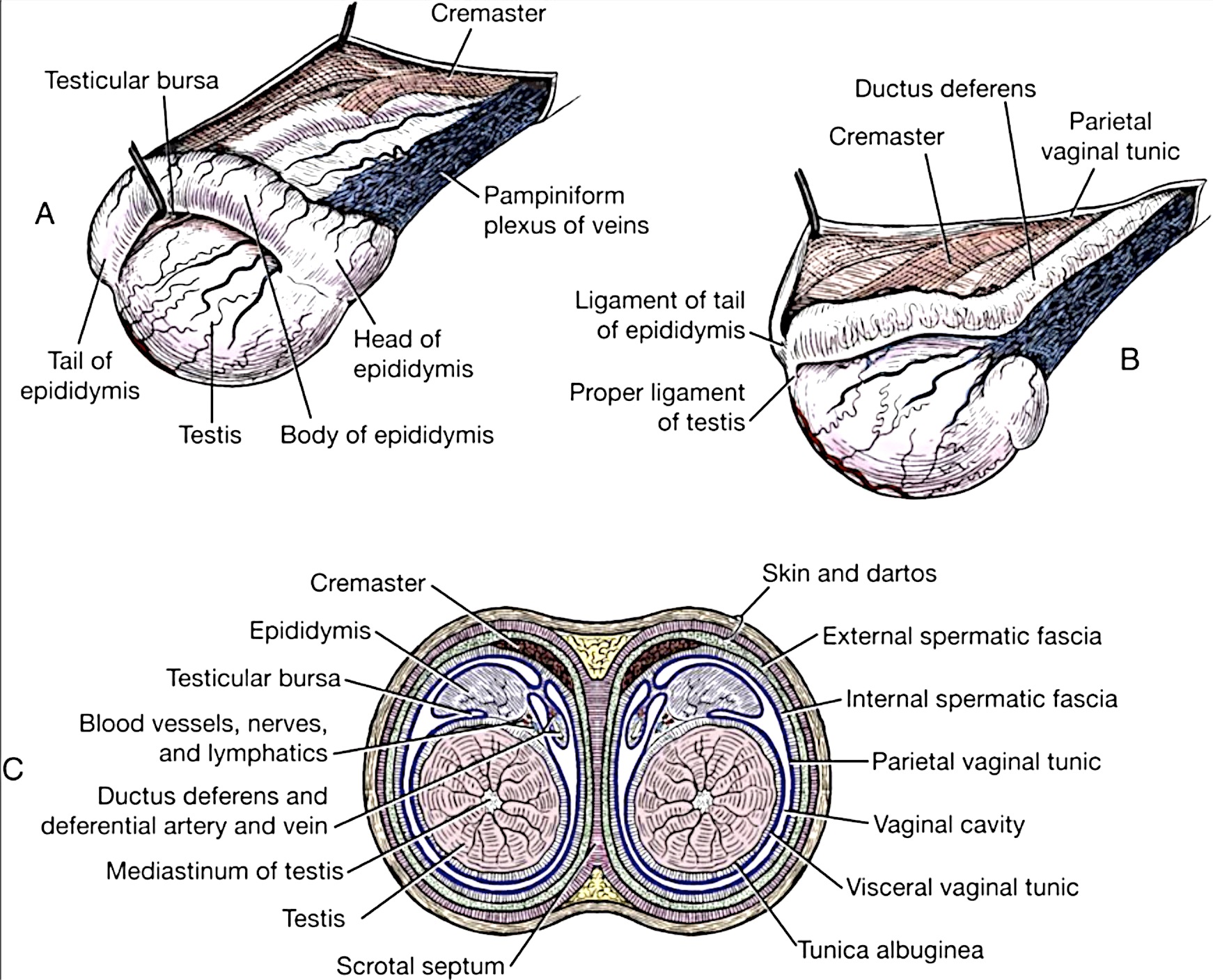
Figure 6-14. Canine testis and scrotum. A, Right testis, lateral aspect. B, Left testis, medial aspect. C, Schematic cross section through the scrotum and testes. (Figure 4-7 from Guide to Dissection of the Dog, 7th Ed.)
MALE GENITALIA – EQUINE
3. ALL MALE PONY specimens: Identify both ductus deferens (left and right) and the genital fold, and if present, observe the uterus masculinus. Identify the accessory sex glands (ampullae/ampullary glands, vesicular glands, body of the prostate gland, and bulbourethral glands) and the urethralis m. (See Fig. 6-15)
-
- Identify the ductus deferens on both the left and right sides (one from each testis).
- Identify the genital fold which is the membrane spanning the space in between the two ductus deferens. (In other words, the genital fold is the membrane connecting the bilateral (left and right) ductus deferens.)
-
- Within this fold there may be a remnant of the paramesonephric ducts, the uterus masculinus. If present, it will appear as a midline thickening.
-
- Follow the ductus deferens and note that each one will have a distal enlargement of its wall called the ampulla (ampullary gland).
-
- The ampullary glands/ampullae are accessory sex glands that are paired (i.e., one on each side/one for each ductus deferens).
-
- Identify the vesicular glands (aka seminal vesicles), which are also paired accessory sex glands.
-
- In the equine, these are smooth, sack-like structures lateral to the ampullae.
-
- Identify the body of the prostate gland.
-
- The prostate gland is an unpaired accessory sex gland and surrounds the urethra in the region caudal to the ampullae and vesicular glands.
- In the equine, the shape of the body of the prostate resembles a butterfly.
-
- Identify the urethralis muscle surrounding the (post-prostatic) pelvic urethra, caudal to the prostate gland.
-
- This is a tough sphincter surrounding the urethra that is responsible for urinary continence (prevention of urine flow).
-
- Identify the bulbourethral glands, which are another paired accessory sex gland.
-
- The bulbourethral glands are partially covered with skeletal muscle and are found at the ischial arch, just proximal to the root of the penis.
-
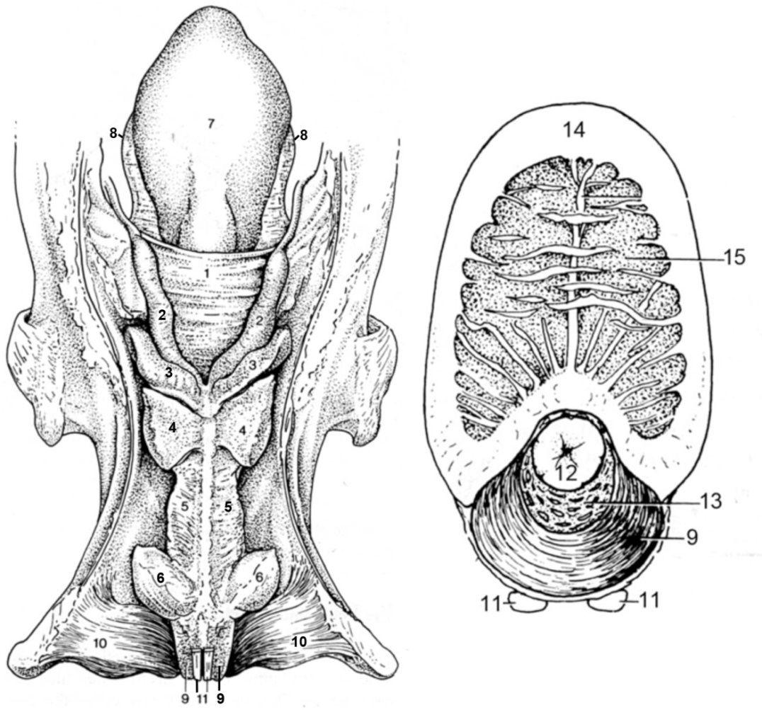
Figure 6-15. Equine male genitalia. Left: dorsal view of genital tract; Right: cross section of penis. 1, genital fold; 2, ampullary glands/ampullae of the ductus deferens; 3, vesicular glands; 4, body of the prostate gland; 5, urethralis muscle; 6, bulbourethral glands; 7, urinary bladder; 8, lateral ligament of the bladder; 9, bulbospongiosus m.; 10, ischiocavernosus m. (covering the crus of the penis); 11, retractor penis m.; 12, urethral mucosa; 13, corpus spongiosum; 14, tunica albuginea; 15, corpus cavernosum.
4. ALL MALE PONY specimens: Identify the prepuce and penis; open the prepuce to expose the penis and identify its parts. Identify the preputial orifice and preputial ring. (See Fig. 6-16) On the penis, identify the glans penis (w/corpus spongiosum glandis), urethral process, fossa glandis, and urethral sinus.
-
- Identify the prepuce and penis.
-
- The prepuce is the sheath/fold of skin covering the glans of the penis.
- If needed, open the prepuce with a ventral midline incision to expose the penis and identify its parts.
-
- Identify the preputial orifice.
-
- This is the external opening of the prepuce where hairy external skin meets the preputial lining.
-
- Identify the preputial ring.
-
- This is the free folded edge of the inner layer of the prepuce.
- When the penis is erect, the preputial ring appears as a raised ridge going around the penis.
-
- Identify the glans penis.
-
- The glans penis is the distal end of the penis which contains erectile tissue (corpus spongiosum glandis) and is shaped like a ‘bell’ when engorged.
-
- At the tip of the penis, identify the urethral process.
-
- This is a protrusion on the tip of the penis that is surrounded by ‘moat’ called the fossa glandis.
- Identify the urethral sinus; the urethral sinus is a dorsal diverticulum of the fossa glandis. This is where you would find what is known as the ‘bean’. The ‘bean’ is a hardened accumulation of smegma (secretion produced by glands in the preputial lining).
-
- Identify the prepuce and penis.
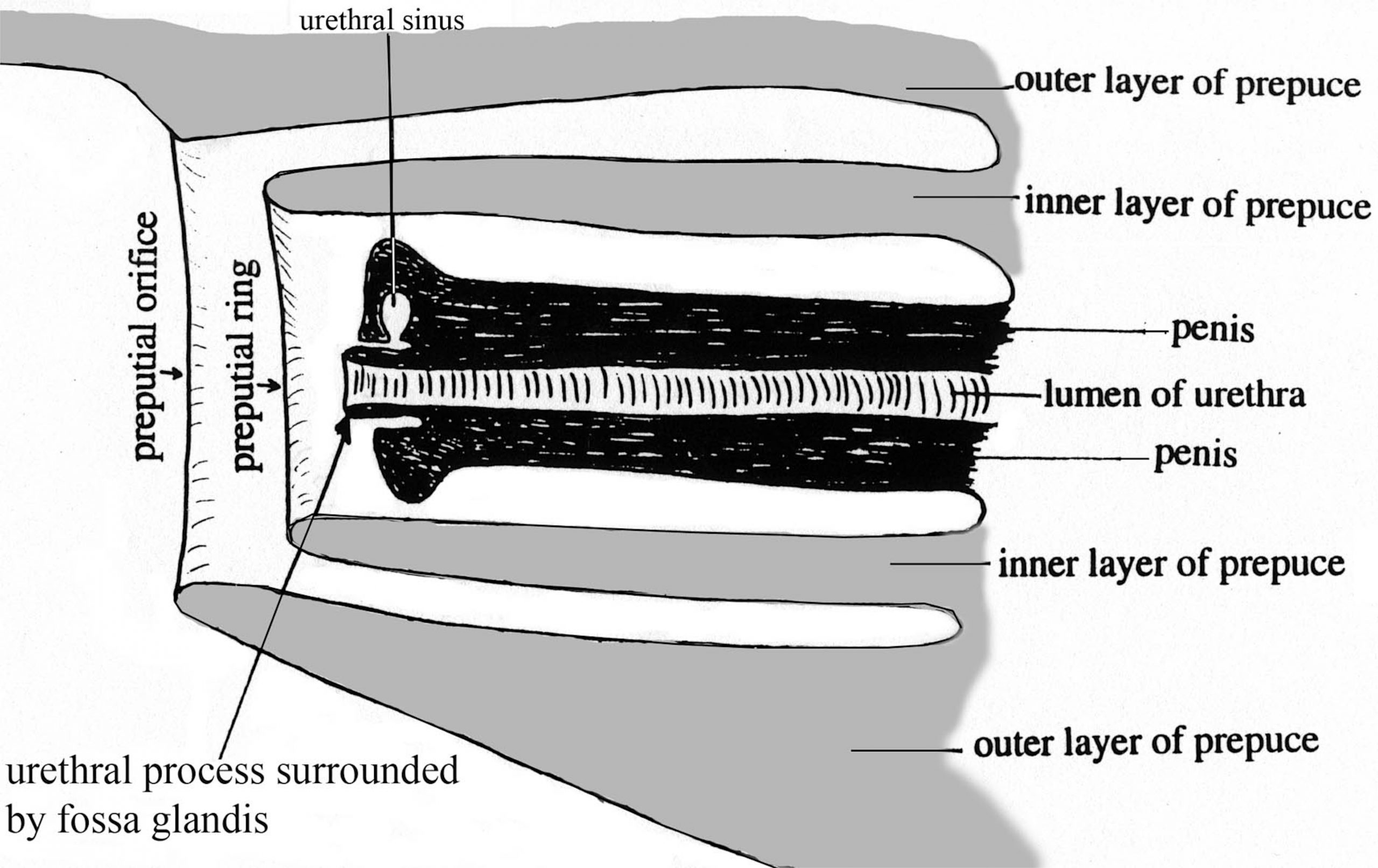
Figure 6-16. Schematic median section of equine penis and prepuce.
5. ALL MALE PONY specimens: Identify the three regional parts of the penis: root, body & free part.
-
- Identify the root of the penis.
-
- The root is the proximal part of the penis with laterally positioned crura (left and right).
- Recall that the root of the penis is formed by the left and right crura (anchored to the ischial arch) and the bulb of the penis. Where the crura come together on the midline of the penis marks the end of the root and beginning of the body.
-
- Identify the body of the penis.
-
- The body begins at the region where the bilateral crura meet and then extends distally to the free part.
-
- Identify the free part of the penis.
-
- The free part is the portion of the penis within the prepuce, including the distal glans penis (with corpus spongiosum glandis), which was previously identified.
-
- Identify the root of the penis.
6. ALL MALE PONY specimens: Identify the crura (left crus and right crus) of the penis. Identify the corpus cavernosum penis, ischiocavernosus m., and tunica albuginea. Identify the corpus spongiosum penis and the bulbospongiosus m. Identify the retractor penis m. as well as the dorsal n. and dorsal a. of the penis.
-
- Identify the crura of the penis (left crus and right crus) anchored to the ischial arch at the root. Make a transverse cut across one of the crura to identify the internal features.
-
- Each crus is composed of erectile tissue called corpus cavernosum penis, encased with a fibrous tunic called tunica albuginea. The crura serve to anchor each side of the penis to the ischial arch.
-
- The crura are covered by ischiocavernosus m.; identify this striated muscle covering each crus.
- Medial to the crura, in the region referred to as the bulb of the penis, the expanded erectile tissue called corpus spongiosum penis is found.
-
- Make an incomplete transection through the penis to identify the corpus spongiosum penis.
- This erectile tissue is also surrounded by muscle, the bulbospongiosus m., which you should identify. In the horse, this muscle extends most of the length of the penis.
-
- Identify the more superficial, paired, retractor penis muscles extending the length of the penis.
- Make another incomplete transverse section through the body of the penis.
-
- Observe/identify the tunica albuginea surrounding the corpus cavernosum penis.
- Observe/identify the corpus spongiosum penis partially surrounding the penile urethra. (Fig.6-15)
-
- Find and identify the dorsal nerve of the penis running along the dorsal midline of the penis. Note that this nerve is a continuation of the pudendal n.
- Also running along the dorsal midline of the penis, identify the dorsal a. of the penis.
-
- Pony Dissection Note: Note that in the equine, this artery is supplied by more than one parent artery (it anastomoses with other arteries) so it has more than one part/section (i.e., it is not one continuous artery, as is seen in the ruminant and in the dog).
-
- Identify the crura of the penis (left crus and right crus) anchored to the ischial arch at the root. Make a transverse cut across one of the crura to identify the internal features.
Important Note: You should be able to identify the parts of the equine penis on the dried and/or plastinated specimens (from the museum collection) as well as the wet equine isolated genitalia specimens available in lab. These supplemental specimens are fair game for lab practical assessments!
MALE GENITALIA – BOVINE
Important Dissection Note: Some of the calf specimens will have had a prior surgery on the penis so it may not be in its original position.
7. ALL MALE CALF specimens: Identify both ductus deferens (left and right) and the genital fold, and if present, observe the uterus masculinus. Attempt to identify the accessory sex glands (ampullae/ampullary glands, vesicular glands, and body of the prostate gland). Note that bulbourethral glands are present in the bovine but are underdeveloped in the calf, so they may not be identifiable.
-
- Important Dissection Note: Even though the accessory glands may be underdeveloped in your calf specimens you should still attempt to find them. You should also identify these structures on available supplemental specimens in the lab.
- Identify the ductus deferens on both the left and right sides (one from each testis).
- Identify the genital fold which is the membrane spanning the space in between the two ductus deferens. (In other words, the genital fold is the membrane connecting the bilateral (left and right) ductus deferens.)
-
- Within this fold there may be a remnant of the paramesonephric ducts, the uterus masculinus. If present, it will appear as a midline thickening.
-
- Follow the ductus deferens and note that each one will have a distal enlargement of its wall called the ampulla (ampullary gland).
-
- The ampullary glands/ampullae are accessory sex glands that are paired (i.e., one on each side/one for each ductus deferens). Like the horse, the ampulla is a distal enlargement (wider diameter portion) of the ductus deferens with a glandular wall.
-
- Identify the vesicular glands (aka seminal vesicles), which are also paired accessory sex glands.
-
- These are lobular glands in the bovine and are adjacent to the insertion of the ampullae. They have a distinctively ‘lumpy’ appearance.
- The vesicular glands produce most of the seminal plasma in the bull. These glands are more than twice as large in a bull as they are in a steer.
-
- Attempt to identify the body of the prostate gland (unpaired).
-
- The prostate gland is an unpaired accessory sex gland and surrounds the urethra in the region caudal to the ampullae and vesicular glands.
- The prostate is ring-shaped and small in the bovine.
- Note that the prostate gland is not grossly visible in small ruminants (i.e., it consists of only internal glandular tissue).
-
- Identify the urethralis muscle surrounding the (post-prostatic) pelvic urethra, caudal to the prostate gland.
-
- This is a tough sphincter surrounding the urethra that is responsible for urinary continence (prevention of urine flow).
-
- Note that ruminants also have bulbourethral glands, which are another paired accessory sex gland.
-
- These glands are small in ruminants and due to their underdeveloped state you do not need to identify them in the calf specimens.
- The bulbourethral glands are partially covered with skeletal muscle and are found at the ischial arch, just proximal to the root of the penis.
-
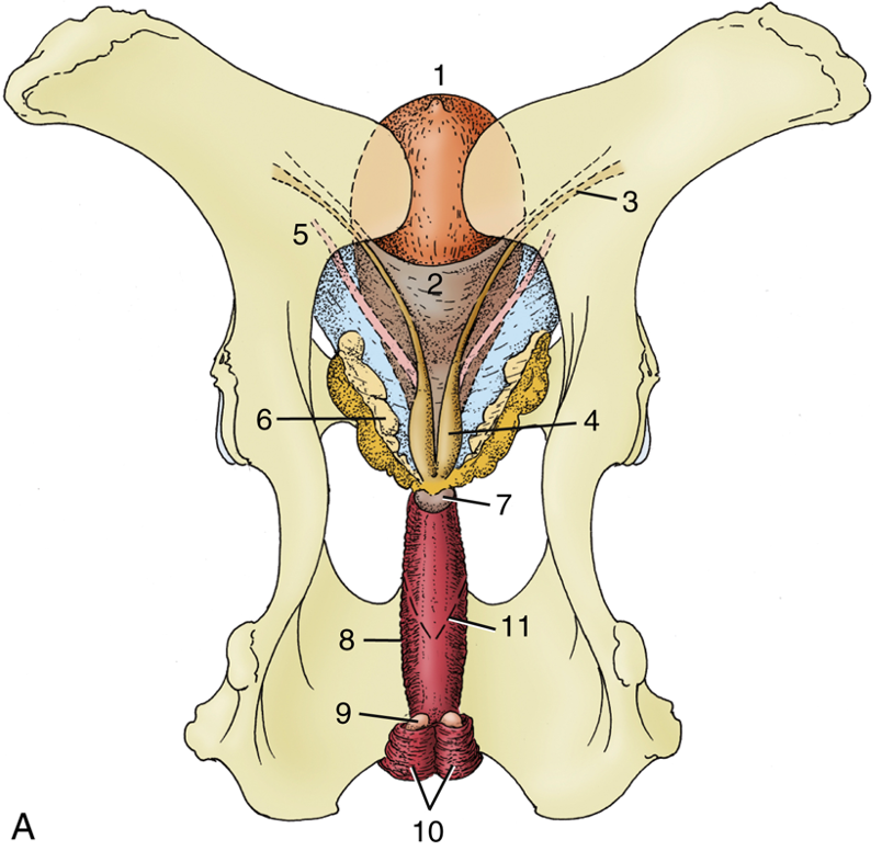
Figure 6-17. Dorsal view of the bull’s pelvis and related urogenital organs. 1, Urinary bladder; 2, genital fold; 3, right ductus deferens; 4, ampulla of deferent duct; 5, left ureter; 6, vesicular gland; 7, body of prostate; 8, urethralis muscle (surrounding urethra); 9, bulbourethral gland; 10, bulbospongiosus muscle; 11, caudal extent of the rectogenital pouch (broken line). (Duplication of TVA Fig. 29.29)
8. ALL MALE CALF specimens: Identify the prepuce and penis; open the prepuce to expose the penis and identify its parts. Identify the preputial orifice. Attempt to identify portions of the preputial mm. Identify the glans penis and the urethral process.
-
- Identify the prepuce and penis.
-
- The prepuce is the sheath/fold of skin covering the glans of the penis.
- If needed, open the prepuce with a ventral midline incision to expose the penis and identify its parts.
-
- Identify the preputial orifice.
-
- This is the external opening of the prepuce where hairy external skin meets the preputial lining.
-
- The mucous membrane of the prepuce is reflected onto the free part of the penis at the fornix. Portions of preputial muscle may be found associated with the sheath; attempt to identify portions of the preputial mm.
-
- Preputial muscles are derived from the cutaneous trunci m. (The cranial preputial muscle, (Fig. 6-18) causes a protrusion (prolapse) of the preputial lining, while the caudal preputial muscle is a retractor of the prepuce.)
-
- Identify the glans penis and the urethral process.
-
- Examine the distal end of the penis and note the small glans penis and the tubular urethral process, which is fused to the glans.
- Comparative Species Note: The urethral process of the ram (ovine) and billy (caprine) are elongated and free (see museum specimens in the demo area)
-
- Identify the prepuce and penis.
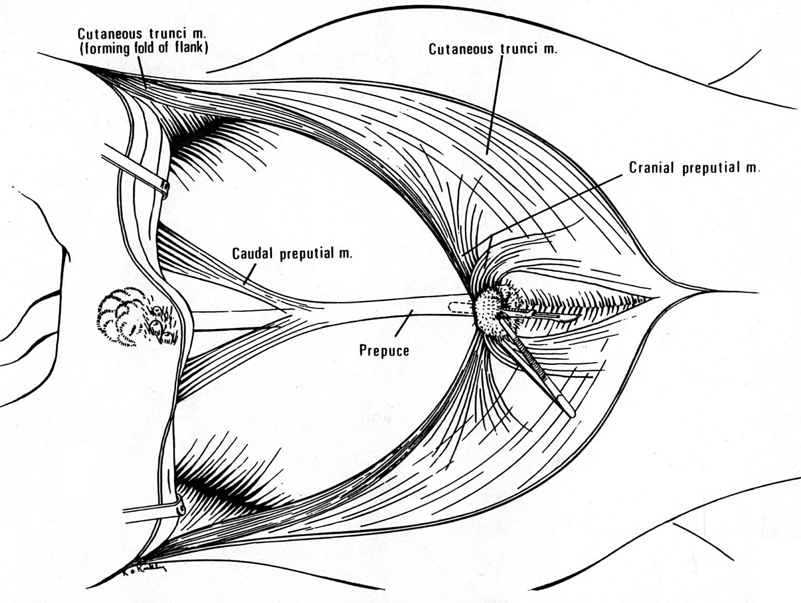
Figure 6-18. Bovine preputial muscles, ventral view.
9. ALL MALE CALF specimens: Identify the three regional parts of the penis: root, body & free part. Identify the sigmoid flexure; identify its proximal and distal loops. Identify the retractor penis muscles.
-
- Identify the root of the penis.
-
- The root is the proximal part of the penis with laterally positioned crura (left and right).
- Recall that the root of the penis is formed by the left and right crura (anchored to the ischial arch) and the bulb of the penis. Where the crura come together on the midline of the penis marks the end of the root and beginning of the body.
-
- Identify the body of the penis.
-
- The body begins at the region where the bilateral crura meet and then extends distally to the free part.
- In ruminants, the body of the penis is curved into the sigmoid flexure which is composed of a proximal loop and a distal loop; identify these structures in the calf and any supplemental bovine specimens available in the lab.
-
- Observe the smooth curvature of the proximal loop and the acute bend that forms the distal loop.
- Note that the retractor penis m. inserts beyond the distal loop, but is free from the proximal loop (Fig. 6-19).
-
-
- Identify the free part of the penis.
-
- The free part is the portion of the penis within the prepuce, including the distal glans penis (with corpus spongiosum glandis), which was previously identified.
-
- Identify the root of the penis.
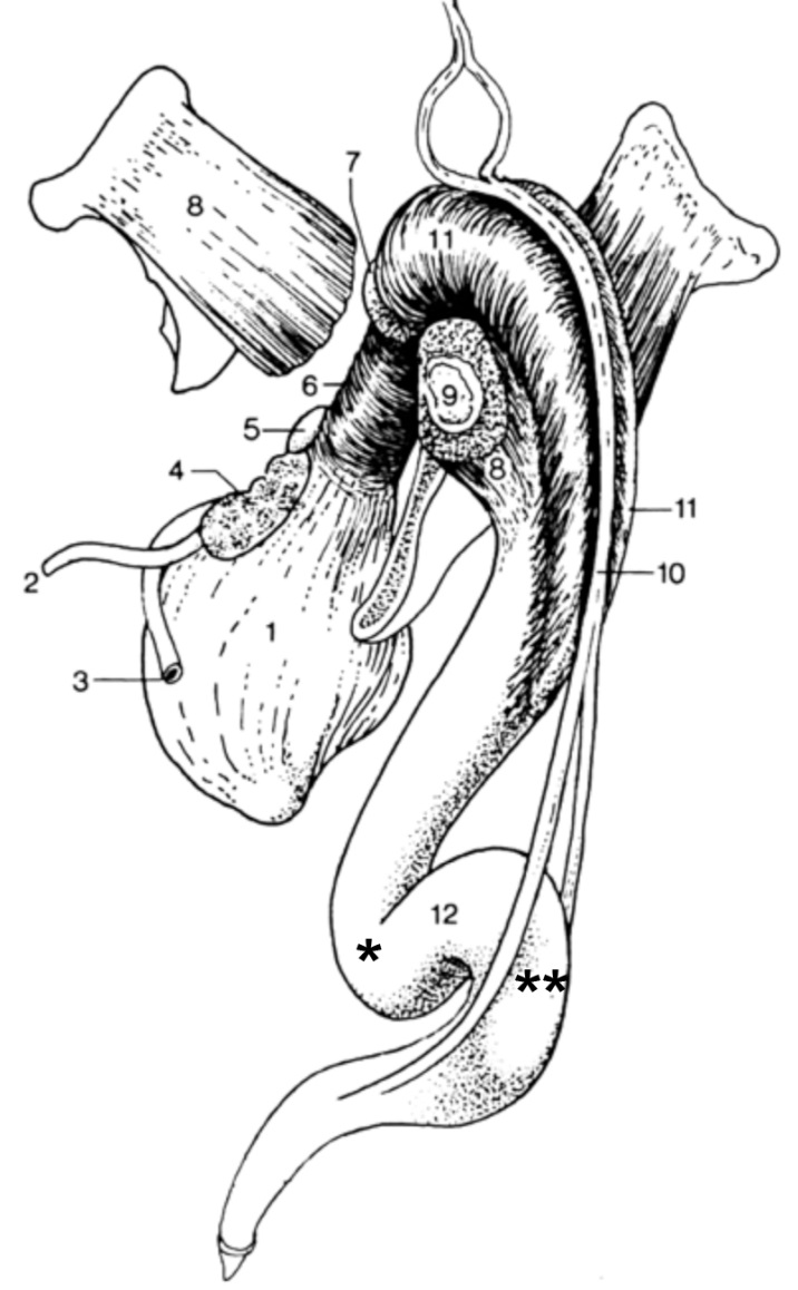
Figure 6-19. Bovine male genitalia, caudal lateral view; part of the left pelvis has been removed. 1, urinary bladder; 2, ureter; 3, ductus deferens; 4, vesicular gland; 5, prostate gland; 6, urethralis m.; 7, bulbourethral gland; 8, ischiocavernosus m., covering the crus of the penis (9); 10, retractor penis muscle; 11, bulbospongiosus m.; 12, proximal loop (*) and distal loop (**) of sigmoid flexure of penis. (Duplication of TVA Fig. 29-33)
10. ALL MALE CALF specimens: Identify the crura (left crus and right crus) of the penis. Identify the corpus cavernosum penis, ischiocavernosus m., and tunica albuginea. Identify the corpus spongiosum penis and the bulbospongiosus m. Identify the retractor penis m. as well as the dorsal n. and dorsal a. of the penis.
-
- Identify the crura of the penis (left crus and right crus) anchored to the ischial arch at the root. Make a transverse cut across one of the crura to identify the internal features.
-
- Each crus is composed of erectile tissue called corpus cavernosum penis, encased with a fibrous tunic called tunica albuginea. The crura serve to anchor each side of the penis to the ischial arch.
-
- The crura are covered by ischiocavernosus m.; identify this striated muscle covering each crus.
- Medial to the crura, in the region referred to as the bulb of the penis, the expanded erectile tissue called corpus spongiosum penis is found.
-
- Make an incomplete transection through the penis to identify the corpus spongiosum penis.
- This erectile tissue is also surrounded by muscle, the bulbospongiosus m., which you should identify. In the horse, this muscle extends most of the length of the penis.
-
- Identify the more superficial, paired, retractor penis muscles extending the length of the penis.
- Make another incomplete transverse section through the body of the penis.
-
- Observe/identify the tunica albuginea surrounding the corpus cavernosum penis.
- Observe/identify the corpus spongiosum penis partially surrounding the penile urethra. (Fig.6-15)
-
- Find and identify the dorsal nerve of the penis running along the dorsal midline of the penis. Note that this nerve is a continuation of the pudendal n.
- Also running along the dorsal midline of the penis, identify the dorsal a. of the penis.
- Identify the crura of the penis (left crus and right crus) anchored to the ischial arch at the root. Make a transverse cut across one of the crura to identify the internal features.
Important Note: You should be able to identify the parts of the bovine penis on the dried and/or plastinated specimens (from the museum collection) as well as the wet bovine isolated genitalia specimens available in lab. These supplemental specimens are fair game for lab practical assessments!
MALE GENITALIA – PORCINE
11. ALL MALE PIG specimens: Identify the prepuce, penis and testes. Identify the sigmoid flexure of the penis; identify its proximal and distal loops. (See Fig. 6-20)
-
- Comparative Note: While boar testes are similar to other species the tail of the epididymis is very large in boars. Examine the available specimens of boar testes and note the enlarged epididymis.
- Identify the sigmoid flexure of the penis.
-
- The sigmoid flexure of the boar penis usually lies at the level of the testes.
- Similar to the ruminant, the sigmoid flexure of the boar is composed of a proximal loop and a distal loop; identify these in the available specimen(s).
- Note that the end of the boar penis has a curvature which becomes corkscrew shaped when erected.
- Near the preputial orifice find the opening of the preputial diverticulum (aka ‘stink bag’) (Fig. 6-20). On the demonstration boar specimen, be sure to observe the interior of the preputial diverticulum.
-
12. ALL MALE PIG specimens: Identify the accessory sex glands (vesicular glands, prostate gland and bulbourethral glands) in the fixed boar specimen in lab.
-
- Note that the accessory sex glands are large in the boar but small in the barrow.
- Identify the vesicular glands (paired): The vesicular glands are very large in the boar.
- Identify the prostate gland (unpaired). The prostate may be hidden from view by the large vesicular glands.
- Identify the very large bulbourethral glands (paired).
-
- In the caudal part of the pelvic cavity identify the large bulbourethral glands which furnish the watery pre-sperm fraction that washes the urine out of the urethra before the sperm-rich fraction is passed.
-
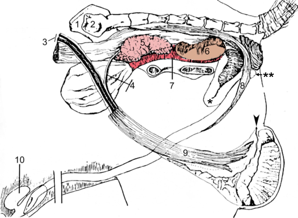
Figure 6-20.Genitalia of a boar (left limb and hemipelvis removed). 1, right wing of ilium; 2, left wing of sacrum; 3, testicular vessels; 4, ductus deferens and associated vessels; 5, vesicular gland (obscures view of prostate gland); 6, bulbourethral gland; 7, urethralis m. (skeletal muscle); 8, retractor penis m. (smooth muscle); 9, cremaster m. (skeletal muscle) of left testis; 10, preputial diverticulum (‘stink bag’), arrowhead, cut edge of vaginal tunic; proximal loop (*) and distal loop (**) of sigmoid flexure of penis.
Dissection Videos for this Section of Material
Genitalia
- Pony
- Male Pony:
- Internal Pelvis & Genitalia: https://youtu.be/iIfO7UkxxfA
- Male Pony:
- Calf
- Male Calf:
- Internal Pelvis & Genitalia: https://youtu.be/e6SDKnCRbgI
- Male Calf:
- Isolated specimens
- Male:
- Boar, Ram & Billy (Goat) Genitalia: https://youtu.be/ntEKF-aPB8w
- Male Repro Specimens: https://youtu.be/149mzQ-rVns
- Male:
