
Part 2: Proximal Pelvic Limb
Abby Brown
IMPORTANT NOTE: For this part of the chapter, the Guide directions will refer to either “ALL specimens” (meaning dissect pony and calf specimens in the same way) or to one species in particular, i.e., “ALL (PONY) specimens” or “ALL (CALF) specimens.” This added instructional note is needed because the dissection may differ slightly according to species.
CAUDAL THIGH MUSCLES (aka ‘hamstring’ muscles)
- ALL (PONY) specimens: Identify the biceps femoris m. on both left and right sides and define its borders by dissection; transect and reflect it distally.
- Identify the biceps femoris m. (in the pony specimen) on both right and left hind limbs. Define its borders as best you can. (Fig. 2-1)
- Transect the biceps femoris m. proximally, near its origin, and reflect it distally toward the stifle joint, but do not cut through its attachment to the fascia lata.
- ALL (CALF) specimens: Identify the gluteobiceps m. on both left and right sides; transect and reflect it distally.
- Identify the gluteobiceps m. (in the calf specimens) on both right and left hind limbs. (Fig. 2-2/4)
- In the bovine, the superficial gluteal muscle is fused to the biceps femoris m. to form the gluteobiceps m.
- Transect the gluteobiceps m. proximally, near its origin, and reflect it distally toward the stifle joint but do not cut through its attachment to the fascia lata.
- ALL specimens: Identify the semitendinosus and semimembranosus mm. on both left and right sides; transect and reflect them distally.
- Identify the semitendinosus m. caudal to the biceps femoris m./gluteobiceps m. on both left and right hind limbs. (Figs. 2-1, 2-2)
- Transect the semitendinosus m. proximally, near its origin, and reflect it distally, similar to your reflection of the biceps femoris m./gluteobiceps m. (Do this on both left and right sides.)
- Identify the semimembranosus m. medial to the semitendinosus m. on both left and right hind limbs, where it lies adjacent to the anus.
- In the pony, this muscle has two heads of origin; one from the caudal border of the sacrosciatic ligament, and the other from the tuber ischii. In the calf, this muscle originates only from the tuber ischii.
- Note that the semimembranosus m. will be VERY thick in the calf.
- Pony specimen: Transect the semimembranosus m. proximally, near its origin, and reflect it distally. Do this on both left and right sides.
- Calf specimen: Attempt to transect the entirety of the semimembranosus m. on the left side and reflect it distally. (Note that due to the large quantity of muscle mass, this muscle will not reflect very far distally.)
-
- At this time, you do not need to transect the semimembranosus m. on the right side of the calf.
-
- Identify the semitendinosus m. caudal to the biceps femoris m./gluteobiceps m. on both left and right hind limbs. (Figs. 2-1, 2-2)
- ALL specimens: Identify the tensor fasciae latae m. and the attached fascia lata; isolate the tensor fasciae latae m. Transect the caudal portion of the fascia lata and reflect the tensor fasciae latae muscle cranially.
-
- On the lateral side of the limb, identify the tensor fasciae latae m. (Figs. 2-1, 2-2/1) and the attached fascia lata on the cranial aspect of the thigh.
- Isolate the tensor fascia latae m. by dissection, separating the tensor fasciae latae m. and the proximal portion of the attached fascia lata from the underlying muscles.
- Transect the caudal portion of the fascia lata transversely, so that the tensor fasciae latae m. can be reflected cranially, yet will remain attached to the limb.
- Dissection Note: If necessary, the tensor fasciae latae m. could also be transected near its origin and reflected distally on the limb, but try to keep it attached to the limb (i.e., don’t cut all the way through the fascia lata!).
- On the lateral side of the limb, identify the tensor fasciae latae m. (Figs. 2-1, 2-2/1) and the attached fascia lata on the cranial aspect of the thigh.
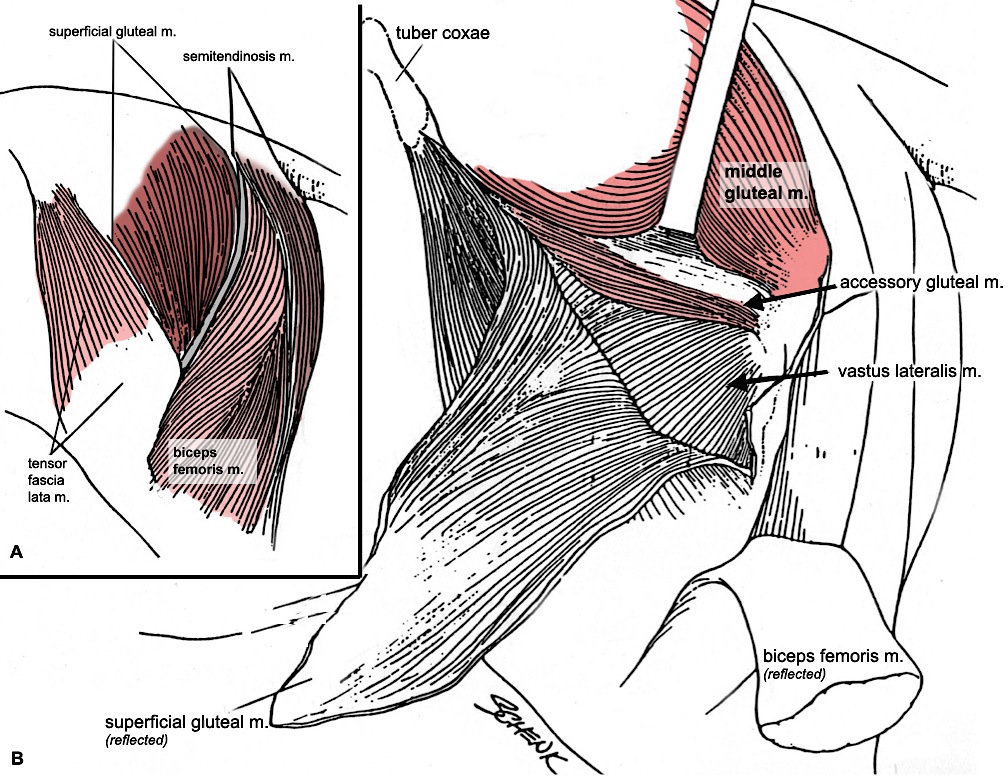
Figure 2-1. Equine hamstring and gluteal muscles, lateral views.
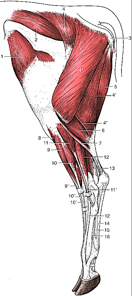 |
Figure 2-2. Bovine left hind limb, lateral view. 1, tensor fasciae latae m.2, middle gluteal m.3, ischial tuber4, 4’, 4’’ gluteobiceps m. (transected at 4’’)5, semitendinosus m.6, lateral head of gastrocnemius m.7, rudimentary soleus m.8, cranial tibial m.9, 9’, peroneus tertius m.10, 10’, 10’’, long digital extensor m.11, 11’, peroneus longus m.12, lateral digital extensor m.13, lateral digital flexor m.14, tendon of superficial digital flexor m.15, combined tendon of deep digital flexor mm.16, interosseous (suspensory ligament) (Modified from TVA Fig. 31-2) |
GLUTEAL MUSCLES
5. ALL (PONY) specimens: Identify the superficial gluteal m. on both left and right sides and then transect and reflect it.
-
- Identify and isolate the V-shaped superficial gluteal m. of the horse (Fig. 2-1) on both left and right sides.
- Notice that the origin of the superficial gluteal consists of two parts joined by an aponeurosis that covers the middle gluteal m.
- Transect the superficial gluteal m. near its origin (extending through the aponeurosis) and reflect it by carefully working between the cranial and caudal edges of the muscle and the underlying middle gluteal. Attempt to keep a portion of the aponeurosis that connects the two parts.
- Once you have separated the superficial gluteal m., reflect it to its insertion on the third trochanter. (Fig. 2-3/4).
- Identify and isolate the V-shaped superficial gluteal m. of the horse (Fig. 2-1) on both left and right sides.
6. Important Dissection Note: Recall that the bovine does not have a distinct superficial gluteal muscle because it is fused to the cranial edge of the biceps femoris m., thereby forming a gluteobiceps muscle (Fig. 2-2/4) (which you have already transected). Also, the bovine does not have a third trochanter. (You should compare the femurs of horse and ox and note the presence and absence of the third trochanter respectively.)
7. ALL specimens: Identify the middle gluteal m. on both left and right sides and then transect and reflect it.
-
- Identify the middle gluteal m. (Figs. 2-1 and 2-2) on both left and right sides.
- Pony specimen: Note that this is a massive muscle in the horse that largely covers the gluteal surface of the ilium and extends from the ilium, sacrum, and surface of the longissimus muscle, to the caudal part of the greater trochanter of the femur.
- Calf specimen: Note the relative underdevelopment of the middle gluteal muscle mass as compared to the pony. (Fig. 2-2/2)
- Elevate the craniolateral edge of the middle gluteal m. to visualize the accessory gluteal muscle underneath (Fig. 2-1, right).
- Work your hand under the middle gluteal to detach it from the underlying attachments, including the accessory gluteal m.
- Carefully transect the middle gluteal m. with an arching, transverse incision, near its origin, and reflect the main muscle mass caudally.
- Dissection Note: In the pony, there is a well-defined separation of the middle gluteal from the accessory gluteal muscle craniolaterally, but medially the middle and accessory gluteals are fused and separation is difficult.
8. ALL specimens: Identify the accessory gluteal m. on both left and right sides and then transect and reflect it; observe the trochanteric bursa in the pony.
-
- Appearing as a deep (craniolateral) portion of the middle gluteal m., identify the accessory gluteal m.
- This is a small muscle with a flat tendon that passes over the cranial part of the greater trochanter to its own separate attachment site (Fig. 2-3/3). The bone is protected by the trochanteric bursa (Fig. 2-4/TB).
- Detach the accessory gluteal m. from the underlying deep gluteal m. by working inward from the cranial border of the accessory gluteal to separate the two.
- Transect the accessory gluteal m. and reflect it distally toward its insertion; in the pony reflect it distal to the cranial part of the greater trochanter.
- Pony specimen: Identify the trochanteric bursa as the accessory gluteal m. is reflected. (Fig. 2-4/TB).
- Calf specimen: You will not be able to reflect the accessory gluteal m. far enough distally to see the trochanteric bursa due to the vastus lateralis (of the quadriceps femoris m.) that is covering the insertion point of the accessory gluteal on the greater trochanter. Hence, you do not need to identify the trochanteric bursa on the calf specimens.
- Appearing as a deep (craniolateral) portion of the middle gluteal m., identify the accessory gluteal m.
9. All specimens: Identify the deep gluteal m. on both left and right sides.
-
- Identify the deep gluteal m. (Fig. 2-4) on both left and right sides, after reflection of the middle and accessory gluteal mm.
- Note that this is a small, triangular shaped muscle that extends from the ischiatic spine to the cranial edge of the greater trochanter.
- Identify the deep gluteal m. (Fig. 2-4) on both left and right sides, after reflection of the middle and accessory gluteal mm.
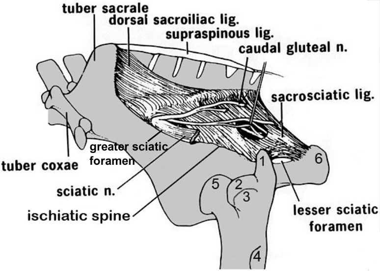
Figure 2-3. Equine pelvis and proximal femur, lateral view. 1, Caudal (middle gluteal) cusp of the greater trochanter; 2, cranial (deep gluteal) cusp of the greater trochanter; 3, facet for insertion of accessory gluteal; 4, third trochanter (superficial gluteal); 5, head of femur (in the acetabulum); 6, tuber ischii.
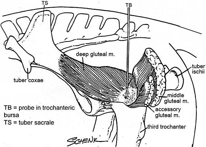
Figure 2-4. Equine gluteal region, deep dissection, lateral view.
SCIATIC NERVE and related Structures
10. ALL (PONY) specimens: Complete the following steps:
-
- Identify the large sciatic n. emerging from the greater sciatic foramen and passing caudally over the dorsal aspect of the hip joint. (Figs. 2-3 and 2-5)
- Dissection Note: The sciatic nerve is the largest nerve in the body.
- Caudal to the hip joint, find a large branch of the sciatic nerve innervating the hamstring (caudal thigh) muscles and then observe the continuation of the sciatic n. as it courses distally through the hind limb.
- Identify the stump of the cranial gluteal a. emerging alongside the sciatic nerve from the greater sciatic foramen. (Note that this artery will be cut due to the reflection of the gluteal muscles.)
- Caudal to the greater sciatic foramen, locate the lesser sciatic foramen and identify the internal obturator m. (tendon) emerging from it.
- Identify the large caudal gluteal a. that passes through the sacrosciatic ligament close to the caudal part of the sacrum.
- Dissection Note: The caudal gluteal artery is large in the horse due to the massive gluteal muscles.
- Be able to identify the greater and lesser sciatic foramina and the sacrosciatic ligament on the demonstration (museum) specimens as well.
- Identify the large sciatic n. emerging from the greater sciatic foramen and passing caudally over the dorsal aspect of the hip joint. (Figs. 2-3 and 2-5)
11. ALL (CALF) specimens: Complete the following steps:
-
- Identify the large sciatic n. emerging from the greater sciatic foramen and passing caudally over the dorsal aspect of the hip joint. (Fig. 2-5)
- Dissection Note: The sciatic nerve is the largest nerve in the body.
- Caudal to the hip joint, find a large branch of the sciatic nerve innervating the hamstring muscles and then observe the continuation of the sciatic n. as it courses distally through the hind limb.
- Identify the stump of the cranial gluteal a. emerging alongside the sciatic nerve from the greater sciatic foramen. (Note that this artery will be cut due to the reflection of the gluteal muscles.)
- Caudal to the greater sciatic foramen, locate the lesser sciatic foramen and identify the caudal gluteal a. emerging from it.
- Dissection Note: The lesser sciatic foramen will be located more dorsally in the calf than in the pony. In the calf, move toward the base of the tail to locate the lesser sciatic foramen and identify the caudal gluteal a. emerging from it.
- Note that in the calf, the caudal gluteal a. does not pass through the sacrosciatic ligament as it does in the pony.
- Dissection Note: Ruminants lack an internal obturator muscle (so you will not look for the tendon of this muscle as you did in the pony).
- Be able to identify the greater and lesser sciatic foramina and the sacrosciatic ligament on the demonstration (museum) specimens as well.
- Identify the large sciatic n. emerging from the greater sciatic foramen and passing caudally over the dorsal aspect of the hip joint. (Fig. 2-5)
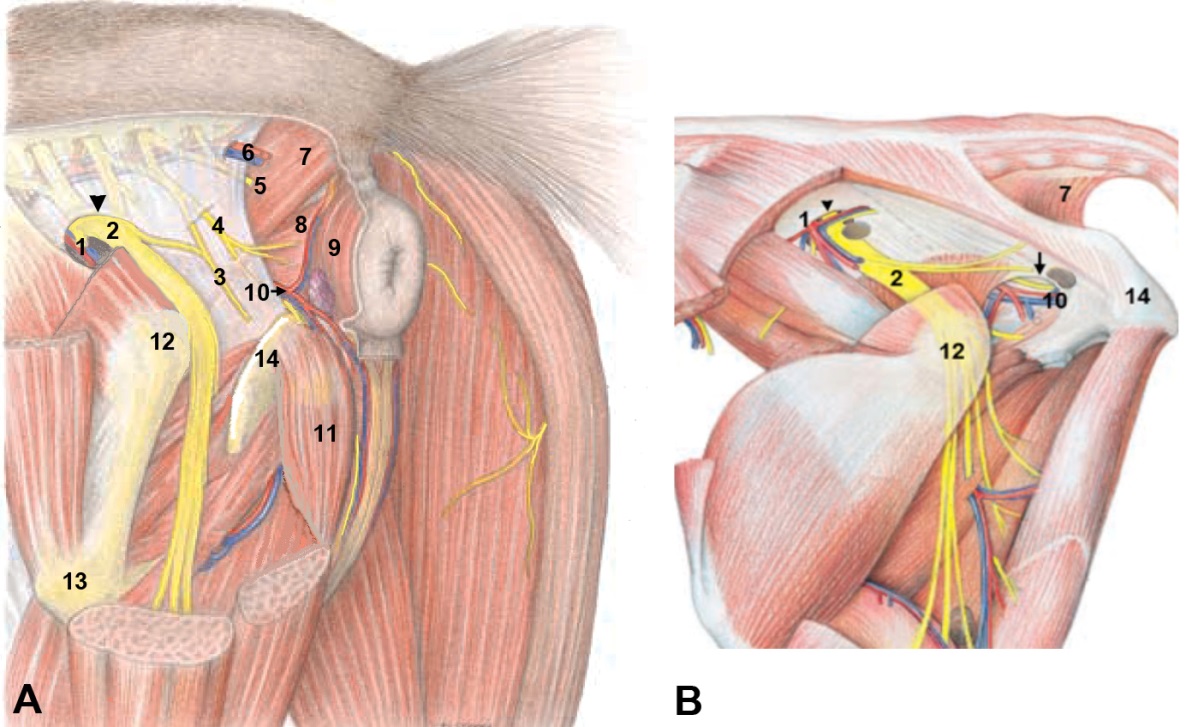
Figure 2-5. (Above) Deep muscles and associated vessels and nerves of the pelvic region (lateral view) in the (A) horse, male and (B) ox. 1, cranial gluteal a., v.; 2, sciatic n.; arrowhead, greater sciatic foramen; arrow, 3, caudal cutaneous femoral n.; 4, pudendal n.; 5, caudal rectal n.; 6, caudal gluteal a., v.; 7, coccygeus m.; 8, levator ani m.; 9, external anal sphincter m.; 10, internal pudendal a., v., and dorsal nerve of penis; 11, ischiocavernosus m.; 12, greater trochanter; 13, third trochanter. Modified from Budras.
REMOVAL OF LEFT HIND LIMB AT HIP JOINT
12. You will remove the LEFT hind limb of the hanging specimens by disarticulating it from the hip joint. (NOTE: Remove only the LEFT hind limb of the hanging specimens by completing transections of muscles as directed.) Note that this process will involve transection of numerous structures and muscles that have not yet been identified. Please proceed with the LEFT hind limb removal instructions as outlined below:
- On the left hind limb, on the dorsolateral side, transect all structures connecting the femur to the pelvis. These include:
- tensor fasciae latae m.
- deep gluteal m.
- internal obturator m. (tendon) (pony)
- sciatic nerve (transect where it emerges from the pelvis)
- Using the chain hoist, lower the specimen to the floor and place it in right lateral recumbency (right side down).
- Abduct (lift) the left hind limb while holding the right limb down on the floor. (Work as a team!)
- With your scalpel, on the medial side of the limb, incise the left hind limb musculature ~1 inch from the ventral midline.
- In males, be careful to avoid damage to the penis; push it over to the right side so it remains with the body and right hind limb.
- In all specimens, be careful not to cut into the abdominal cavity! (Due to the large abdominal cavity in ungulates, there is a more extensive medial attachment to the hind limb.)
- As you cut, elevate (abduct) the left limb further and further away from the body as you progress, to make cutting easier.
- Note that you will cut through numerous structures: skin, gracilis m., adductor m., pectineus m., sartorius m., external obturator m., rectus femoris m., iliopsoas m., and caudal hip muscles.
- Abduct the left limb even further and incise the hip joint capsule to observe the head of the femur.
- All (pony) specimens: Locate the accessory ligament of the femoral head (Fig. 2-7). (Recall only the horse has this ligament.) Identify and transect it 1cm distal to the rim of the acetabulum.
- Abduct the left limb further to increase the rotation of the femoral head and make the ligament of the femoral head visible. Identify and then transect it.
- Keep transected/reflected muscles with the limb.
- Complete the removal of the left hind limb by transecting remaining femoral and gluteal vessels and nerves so that a stump can be found on both parts for later identification.
- This will complete the removal of the left hind limb.
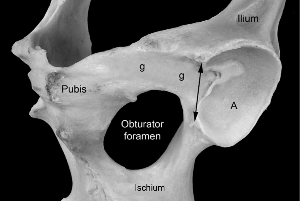
Figure 2-6. Equine left acetabulum, ventral lateral view. A, Articular surface of the acetabulum; g, shallow groove for the accessory ligament of the femoral head; double headed arrow, acetabular notch and the location of the transverse acetabular ligament.
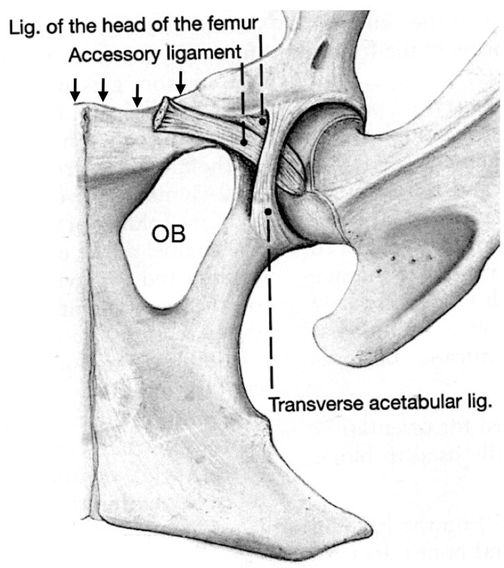
Figure 2-7. Equine left hip joint, ventral view. OB, Obturator foramen; arrows, prepubic tendon attachment points. Note: Accessory ligament is only present in the horse.
IMPORTANT NOTE: The Guide instructions for this section will refer to either “ALL hind limbs” (meaning attached and detached) or to the “DETACHED” or “ATTACHED” hind limbs.” This added instructional note is needed because the dissection of of some regions may be different for attached vs. detached limbs.
Skin reflection and cranial thigh muscles
13. ALL hind limbs: Make a skin incision on the medial side of each hind limb, down to the level of the stifle, and reflect the skin from the medial side of the proximal hind limb.
-
- On the medial side of each hind limb, make a skin incision from the hip region down to the level of the stifle.
- In males, be careful to leave the prepuce intact.
- Make a circular incision around the leg at the level of the stifle.
- Reflect the remaining skin off the proximal hind limb and discard it.
- On the medial side of each hind limb, make a skin incision from the hip region down to the level of the stifle.
14. ALL hind limbs: If needed, transect the tensor fascia latae m. and reflect it to identify the quadriceps femoris m. in the next step.
-
- DETACHED hind limbs: If the tensor fascia latae m. is still attached to the limb, reflect it distally, but do not remove it.
- ATTACHED hind limbs: If needed, transect the fascia lata just distal to the tensor fascia latae muscle. Reflect the caudal portion of the tensor fascia latae m. cranially.
15. ALL hind limbs: After reflection of the tensor fasciae latae m., identify the four heads of the quadriceps femoris m.: rectus femoris, vastus lateralis, vastus intermedius, and vastus medialis
-
- In both the pony and calf, the heads of the quadriceps femoris m. are like those of the dog, having 4 heads of origin. Identify the rectus femoris, vastus lateralis, vastus intermedius, and vastus medialis.
- Recall the ‘hot dog in a bun’ analogy from Anatomy I: The rectus femoris is the ‘hot dog’ inside the ‘bun’ made up of the three vastus heads. Vastus lateralis is the lateral side of the ‘bun’, vastus medialis is the medial side of the ‘bun’, and vastus intermedius is the bottom of the ‘bun’.
- Dissection Note: If necessary, to view the vastus intermedius transect the rectus femoris in the approximate middle of the muscle belly and reflect it.
- Calf specimen: In the calf, note that the vastus lateralis substantially overlaps the rectus femoris on the craniolateral aspect. Also note that the rectus femoris m. is large, while the vastus medialis is quite small.
- In both the pony and calf, the heads of the quadriceps femoris m. are like those of the dog, having 4 heads of origin. Identify the rectus femoris, vastus lateralis, vastus intermedius, and vastus medialis.
16. ATTACHED hind limbs: Confirm by dissection the origin of rectus femoris m. from the pelvis.
-
- On the cranial aspect of the attached hind limb, transect the deep gluteal muscle and confirm by dissection that the rectus femoris m. originates from the pelvis, cranial to the acetabulum.
- Note that all of the ‘vastus’ heads of the quadriceps m. originate from the femur.
Dissection of Medial thigh
17. ALL hind limbs: Identify the gracilis m. If needed, transect the gracilis m. proximally and then reflect it distally. Identify the adductor m. deep to the gracilis m.
-
- On the medial side of the limb, identify the broad, flat gracilis m. (Fig. 2-8/1).
- ATTACHED hind limbs: Transect the gracilis m. proximally (as close to the origin as possible).
- Reflect the gracilis m. distally on the limb but do not remove it.
- After reflection of the gracilis m. identify the underlying adductor m. (Fig. 2-8/2)
- Dissection Note: The adductor m. is relatively small in ungulates.
- Calf specimen: The adductor m. may be difficult to distinguish from the semimembranosus m.
18. ATTACHED hind limbs: Re-identify the semimembranosus m. Identify the sartorius m.; transect and reflect it. Attempt to identify the pectineus m. Identify the region of the femoral triangle and identify the femoral artery and femoral vein within that space.
-
- On the medial aspect, re-identify and isolate the semimembranosus m. caudally.
- Also on the medial side, identify the sartorius m. cranially and attempt to identify the pectineus m. adjacent to the adductor. (Fig. 2-8)
- Note that only a small portion of the pectineus may be seen when the specimen is in the hanging position.
- Identify the region of the femoral triangle formed by the pectineus and sartorius mm. and attempt to identify the femoral artery (Fig. 2-8/11) and femoral vein lying deep within that space.
- Pony specimen: Note that you may see some superficially located deep inguinal lymph nodes in this dissection field, but you do not need to specifically identify/save them.
- Dissection Note: Abducting (lifting) the limb laterally (away from the body) may aid you in viewing the pectineus and femoral triangle region, so be sure to utilize your team members effectively to complete this dissection. However, note that these structures may be difficult to see in the hanging specimens.
19. DETACHED hind limbs: Re-identify the semimembranosus m. Identify the sartorius m.; reflect it distally. Identify the pectineus m. Identify the region of the femoral triangle and identify the femoral artery and femoral vein within that space.
-
- On the medial aspect of the limb, re-identify and isolate the semimembranosus m. caudally.
- Also on the medial side, identify the sartorius m. cranially; reflect it distally.
- Identify and isolate the pectineus m. adjacent to the adductor m. (Fig. 2-8).
- Important Dissection Note: Keep in mind, as you complete this dissection, that the semimembranosus, sartorius, and pectineus muscles have all been transected during the removal of the limb from the body, so you will identify the stumps still associated with the limb.
- Identify the region of the femoral triangle formed by the pectineus and sartorius mm. Then, identify the femoral artery (Fig. 2-8/11) and femoral vein lying deep within that space.
- Pony specimen: Note that you may see some superficially located deep inguinal lymph nodes in this dissection field, but you do not need to specifically identify/save them.
20. ATTACHED hind limbs: Identify the iliopsoas mm.
-
- From the lateral side, identify the iliopsoas mm. (deep to the tensor fasciae latae m.)
- Note: You may notice that the iliopsoas has several parts, but it is not necessary to differentiate them.
- From the lateral side, identify the iliopsoas mm. (deep to the tensor fasciae latae m.)
21. DETACHED hind limbs: Identify the iliopsoas mm.
-
- On the medial side of the limb, locate the iliopsoas mm. cranial to the pectineus m. (Fig. 2-8/8) and if possible, trace it to its insertion on the lesser trochanter of the femur.
- Dissection Note: When removing the left hind limb, the iliopsoas mm. were transected, so you will identify the stump of this muscle group still associated with the limb. You should also identify the rest of the iliopsoas mm. remaining on the body for orientation; this part of the iliopsoas would be found laterally, on the left side of the body. You may notice that the iliopsoas has several parts, but it is not necessary to differentiate them.
22. ALL hind limbs: Be familiar with the location of, and be able to identify, the accessory ligament of the femoral head (eq) and the prepubic tendon. (Note that this includes identification of these structures on the dried equine specimen available in the demo area.)
-
- DEATCHED (PONY) hind limbs: You have already found, and transected, the accessory ligament of the femoral head extending from the hip joint to the prepubic tendon (Fig. 2-7), however you should attempt to identify these structures on the detached limb and/or on the body of the hanging cadaver.
- DETACHED (CALF) hind limbs: You should attempt to identify the prepubic tendon on the hanging cadaver.
- ALL hind limbs: As noted above, you should observe and be able to identify the accessory ligament of the femoral head (eq)and the prepubic tendon on the available dried equine specimen(s) available in the lab.
- Calf specimen: Note that the ruminant does have the prepubic tendon, but there is no dried specimen available to illustrate this structure in the bovine animal. However, recall that the ruminant lacks the accessory ligament of the femoral head.
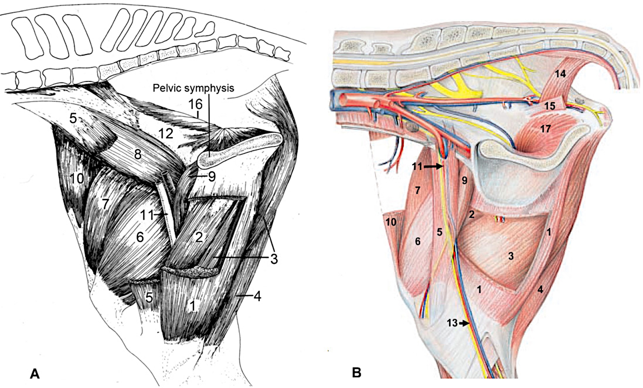
Figure 2-8. Thigh region of the horse (A) and ox (B), medial view. 1, gracilis m. (partially removed to reveal 2,3); 2, adductor m.; 3, semimembranosus m.; 4, semitendinosus m. (positioned lateral to 3); 5, sartorius m. (in the horse image most has been removed to reveal the more lateral muscles: 6,7,8); 6, vastus medialis m.; 7, rectus femoris m.; 8, iliopsoas m.; 9, pectineus m.; 10, tensor fasciae latae m.; 11, femoral artery; 12, iliac shaft, 13, medial saphenous a., v., n. (shown in ox); 14, coccygeus m.; 15, levator ani m. (shown in ox); 16, internal obturator m. (equine only, absent in ox) 17, external obturator m. (intrapelvic part in ox in place of an internal obturator m.).
Dissection Videos for this Section of Material
Proximal Pelvic Limb
- Pony:
- Proximal Pelvic Limb, watch from 1:08-end: https://youtu.be/65hVe2ekzqI
- Proximal Pelvic Limb – Attached, watch from 0:00-6:38: https://youtu.be/B26J-aCqhiU
- Proximal Pelvic Limb – Detached, watch from 0:00-5:30: https://youtu.be/4z1ezvuzkzk
- Calf:
- Proximal Pelvic Limb, watch from 1:17-end: https://youtu.be/e44X-gYwyx4
- Proximal Pelvic Limb – Attached, watch from 0:00-6:19: https://youtu.be/rWwfvDhVWmI
- Proximal Pelvic Limb – Detached, watch from 0:00-5:08: https://youtu.be/lnKg–Y6dxM
