
Part 6: Pelvic Vessels and Nerves
Abby Brown
IMPORTANT NOTE: For this part of the chapter, the Guide directions will refer to either “ALL specimens” (meaning dissect pony and calf specimens in the same way) or to one species in particular, i.e., “ALL (PONY) specimens” or “ALL (CALF) specimens.” This added instructional note is needed because the dissection may differ slightly according to species. The dissection will also vary based on the sex of the animal.
PELVIC VESSELS
- ALL specimens: Trace the descending abdominal aorta and attempt to find where it gives rise to the right and left testicular aa. (male) or ovarian aa. (female). These may be difficult to identify, depending on the specimen cuts/preparation.
- Dissection Note for ALL FEMALE specimens: The satellite vein of the uterine a. is very small, so the main drainage vessel of the uterus is the ovarian vein. (TVA, 570, mare); identify the ovarian v. after you identify the ovarian a. (The ovarian vein will satellite the ovarian artery.)
- ALL specimens: Trace the terminal branches of the descending abdominal aorta. The terminal branches of the aorta we will identify include the deep circumflex iliac aa. (left and right), external iliac aa. (left and right), and internal iliac aa. (left and right). Use blunt dissection to trace these arteries (and their branches) as directed. As always, there can be variation in the patterns described. Those described are provided as the most common arterial branching patterns.
- Note that the calf (ruminants) will also have a median sacral artery as the distal continuation/final branch of the aorta, but this artery is absent in the pony (equine). You do not need to dissect/identify this artery in your calf specimens.
- Dissection note: Near the termination of the abdominal aorta and the iliac aa., there may be several lymph nodes (e.g., external iliac/deep inguinal, sacral) but you do not need to identify them in your specimens.
- ALL specimens: Identify the deep circumflex iliac aa. (left and right) arising from the terminal portion of the aorta, just cranial to the origin of the external iliac arteries (most often seen in equines) OR from the external iliac arteries near their origin from the aorta (most often seen in ruminants).
- ALL specimens: Identify the origins of the external iliac arteries (left and right) as they are given off of the aorta. These arteries run caudoventrally to supply the pelvis and pelvic limbs.
- Each external iliac a. will then leave the abdomen through the vascular lacuna and become the femoral artery supplying each hind limb, which was previously dissected in Chapter 2.
- Trace the external iliac a. caudoventrally and identify the deep femoral artery branching from it before it exits the abdomen through the vascular lacuna.
-
- Dissection Note: Note that the deep femoral a. branches from the external iliac INSIDE the abdomen, before the external iliac leaves the abdomen through the vascular lacuna to become the femoral a.
-
- ALL FEMALE (PONY) Specimens: As you trace the external iliac a., find and identify the uterine a. arising from the external iliac a. just cranial to the origin of the deep femoral a. This is the primary blood supply to the uterus. Attempt to trace this artery to the uterus.
-
- In the male horse, this would be the cremasteric a. instead of uterine.
- Note that in the pony, the uterine artery arises from the external iliac a. instead of as a branch associated with the internal pudendal a. (as in other species.)
-
- ALL specimens: Trace the deep femoral a. caudally and identify the short pudendoepigastric trunk branching from it.
-
Two branches are given off of the pudendoepigastric trunk, the external pudendal a. and the caudal epigastric a.
-
-
Identify the external pudendal a. leaving the pudendoepigastric trunk and passing through the inguinal canal. Note that this artery forms an important supply to the penis of the stallion and is the major arterial supply of the mammary gland.
-
-
Trace the external pudendal a. into the deep inguinal ring; it will then pass through the inguinal canal and out through the superficial inguinal ring.
-
-
- Identify the caudal epigastric a. leaving the pudendoepigastric trunk and coursing cranially on the deep surface of the rectus abdominis m.
-
-
-
-
ALL specimens: On the RIGHT side of your specimen, trace the external iliac a. distally (into the hind limb) where, as it passes through the vascular lacuna, it changes names to become the femoral a. Identify the femoral a. as it courses distally in the (proximal) hind limb.
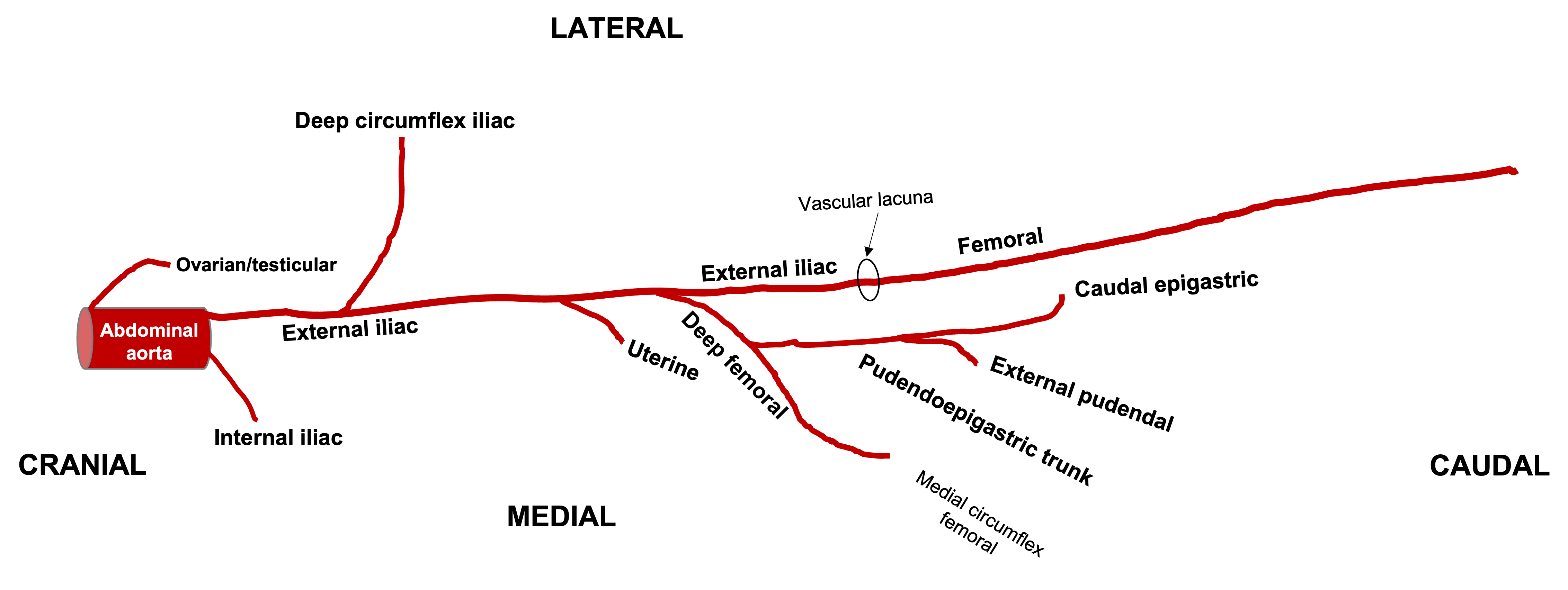
Figure 6-21. Ventral view onto the left external iliac artery and branches in the equine. Labeled branches: abdominal aorta, ovarian/testicular, internal iliac, external iliac, deep circumflex iliac, uterine (if female), deep femoral, pudendoepigastric trunk, external pudendal, caudal epigastric, femoral. All arteries are bilateral except for the aorta. (RLarsen)

Figure 6-22. Ventral view onto the left external iliac artery and branches in the bovine. Labeled branches: abdominal aorta, ovarian/testicular, internal iliac, external iliac, deep circumflex iliac, deep femoral, pudendoepigastric trunk, external pudendal, caudal epigastric, femoral. All arteries are bilateral, except for median sacral and the aorta. (RLarsen)
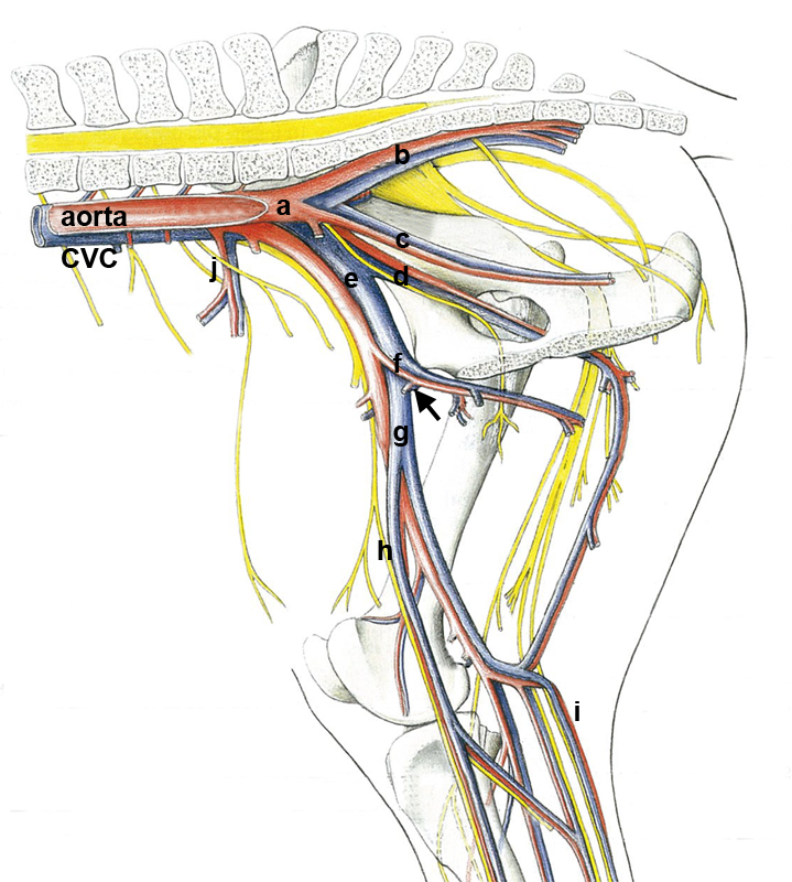
Figure 6-23. Medial vessels of the right internal pelvic region of the horse. a, internal iliac vessels; b, caudal gluteal vessels; c, internal pudendal vessels; d, obturator vessels and nerve; e, external iliac vessels; f, deep femoral vessels; arrow, pudendoepigastric trunk; g, femoral vessels; h, saphenous artery and nerve and medial saphenous vein; i, lateral saphenous vein and caudal cutaneous sural nerve (tibial branch); j, deep circumflex a./v. (Modified from p. 21 K. Budras, et.al., Anatomy of the Horse, (5thed.) 2005)
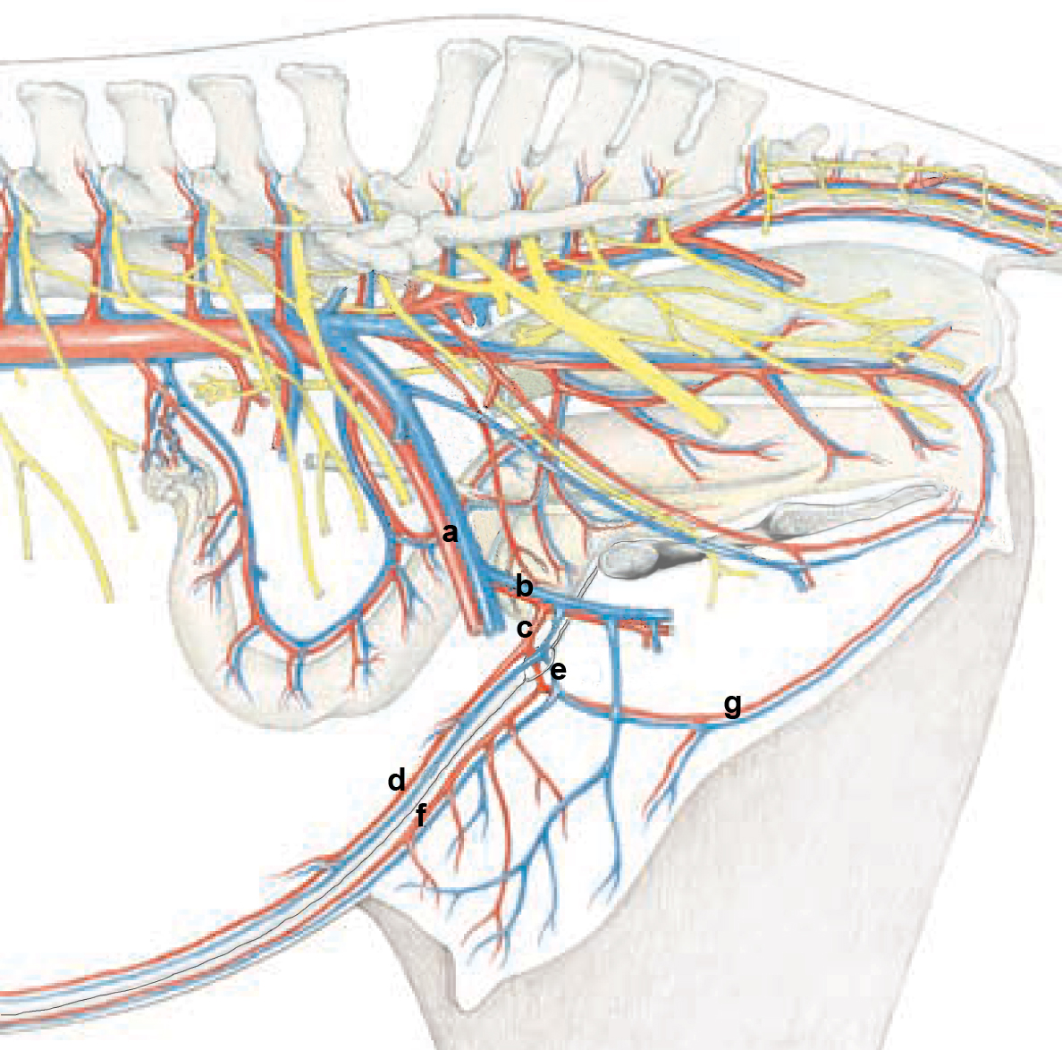
Figure 6-24. Medial vessels of the right internal pelvic region of the mare. a, external iliac a./v.; b, deep femoral a./v.; c, pudendoepigastric a./v.; d, caudal epigastric a./v.; e, external pudendal a./v. exiting through the superficial inguinal ring; f, caudal superficial a./v.; g, ventral labial a./v.(Modified from p. 21 K. Budras, et.al., Anatomy of the Horse, (5thed.) 2005)
INTERNAL PELVIC VESSELS
7. ALL specimens: Return to the terminal aorta and identify the internal iliac aa. (left and right) and carefully trace them to identify their main branches. With few exceptions, pelvic arteries are derived from the internal iliac arteries. Each internal iliac a. will give off an umbilical artery and then terminate as the internal pudendal artery and caudal gluteal a. As always, there can be variation in the patterns described. Those described are provided as the most common arterial branching patterns.
-
- Summary of branching in the PONY, the internal iliac a. gives rise to two main branches the “wall branch” and the “visceral branch” (caudal gluteal and internal pudendal aa., respectively) at a relatively proximal point in the pelvic cavity.
-
- The caudal gluteal a. gives rise to the cranial gluteal a. The cranial gluteal usually gives rise to the obturator a., which is only in the HORSE. It follows the obturator n. through the obturator foramen and is an important vascular supply to the equine penis.
- The internal pudendal a. usually gives rise to the umbilical a. (going towards the bladder and ventral body wall) first, then the urogenital (prostatic/vaginal), and terminates as the dorsal a. of the penis/clitoris.
-
- Summary of branching in the CALF, there is a relatively long internal iliac, which gives rise to the umbilical a. first (which may also give rise to the uterine a.), then the cranial gluteal a., then lastly the urogenital a. (prostatic/vaginal) before splitting into the caudal gluteal and internal pudendal aa. In BOVINE, the split of the caudal gluteal and internal pudendal aa. occurs more distally than in EQUINE, and is near the lesser sciatic foramen. The internal pudendal terminates as the dorsal a. of the penis/clitoris.
- Summary of branching in the PONY, the internal iliac a. gives rise to two main branches the “wall branch” and the “visceral branch” (caudal gluteal and internal pudendal aa., respectively) at a relatively proximal point in the pelvic cavity.
8. ALL (CALF) Specimens: Trace the internal iliac a. and identify the umbilical a. running to the apex of the urinary bladder. This artery is the first branch of the internal iliac; it arises near the origin of the internal iliac a. from the aorta.
-
- Remove peritoneum and pelvic fascia as needed to identify/trace the umbilical a.
- Comment: In the fetus, the umbilical a. carries deoxygenated blood to the placenta for oxygenation. In the adult, the umbilical a. ends as the round ligament of the bladder (located in the lateral ligament of the bladder).
- In the calf specimens, the umbilical a. may look abnormally thickened due to their young age.
- ALL FEMALE (CALF) Specimens: As you trace the umbilical a., find and identify the uterine a. arising from it. This is the primary blood supply to the uterus. Attempt to trace the uterine a. to the uterus.
-
- Note that the uterine artery is sometimes referred to as the ‘middle uterine artery’ in ruminants due to the presence of other uterine branches supplying the uterus from other parent arteries. (There is a uterine branch of the ovarian a. and a uterine branch from the vaginal artery as well.)
- In the male bovine, this would be the artery of the ductus deferens instead of the uterine artery.
-
9. ALL specimens: Trace the internal iliac artery to where it divides into two branches; a parietal branch, the caudal gluteal artery, and a visceral branch, the internal pudendal a.; identify these branches as described. As you proceed with the dissection, try to identify the entire vessel (if possible) on the right side of the internal pelvis and then attempt to identify as much of the vessel as possible on the left side of the internal pelvis as well.
-
- Important Dissection Notes: The internal iliac a. is called the internal iliac up until it splits into the caudal gluteal and internal pudendal arteries. There are species differences as to when this split occurs; for example, in cattle the split occurs further distally/caudally than in the horse, near the lesser sciatic foramen (i.e., there is a long internal iliac artery which gives off the urogenital branch, the cranial gluteal branch, etc. before terminating as the caudal gluteal and internal pudendal arteries). In horses, the split occurs more cranially than in the ruminant (i.e., there is a short internal iliac artery which branches almost immediately into the caudal gluteal and internal pudendal arteries).
- ALL (PONY) specimens: In your pony dissections, note the large size and proximal location of the caudal gluteal a. branch which relates to the substantial rump muscle mass and the more proximal origin of the hamstring muscles.
- Summary of species differences in branching pattern of internal iliac arteries:
-
- Horse and Dog: proximal parietal/ visceral branching pattern, i.e., there is a short internal iliac a. before branching into caudal gluteal and internal pudendal aa.
- Cattle: distal parietal/visceral branching, i.e., there is a long internal iliac a. with terminal branching into caudal gluteal and internal pudendal aa. at the level of the lesser sciatic foramen.
-
10. ALL (PONY) Specimens: Re-trace the internal iliac a. and identify the internal pudendal a. and caudal gluteal a. branches. Branching from the caudal gluteal artery, identify the cranial gluteal a. and the obturator a.
-
- Trace the caudal gluteal a. and identify the cranial gluteal a. branching from it.
-
- Note that there are several branches arising from the caudal gluteal a., but the largest branch is the cranial gluteal a.
- Recall that the caudal gluteal artery passes laterally through the sacrosciatic ligament to supply muscles of the tail (as seen/dissected in Chapter 2).
-
- Trace the cranial gluteal a. and identify the obturator a. (pony only) branching from it before the cranial gluteal artery courses laterally through the greater sciatic foramen to supply the gluteal muscles (as seen/dissected in Chapter 2).
-
- Trace the obturator a. to the obturator foramen. The obturator a. courses through the obturator foramen alongside the obturator n.
- Note that the obturator artery is unique to the horse and is an important vascular supply to the equine penis.
-
- Trace the caudal gluteal a. and identify the cranial gluteal a. branching from it.
11. ALL (CALF) Specimens: Re-trace the internal iliac a. and identify the cranial gluteal a. and urogenital (prostatic or vaginal) a. branching from it. Continue to trace the internal iliac a. to identify the internal pudendal a. and caudal gluteal a. branches.
-
- Identify the cranial gluteal a. branching from the internal iliac a. This branching typically occurs after the umbilical a. but before the urogenital a. (which will be discussed in the following steps).
-
- Trace the cranial gluteal a. to see where it courses laterally through the greater sciatic foramen to supply the gluteal muscles (as seen/dissected in Chapter 2).
-
- The next artery arising from the internal iliac a. is the vaginal or prostatic artery, dependent on sex of the animal. Identify the urogenital (vaginal a. or prostatic a., based on sex) a. branching from the internal iliac a.
-
- In male bovine (bulls, steers), the prostatic artery supplies blood to the prostate, ductus deferens and urethra.
- In female bovine (cows), the vaginal artery supplies blood to the uterus, vagina, rectum and perineum.
-
- Continue tracing the internal iliac a. to identify the caudal gluteal a. and the internal pudendal a. branches.
-
- Recall that the caudal gluteal a. will pass through the lesser sciatic foramen in the calf (as seen/dissected previously in Chapter 2) to supply the ventral and lateral muscles of the tail.
- The internal pudendal a. will supply the rectum and perineum; after giving off branches to those regions, the internal pudendal a. is continued as either the artery of the penis in the male or artery of the clitoris in the female.
-
- Identify the cranial gluteal a. branching from the internal iliac a. This branching typically occurs after the umbilical a. but before the urogenital a. (which will be discussed in the following steps).
12. ALL (PONY) Specimens: Trace the internal pudendal a. as it courses toward the pelvic viscera and gives off the umbilical a. and urogenital a. (prostatic a. or vaginal a.).
-
- Trace the internal pudendal a. and identify the umbilical a. running to the apex of the urinary bladder. This artery is the first branch of the internal pudendal.
- The next artery arising from the internal pudendal a. is the vaginal or prostatic artery, dependent on sex of the animal. Identify the urogenital a. (vaginal a. in female or prostatic a. in male) which supplies blood to many pelvic and reproductive organs.
-
- In male horses (stallions, geldings), the prostatic artery supplies blood to the prostate, urethra, rectum, urinary bladder, ureter, ductus deferens and accessory sex glands.
- In female horses (mares), the vaginal artery supplies blood to the uterus, vagina, rectum, urinary bladder, perineum and clitoris.
-
- The internal pudendal a. will then continue on to supply the perineum; after giving off the ventral perineal artery, the internal pudendal a. is continued as either the artery of the penis in the male or artery of the clitoris in the female.
13. ALL MALE (CALF) Specimens: Identify the dorsal artery of the penis (the final terminating branch of the artery of the penis). Re-identify the dorsal nerve of the penis.
-
- The dorsal artery of the penis supplies the glans of the penis and the prepuce and was previously discussed with the male genitalia section of this chapter.
- Re-identify the dorsal nerve of the penis running along the dorsal midline of the penis (next to the dorsal artery of the penis), as was previously discussed with the male genitalia section of this chapter.. Note that this nerve is a continuation of the pudendal n.
14. ALL MALE (PONY) Specimens: Identify the dorsal artery of the penis (the final terminating branch of the artery of the penis). Re-identify the dorsal nerve of the penis.
-
- The dorsal artery of the penis anastomoses with the middle and cranial arteries of the penis to supply the glans of the penis and the prepuce and was previously discussed with the male genitalia section of this chapter.
- Re-identify the dorsal nerve of the penis running along the dorsal midline of the penis (next to the dorsal artery of the penis), as was previously discussed with the male genitalia section of this chapter.. Note that this nerve is a continuation of the pudendal n.
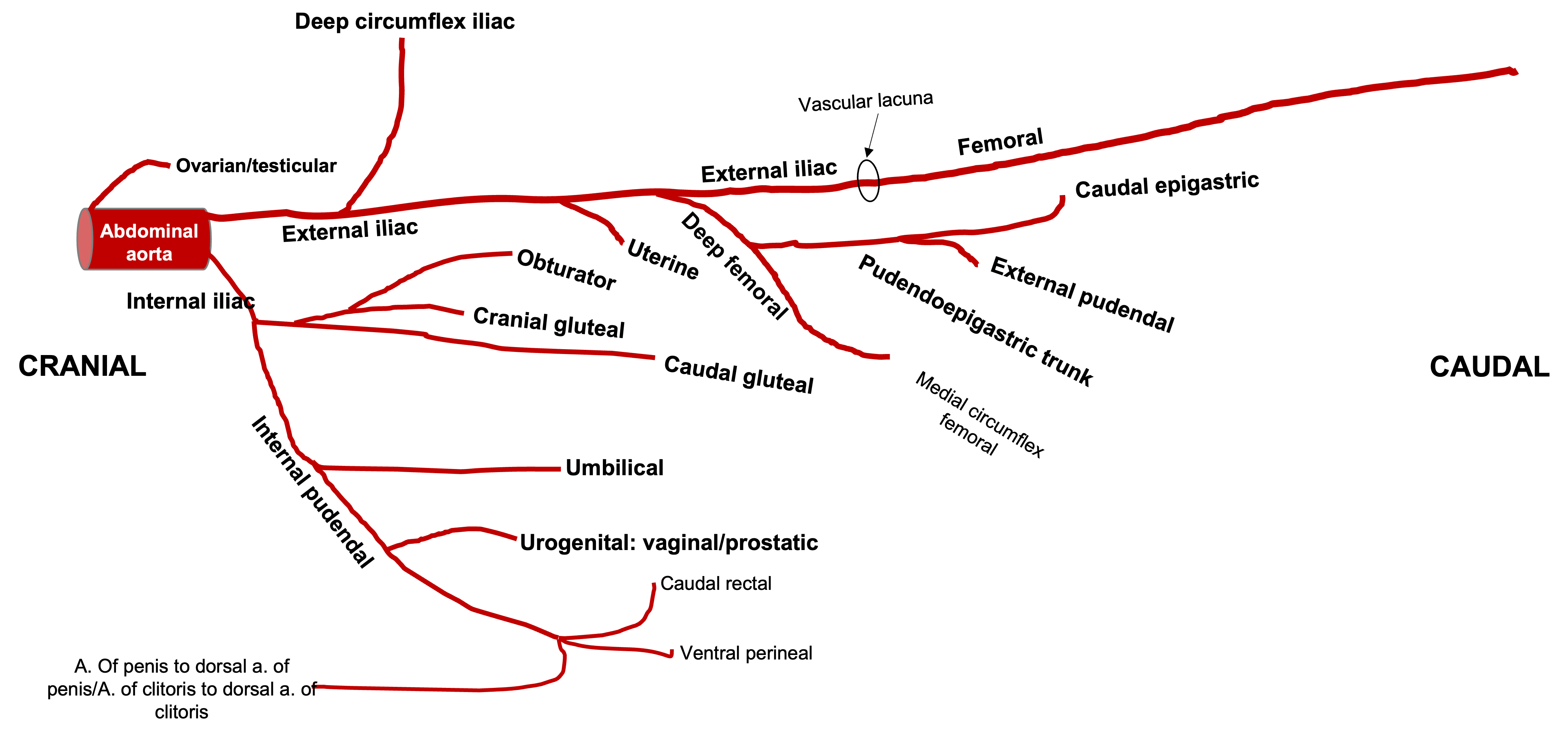
Figure 6-25. Ventral view onto the left external and internal iliac arteries and branches in the equine. Labeled branches: abdominal aorta, ovarian/testicular, external iliac, deep circumflex iliac, uterine (if female), deep femoral, pudendoepigastric trunk, external pudendal, caudal epigastric, femoral; internal iliac, caudal gluteal, cranial gluteal, obturator, internal pudendal, umbilical, urogenital (vaginal/prostatic), dorsal a. of the clitoris/penis. All arteries are bilateral except for the aorta. (RLarsen)

Figure 6-26. Ventral view onto the left external and iliac arteries and branches in the bovine. Labeled branches: abdominal aorta, ovarian/testicular, external iliac, deep circumflex iliac, deep femoral, pudendoepigastric trunk, external pudendal, caudal epigastric, femoral; internal iliac, umbilical, uterine, cranial gluteal, urogenital (vaginal/prostatic), internal pudendal, caudal gluteal, dorsal a. of the clitoris/penis. All arteries are bilateral, except for median sacral and the aorta. (RLarsen)
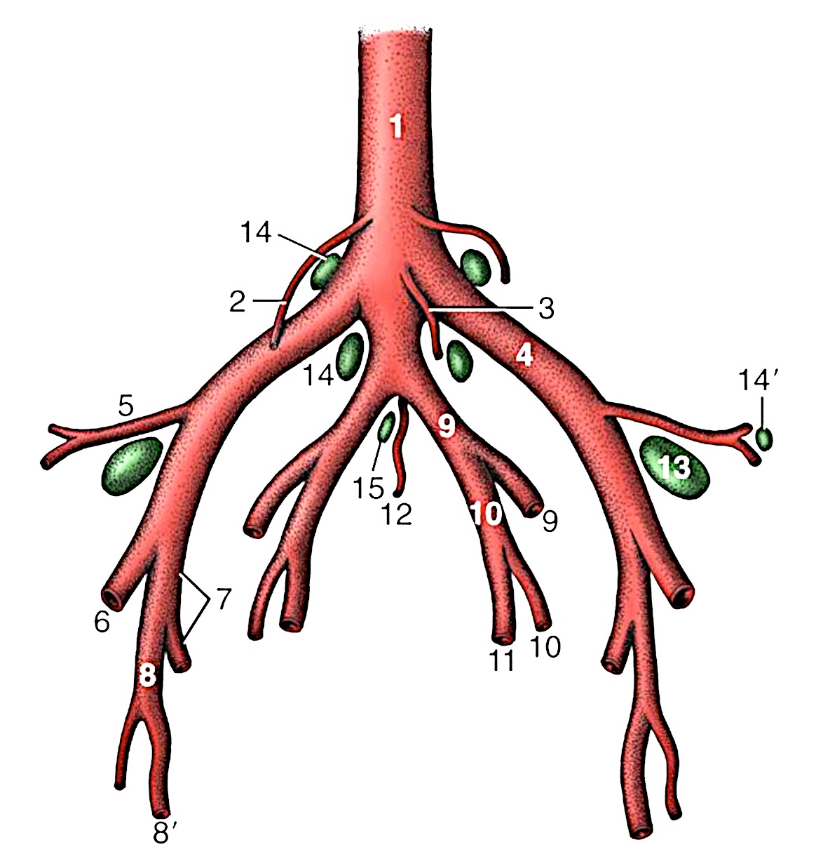
Figure 6-27. Branching pattern of the caudal part of the bovine abdominal aorta. 1, Aorta; 2, ovarian a.; 3, caudal mesenteric a.; 4, external iliac artery; 5, deep circumflex iliac artery; 6, femoral a.; 7, deep femoral a.; 8, pudendoepigastric trunk; 8’, external pudendal a.; 9, internal iliac a.; 10, umbilical a.; 11, uterine a.; 12, median sacral a.; 13, deep inguinal (external iliac/iliofemoral) lymph node; 14, 14’, medial and lateral iliac lymph nodes; 15, sacral lymph nodes. (Duplication of TVA Fig. 29-4).
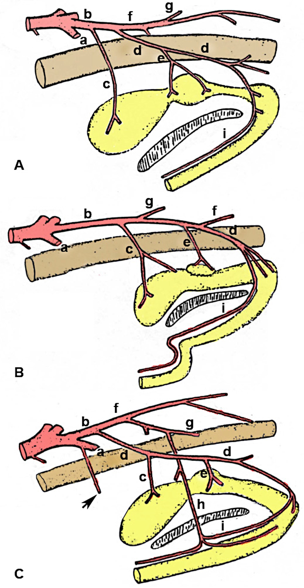
Figure 6-28. Comparative anatomy of the pelvic arteries, schematic lateral view. A, Canine. B, Bovine. C, Equine. a, External iliac a.; b, internal iliac a.; c, umbilical a.; d, internal pudendal a.; e, urogenital a. (prostatic, vaginal for male, female) f, caudal gluteal a. (note the long common trunk which distally terminates into identifiable caudal gluteal/internal pudendal branches in the bovine); g, cranial gluteal a.; h, obturator a. (horse only); i, dorsal artery of the penis. In the mare, the location of the (main) uterine arterial blood supply is indicated by the arrow (a branch off the external iliac artery) in contrast to the main uterine artery of the bitch and cow which come off the vaginal and umbilical arteries, respectively. (Drawing is a modification of original artwork by Dr. Alvin Weber.)
INTERNAL PELVIC NERVES
15. ALL specimens: On the RIGHT side, identify the origin (ventral roots) of the sciatic n., femoral n., and obturator n.; trace these nerves as far as possible as they continue to the pelvic limb.
-
- Identify the origin of the sciatic nerve (next to the caudal gluteal a.) (L6, S1, & S2 roots) and trace it out to the pelvic limb.
- Identify the origin of the femoral nerve (L4-6) and trace it to the pelvic limb.
- Identify the obturator nerve; trace it along the shaft of the ilium and distally through the obturator foramen.
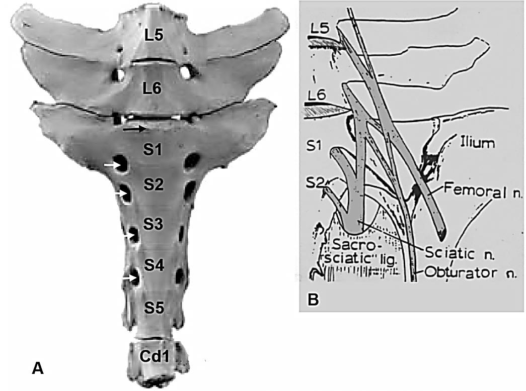
Figure 6-29. Bovine lumbosacral plexus, ventral view. A. Regional osteology on left side; B. Overlay of nerves and ligamentous structures. White arrows, ventral sacral foramina; black arrow, sacral promontory. Image source: A: Budras; B: Cox, VS, et. al. AJVR 36:427-430, 1975.
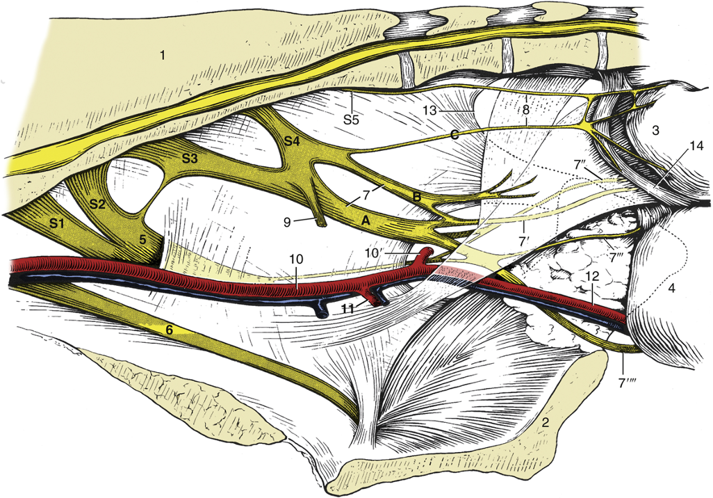
Figure 6-30. Nerves and vessels on the medial surface of the bovine pelvic wall. Local anesthesia of the pudendal nerve (n.) can be obtained by injections at points A and B; anesthesia of the caudal rectal nerves is possible by an injection at point C. 1, Sacrum; 2, pelvic symphysis; 3, rectum (reflected); 4, vagina (reflected); 5, sciatic n.; 6, obturator n.; 7, pudendal n.; 7′, distal cutaneous branch of pudendal n.; 7″, proximal cutaneous branch of pudendal n.; 7′″, deep perineal n.; 7″″, continuation of pudendal n. to clitoris; 8, caudal rectal nerves; 9, pelvic n.; 10, internal iliac artery (a.); 10′, caudal gluteal a.; 11, vaginal a.; 12, internal pudendal a.; 13, caudal border of sacrosciatic ligament; 14, retractor clitoridis; S1 to S5, sacral nerves 1 through 5. (TVA Fig. 29.5)
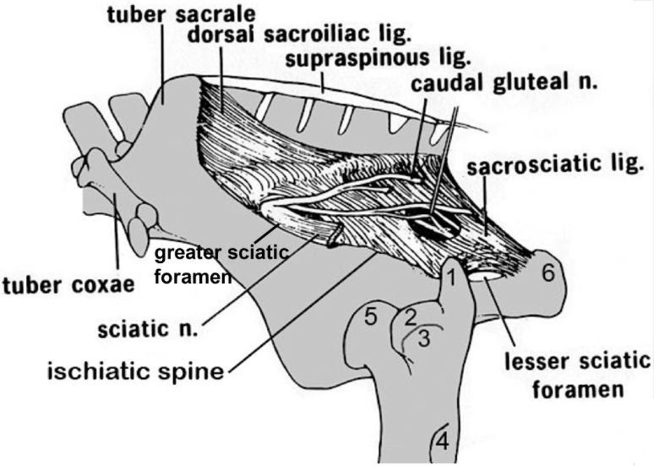
Figure 6-31. Equine pelvis and proximal femur, lateral view. 1, Caudal (middle gluteal) cusp of the greater trochanter; 2, cranial (deep gluteal) cusp of the greater trochanter; 3, facet for insertion of accessory gluteal; 4, third trochanter (superficial gluteal); 5, head of femur (in the acetabulum); 6, tuber ischii. Labeled structures: tuber sacrale, tuber coxae, ischiatic spine, greater and lesser sciatic foramen, sacrosciatic ligament, sciatic n.
Dissection Videos for this Section of Material
Pelvis & Genitalia
- Pony
- Male Pony:
- Internal Pelvis & Genitalia: https://youtu.be/iIfO7UkxxfA
- Female Pony:
- Internal Pelvis & Genitalia: https://youtu.be/KFFRO7oT1-k
- Male Pony:
- Calf
- Male Calf:
- Internal Pelvis & Genitalia: https://youtu.be/e6SDKnCRbgI
- Male Calf:
