
Part 3: Perineal Region
Abby Brown
IMPORTANT NOTE: For this part of the chapter, the Guide directions will refer to either “ALL specimens” (meaning dissect pony and calf specimens in the same way) or to one species in particular, i.e., “ALL (PONY) specimens” or “ALL (CALF) specimens.” This added instructional note is needed because the dissection may differ slightly according to species. The dissection will also vary based on the sex of the animal.
- ALL specimens: Use a hand saw to cut the tail off approximately 5cm from its base and discard it. This will aid in visualizing the perineal area as you progress through the dissection.
- ALL specimens: On the RIGHT side, re-identify the proximal portion of any remaining muscles in the gluteal, proximal thigh, and ischial arch regions (previously identified in Chapter 2).
- In the pony, this will include the superficial, middle and deep gluteal mm., biceps femoris m., semitendinosus, and semimembranosus mm. (Fig. 6-2)
- In the calf, this will include the gluteobiceps m., middle and deep gluteal mm., semitendinosus, and semimembranosus mm. (Fig. 6-2)
- Comparative Note: As mentioned previously in Chapter 2, note that the pony is far more muscular, giving the entire region a smooth, rounded convex shape while the calf, by comparison, is bony and concave. The hamstring mm. originate on (or near) the tuber ischii in the dog and ox, but in the horse they also originate from the sacrum or caudal vertebrae. As a result, there is a thick mass of hamstring muscle on either side of the anus on the equine specimens.
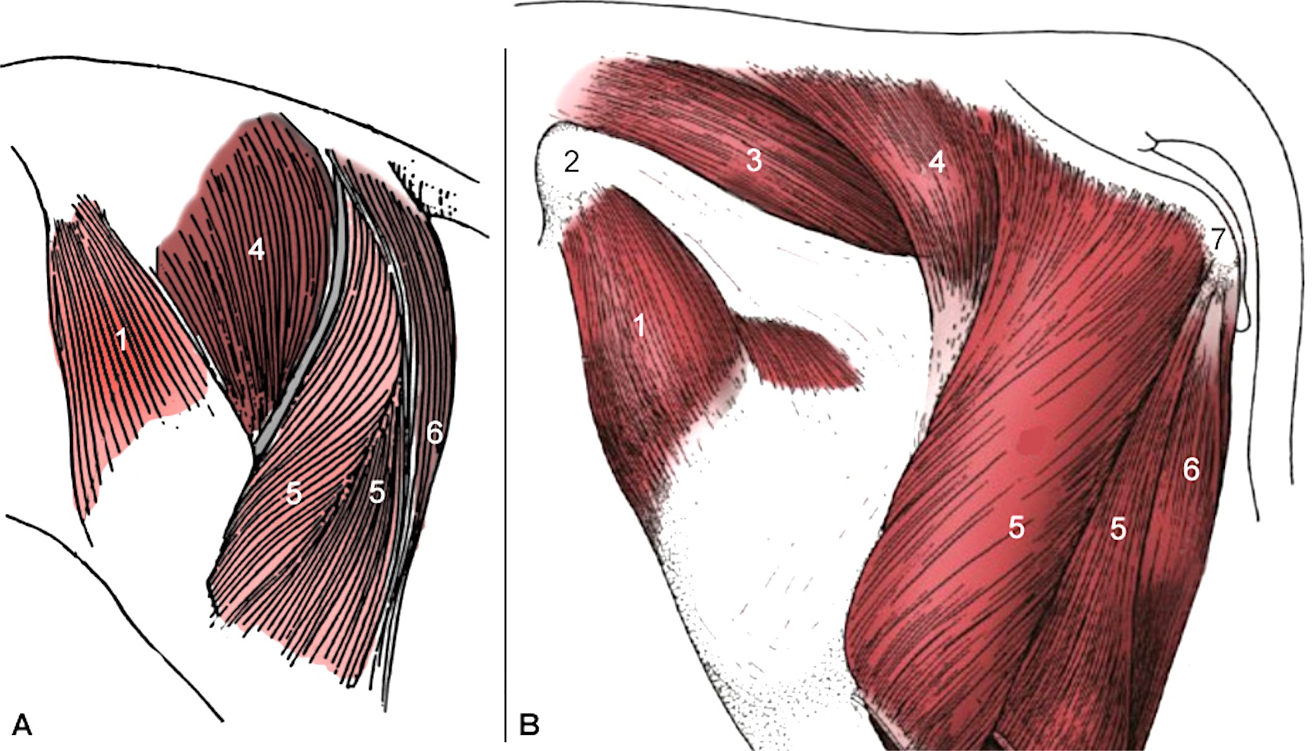 Figure 6-2. Equine (A) and Bovine (B) rump (hind end) region. 1, tensor fasciae latae m.; 2, tuber coxae; 3, middle gluteal m. (not drawn in horse diagram); 4, superficial gluteal m.; 5, biceps femoris m.; gluteobiceps m. in bovine = 4+5; 6, semitendinosus m.; 7, tuber ischii (not identified in horse diagram). (Image B is modified from TVA Fig. 31-2)
3. ALL specimens: Observe the ischial arch. (In order to observe the ischial arch, additional dissection of muscles may be necessary.)
-
- In the calf specimen, transect and reflect the semimembranosus m. on the right side.
- Dissection Note: You may need to transect and/or further reflect the “semi” muscles (semimembranosus and semitendinosus mm.) off of this ischial region (especially in the pony) in order to visualize the bony ischial arch.
- After reflection of muscles as needed, observe the bony caudal edge of the pelvis in your specimen; this is the ischial arch (aka ischiatic arch). The ischial arch is the bony part of the pelvis located caudally between the left and right ischiatic tuberosities (aka tuber ischii). The ischial arch contributes to the formation of the pelvic outlet (caudal opening of the pelvis).
- ALL (CALF) specimens: In the bovine animals, there is a notable depression between the anus and the tuber ischii called the ischiorectal fossa, which is prominent in dairy cows but filled with fat in bulls and beef cows. Identify the ischiorectal fossain your specimen.
4. ALL specimens: Identify the boundaries of the perineum (aka perineal region).
-
- The perineum/perineal region is the region of the body that extends from the base of the tail all the way to the scrotum (male) or udder (female), and laterally as far as the tuber ischii. The perineum includes the anus and the pelvic diaphragm. In this area the digestive and urogenital systems are tightly fused together with connective tissue (especially between the anus and vulva of the female).
- Summary of attachment points for perineal structures (i.e., important ‘anchors’ of the perineal region which help to prevent displacement caused by the straining of defecation, parturition, urination, and to counteract the forward pull of a gravid uterus):
-
- Perineal structures that attach to caudal vertebrae:
-
- anus
- vulva (indirectly via the anus)
- pelvic diaphragm (coccygeus m. portion)
-
- Perineal structures that attach to the external anal sphincter m.:
-
- pelvic diaphragm (levator ani m. portion)
-
- Comparative Note: While the coccygeus muscle is similar to that of the dog, the levator ani m. of ungulates differs markedly by attaching to the external anal sphincter muscle rather than caudal vertebrae (Fig. 12-6).
-
- pelvic diaphragm (levator ani m. portion)
-
- Perineal structures that attach to the anus, vestibule, and rectum:
-
- ‘perineal body’ – a mass of collagenous connective tissue and muscle between the anus and urogenital tract
-
- This musculofibrous node of tissue forms multiple muscular connections (between the external anal sphincter, constrictor vestibuli, and internal anal sphincter mm., and the retractor clitoris (subanal decussation) mm.) and forms a fibrous plate (perineal septum) between the vestibule and rectum, extending craniodorsally from the vestibule.
-
- ‘perineal body’ – a mass of collagenous connective tissue and muscle between the anus and urogenital tract
-
- Perineal structures that attach to the ischial arch:
-
- vestibule
- urogenital diaphragm
-
- Perineal structures that attach to caudal vertebrae:
-
5. ALL specimens: Carefully reflect/remove the skin around the anus (and surrounding the vulva in the female); identify the external anal sphincter m. and the muscles of the pelvic diaphragm: levator ani m. and coccygeus m.
-
- In all specimens, first identify the external opening of the anal canal, which is the anus. Next, reflect and remove the skin covering/surrounding the anus (and surrounding the vulva in the female).
-
- Dissection Note: Note that the skin is VERY thin in this region, so dissect carefully!
-
- Identify the external anal sphincter m., which is the striated muscle surrounding the anus. (Figs. 6-3, 6-4)
- Just cranial to the external anal sphincter m. carefully expose and identify the muscles that make up the pelvic diaphragm: the levator ani m. (Figs. 12-5/4; 12-6/L, L’) and the coccygeus m. (Figs. 12-5/3; 12-6/C) (TVA, 704)
-
- Caudally, the levator ani m. may appear to go deep to the external anal sphincter m.
- The coccygeus m. extends dorsally to attach to the ventral/lateral aspect of the tail.
- Dissection Note: Some of the ventral tail muscles may partially obscure your view of these muscles.
-
- In all specimens, first identify the external opening of the anal canal, which is the anus. Next, reflect and remove the skin covering/surrounding the anus (and surrounding the vulva in the female).
6. ALL FEMALE (PONY) specimens: Attempt to identify the constrictor vulvae m.
-
- In the mare (female equine) specimens, attempt to identify the constrictor vulvae m. by carefully reflecting more of the skin covering the anus and vulva.
- Trace the fibers of the external anal sphincter muscle ventrally to the point where they are continuous with the constrictor vulvae muscle (Fig. 6-4).
7. ALL specimens: Identify the rectococcygeus m. Identify the retractor penis m. (male) or retractor clitoris m. (female).
-
- In all specimens, uncover the vertebral origin of the rectococcygeus m. by dissecting dorsal to the levator ani m. (Fig. 6-4).
-
- The rectococcygeus m. extends from the dorsal surface of the rectum to the ventral surface of the tail (ventral midline). Observe the extension of the rectococcygeus m. onto the outer (longitudinal) muscle coat of the rectum.
-
- In all specimens, as you look for the rectococcygeus m., also uncover the vertebral origin of the retractor penis m. (male) or retractor clitoris m. (female) (the name is respective of sex).
-
- The retractor penis/clitoris m. will extend distally, running deep to/underneath the levator ani m., and will reemerge further distally in the perineal region.
- Dissection Note: In the PONY, the majority of the fibers of the retractor penis or retractor clitoris muscle form a subanal loop that supports the anus in addition to the typical insertion of its muscle fibers onto the clitoris or penis.
-
- Important Dissection Note: Both of these muscles are examples of smooth muscle attaching to bone, a highly unusual situation.
- In all specimens, uncover the vertebral origin of the rectococcygeus m. by dissecting dorsal to the levator ani m. (Fig. 6-4).
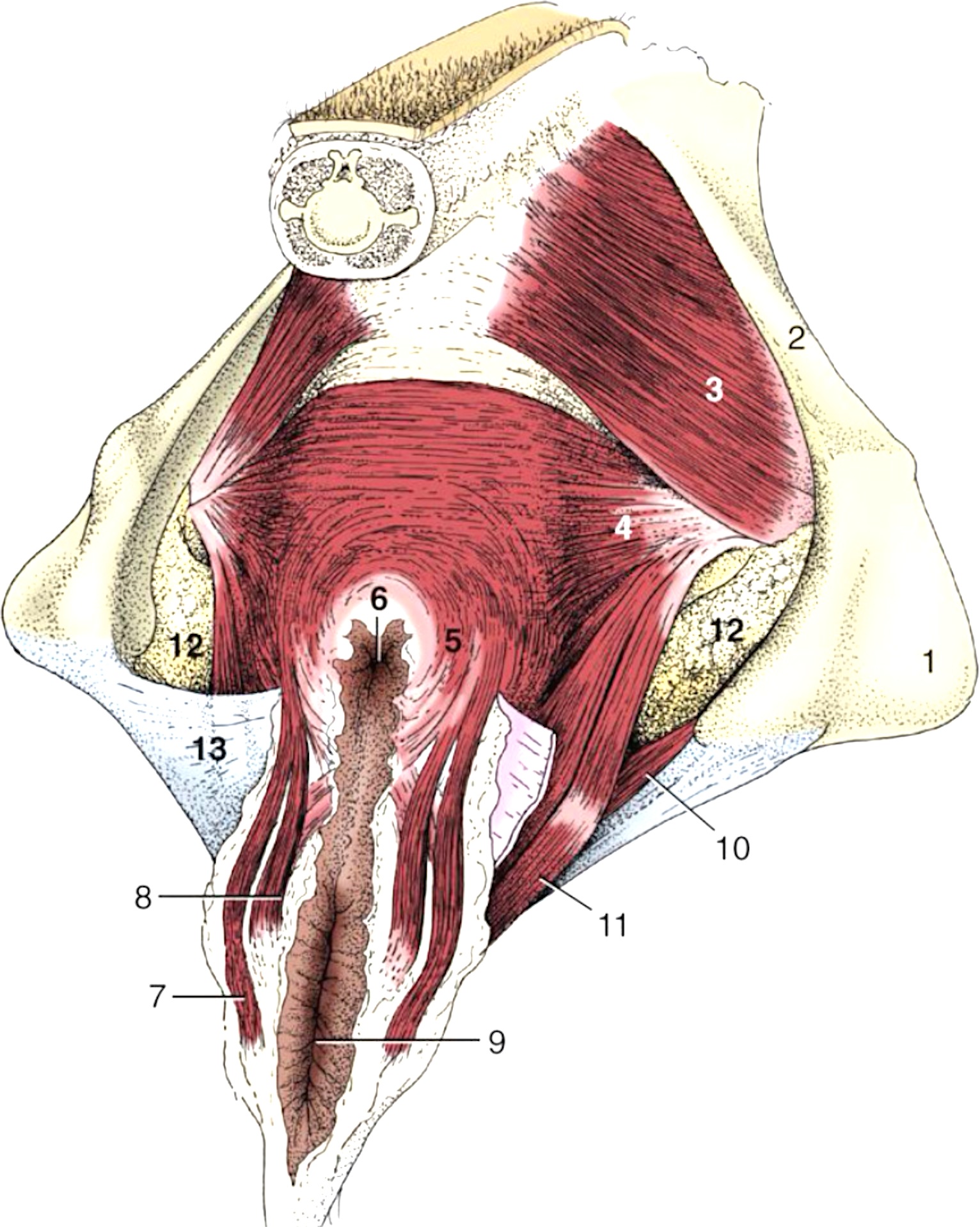
Figure 6-3. (Left) The perineal muscles of a cow. 1, ischial tuber; 2, sacrosciatic ligament; 3, coccygeus m.; 4, levator ani m.; 5, external anal sphincter m.; 6, anus; 7, retractor clitoris m.; 8, constrictor vulvae m.; 9, vulva; 10, urogenital diaphragm; 11, constrictor vestibuli m.; 12, (fat within) ischiorectal fossa; 13, perineal fascia (partly removed on the right side). (Modified from TVA Fig. 29-10.)
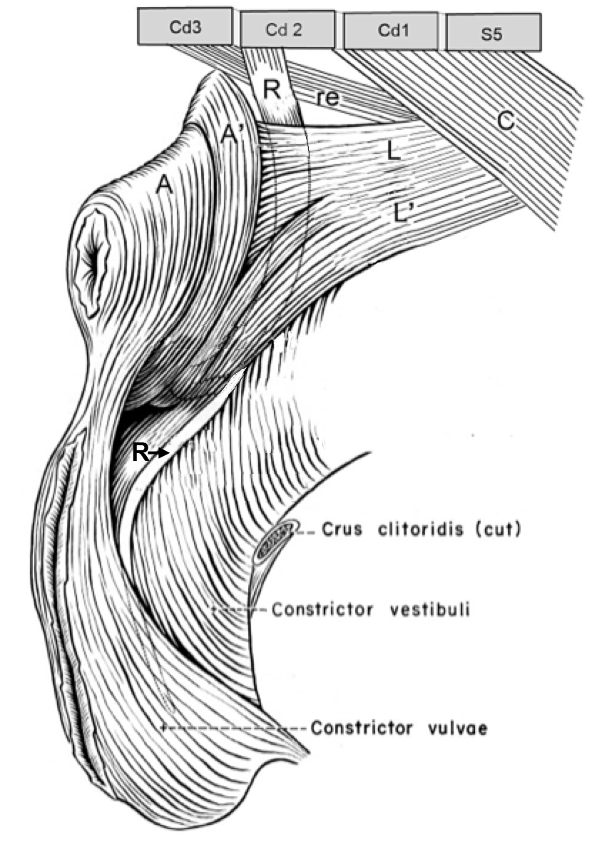
Figure 6-4. (Left) Isolated perineum of a mare. A, A’, external anal sphincter m., caudal and cranial parts; C, coccygeus m.; Cd, caudal vertebrae; L, L’, levator ani m., anal and subanal parts; R, retractor clitoris m.; re, rectococcygeus m.; S5, last sacral vertebral segment.
IMPORTANT DISSECTION NOTE: In the following dissection, you will be cutting only the LEFT CRUS of the clitoris/penis to facilitate removal of the LEFT hemi-pelvis.
8. ALL FEMALE specimens: Expose the left crus of the clitoris and cut/detach it from the ischial arch.
-
- Re-identify the region of the ishcial arch, which is the bony part of the pelvis located caudally between the left and right ischiatic tuberosities (aka tuber ischii). Near the ischial arch, in the female, the constrictor vestibuli muscles cover the crura of the clitoris.
-
- Dissection Note: Recall that ‘crura’ is the plural of ‘crus’. There will be a left crus of the clitoris and a right crus of the clitoris.
-
- On the LEFT side only, scrape the constrictor vestibuli m. off of the left crus of the clitoris to expose its attachment to the ischial arch.
-
- Detach the LEFT crus of the clitoris from the ischial arch by cutting close to the bone, noting any resultant exposure of erectile tissue on the cut surface of the crus.
-
- Re-identify the region of the ishcial arch, which is the bony part of the pelvis located caudally between the left and right ischiatic tuberosities (aka tuber ischii). Near the ischial arch, in the female, the constrictor vestibuli muscles cover the crura of the clitoris.
9. ALL MALE specimens: Expose/identify the attachments of the crura of the penis; transect the left crus of the penis to detach it from the ischial arch.
-
- Re-identify the region of the ishcial arch, which is the bony part of the pelvis located caudally between the left and right ischiatic tuberosities (aka tuber ischii). In the male specimens, expose and identify the attachment of the crura of the penis to the ischial arch (TVA, 717, 8/9).
-
- Dissection Note: Recall that ‘crura’ is the plural of ‘crus’. There will be a left crus of the penis and a right crus of the penis.
-
- After the crura of the penis are exposed, detach the LEFT crus of the penis from the ischial arch by cutting close to the bone.
-
- Dissection Note: Transect only the LEFT crus of the penis from where it attaches to the left side of the ischial arch.
- Note any resultant exposure of erectile tissue on the cut surface of the crus.
-
-
- If the cut is more than a few millimeters from the bone, the erectile tissue will be exposed, and should be recognizable due to its characteristic texture; this is referred to as the ‘pizzle eye’ on a bovine carcass.
-
-
-
- Re-identify the region of the ishcial arch, which is the bony part of the pelvis located caudally between the left and right ischiatic tuberosities (aka tuber ischii). In the male specimens, expose and identify the attachment of the crura of the penis to the ischial arch (TVA, 717, 8/9).
10. ALL specimens: Review the indicated structures on the left side of the specimen prior to removal of the LEFT hemi-pelvis: sacrosciatic ligament, sciatic n., cranial gluteal a., tendon of internal obturator m. (pony only), caudal gluteal a., and pelvic diaphragm.
-
- Observe the left sacrosciatic ligament as it attaches to the ischiatic spine and tuber sacrale.
- Find the (left) sciatic nerve and cranial gluteal artery as they emerge from the greater sciatic foramen.
- PONY specimens: If possible, identify the cut edge of the tendon of the (left) internal obturator m. where it passes through the lesser sciatic foramen. Identify the caudal gluteal a. piercing through the sacrosciatic ligament.
- CALF specimens: Identify the caudal gluteal a. emerging from the lesser sciatic foramen.
- Re-identify the left pelvic diaphragm, which is made up of the levator ani and coccygeus mm.).
11. ALL specimens: On the LEFT side, cut the shaft of the ilium and then cut through the pubic symphysis. Transect/reflect soft tissue attaching to the pelvis, and remove the LEFT hemi-pelvis. Retain the pelvic viscera within the right half of the specimen for in situ study.
-
- On the LEFT side, cut the shaft of the ilium just ventral to the sacroiliac joint (ventral to the wing of the ilium) with a Stryker bone saw.
- Expose the pelvic symphysis by removing portions of muscle as needed from the left side of the pelvis near the ventral midline.
- Make a cut ventrally along the entire length of the pelvic symphysis (ventral midline of the pelvis where the two sides fuse together) with a Stryker bone saw.
-
- Important Dissection Note: Do this very carefully – don’t damage the reproductive tract! Push all reproductive tract parts over to the right side so they are not damaged in the process of cutting the pelvic symphysis!
-
- Transect and remove/reflect the soft tissue structures attaching to the left hemi-pelvis (e.g., sacrosciatic ligament and associated nerves and vessels).
-
- Dissection Note: The attachment of the abdominal wall to the tuber coxae will be preserved; however, the attachment of the rectus abdominis m. to the pelvic brim will be transected on the left side.
-
- ALL FEMALE specimens: If an udder is present, reflect the left portion of the udder from the ventral aspect of the pelvis, retaining the udder with the right side of the specimen.
- Retain all reproductive tract parts (including the parts from the left side) with the right side of the specimen’s pelvis.
- ALL MALE specimens: Retain the left inguinal canal and associated testicular structures with the right side of the specimen.
- Reflect or remove the LEFT hemi-pelvis from the specimen.
Dissection Videos for this Section of Material
Perineal Region
- Pony
- Male Pelvic Inlet & Perineum: https://youtu.be/sLf20VYjmXU
- Female Pelvic Inlet & Perineum: https://youtu.be/KUBAoRBIp1Q
- Calf
- Male Pelvic Inlet & Perineum: https://youtu.be/FggEeCp4qIc
