
2 Distal Limb
Revised Spring 2024 by T. Clark
Additional Resources for this Chapter:
- Valuable Supplemental Large Animal Surgery (LAS) course eBook links (starting with Equine Lameness and going through Swine, Small Ruminant, and Poultry Lameness): LAS Bovine Foot Anatomy, Lameness, Approaches; LAS Nerve and Joint Blocks; LAS Corns; LAS Thrush; LAS Hoof abscesses; LAS Bovine Anatomy; LAS Tendonitis and bowed tendons; LAS Flexural limb deformities; LAS Inferior check desmotomy; LAS Foot Rot; LAS Laminitis; LAS Navicular syndrome; LAS Palmar digital neurectomy; LAS Digital dermatitis; LAS Sole ulcers; LAS White line disease; LAS Foot sepsis
- Veterinary Gross Anatomy Ungulate Dissection (Limb Dissection Labs 1-7)
- Associated Video Links:
- Joints of the Distal Limb (2:09)
- Phalanges (3 videos, 14:14)
Objective
A2.1 Describe and differentiate regions and bones of the (distal) thoracic limb by using correct directional/regional terminology.
Metacarpal bones (Figs. 1-6, 2-1, 2-3)
HORSE – there are three metacarpal bones in the horse (Figs. 1-6, 2-1)
- Only metacarpal 3 (Mc3) is complete and articulates with the digit. The common name for this bone is the cannon bone.
- The proximodorsal surface of the cannon bone bears the metacarpal tuberosity, where the extensor carpi radialis m. tendon inserts.
- The distal end of the cannon bone bears a condyle, with an extensive cylindrical articular surface, and a prominent sagittal ridge that articulates with a complementary sagittal groove of the proximal phalanx (P1). (see Fig. 7 image below of fracture related to these sagittal structures)
- Together, the ridge and groove provide lateral stability to the fetlock (aka metacarpophalangeal/MCP) joint.
- The Mc3 condyle also articulates with the proximal sesamoid bones; the sagittal ridge separates the two proximal sesamoids (Fig. 2-2).
- The 2nd (Mc2) and 4th (Mc4) metacarpal bones are small and commonly called the medial and lateral splint bones, respectively.
- The bulbous ends of the splint bones can be palpated easily on the live animal.
- In the EQUINE, the second carpal bone (C2) rests entirely on the medial splint bone (Mc2) but the fourth carpal bone (C4) rests on both the cannon bone (Mc3) and the lateral splint bone (Mc4).
- Clinical Notes: The direct impact of C2 on Mc2 is thought to contribute to a downward displacement (shearing) of the attachment between Mc2 and Mc3, tearing the interosseous ligament that binds the medial splint bone (Mc2) to the cannon bone (Mc3). The result is an inflammatory swelling that leaves a permanent blemish after it ossifies, called a splint lesion or splints.
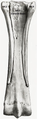
Figure 2-1 Equine metacarpal (Mc) bones of right forelimb, palmar view. Mc2, second metacarpal bone/medial splint bone; Mc3, third metacarpal bone/cannon bone; Mc4, fourth metacarpal bone/lateral splint bone; SR, sagittal ridge.
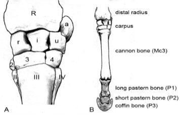 Figure 1-6 Equine left forelimb, dorsal view. A, carpus; B, carpus, metacarpus, and digit.
OX
- In the OX, the 3rd and 4th metacarpals (Mc3, Mc4) are complete bones but fused to each other to form the bovine cannon bone (Fig. 2- 3).
- At the distal end they separate into two condyles (aka trochleas) separated by an intertrochlear notch.
- The 5th metacarpal bone (Mc5) remains vestigial in the proximolateral region of the metacarpus (Fig. 2- 3/5).
Sesamoid bones (Figs. 2-2, 2-3)
HORSE – there are three sesamoid bones in the distal limb, the paired proximal sesamoid bones and the singular distal sesamoid bone (aka the navicular bone) (Fig. 2-2).

Figure 2-2 Bones of the equine digit, palmar surface. e, Extensor process; n, flexor surface of navicular bone (distal sesamoid bone); s, flexor surface of proximal sesamoid bones; sf, solar foramen; sr/sg, sagittal ridge (Mc3) and sagittal groove (P1).
- Proximal sesamoid bones or simply ‘sesamoid bones’:
- The two proximal sesamoid bones are found on the palmar (or plantar) surface of the metacarpophalangeal/MCP/fetlock (or metatarsophalangeal/MTP/fetlock) joint and are very mobile as a unit. These are differentiated as medial and lateral sesamoids.
- The proximal sesamoids are bound together by a fibrocartilaginous intersesamoidean ligament.
- A small groove on the dorsal surface of the sesamoids interdigitates with the sagittal ridge on the distal end of the cannon bone (Mc3). The distal articular surface of the cannon bone exceeds 200 degrees and thus the fetlock joint is capable of a wide range of motion.
- The sesamoid bones have a pointed apex proximally, and a wide base on the distal end.
- The abaxial surfaces of the sesamoids are rough and depressed for insertion of the thick medial and lateral branches (not extensor branches) of the suspensory ligament (interosseous tendon).
- The smooth articular surface articulates with the metacarpal condyle (of Mc3).
- The flexor surface faces the palmar (or plantar) aspect of the MCP (or MTP) joint. The flexor surface in combination with their intersesamoidean ligament forms a groove for the superficial and deep digital flexor tendons. These digital tendons are held in place by the palmar annular ligament (Fig. 2-4).
- Clinical Notes: Fractures of the proximal sesamoid bones can occur (see below, radiograph – lateral view at fetlock). When there is continual repetitive motion, the suspensory apparatus is constantly straining against the proximal sesamoid bones and these sesamoids can be pulled apart.
- The two proximal sesamoid bones are found on the palmar (or plantar) surface of the metacarpophalangeal/MCP/fetlock (or metatarsophalangeal/MTP/fetlock) joint and are very mobile as a unit. These are differentiated as medial and lateral sesamoids.
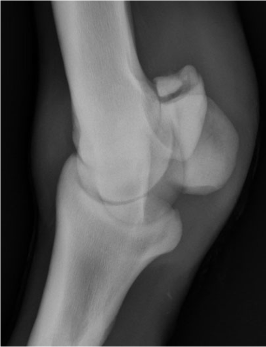
Figure (above) – lateral view at fetlock; sesamoid fracture.
-
- Sesamoid fractures are often referred to as apical or basilar when the fracture line is horizontal. The most common sesamoid fractures in Standardbreds and Thoroughbreds are apical. They are caused by overextension and often are associated with suspensory ligament damage. Other fractures include mid-body, basilar, abaxial, axial, or comminuted, and they may involve one or both sesamoids.
- Navicular bone (aka distal sesamoid bone) – named because of its boat shape (Latin, nave = boat). There is one navicular bone per digit.
- The proximal surface is the narrow flat edge. The distal surface is the narrow curved edge.
- The palmar flexor surface faces the deep digital flexor tendon (DDFT).
- A navicular bursa sits between the navicular bone and the DDFT. More about this clinically relevant structure below.
- The articular (dorsal) surface articulates with P2/middle phalanx/short pastern bone proximally, while a small portion of the dorsal and distal edge articulates with P3/distal phalanx/coffin bone.
- Between P3 and the navicular bone there is a single distal sesamoidean impar (meaning unpaired) ligament (aka distal navicular ligament). The ligament firmly attaches P3 to the navicular bone, so they can move as a unit.
- Medial and lateral edges of the navicular bone each have an associated collateral sesamoidean ligament that attaches to distal portions of P1.

Fig above – Navicular bone placement and surfaces. Right: bones of the distal forelimb from a palmar view (proximal to distal): 2 proximal sesamoids, P1, P2, navicular (blue arrow), P3 wraps around medially and laterally. This model is in the anatomy lab and will be very helpful to look at in person. Left top: looking at the proximal edge and can see part of the articular surface. Left bottom: looking at the flexor surface and the bottom edge of the picture shows the curved distal surface.
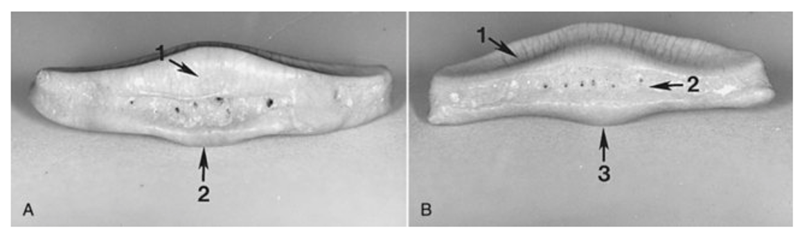
FIG. 24-1 The navicular bone. A, view of the distal surface with synovial fossae (small holes). 1, The small articular surface with the distal phalanx (P3); 2, the projection off the distal border where the impar ligament attaches. B, view of the proximal surface. 1, The articular surface with the middle phalanx (P2); 2, the proximal border itself where the suspensory navicular ligament attaches; 3, the central eminence. The view in B is analogous to that obtained in a palmaroproximal-palmarodistal radiograph. (https://veteriankey.com/the-equine-navicular-bone/)

Fig – Navicular bone (yellow) shown with the hoof and soft tissues removed. Adapted from the Glass Horse Distal Limb (https://horsesidevetguide.com/Navicular+Syndrome+%26+Heel+Pain+in+Horses+).
OX
- The sesamoid bones in the BOVINE are similar to those described in the EQUINE, except the number is doubled in the ox because it has two digits instead of one (Fig. 2-3).
- Each digit has two proximal sesamoid bones (axial and abaxial) for a total of four proximal sesamoid bones per limb. The axis of the limb in the ox courses between digits 3 and 4. The sesamoid bones closest to the axis are the axial sesamoids. Those farthest from the axis are the abaxial sesamoids.
- Each digit has one distal sesamoid (navicular) bone for a total of two per limb.
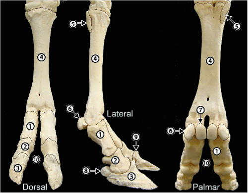
Figure 2-3 Bovine distal forelimb bones. 1, Proximal phalanx (P1); 2, middle phalanx (P2); 3, distal phalanx (P3, pedal bone); 4, fused metacarpal 3+4 (Mc 3+4 = bovine cannon bone); 5, rudimentary Mc5; 6, abaxial proximal sesamoid bone; 7, axial proximal sesamoid bone; 8, distal sesamoid (navicular) bone; 9, extensor process of pedal bone; 10, interdigital space.
Phalanges (Figs. 1-6, 2-2 through 2-6)
EQUINE bear weight on one digit (3rd digit) in their forelimbs and hind limbs, while BOVINE bear weight on two digits (3rd and 4th digits) in their forelimbs and hind limbs.
DIGIT 3 OF THE HORSE (digit 3 of the equine contains two proximal sesamoids and one distal sesamoid (aka navicular bone).
- Proximal phalanx (aka P1, long pastern bone):
- P1 has a wide concave proximal articular surface where it articulates with the cannon bone (Mc3) (Fig. 2-2).
- P1 has a sagittal groove which interdigitates with the sagittal ridge of the cannon bone.
- A triangular rough area on the mid palmar surface of P1 is where the middle (oblique) sesamoidean ligament attaches. This ligament is called oblique because it extends from the distal outer edges of the proximal sesamoid bones to the center palmar surface of P1 (Fig. 2-4/5).
- Note the medial and lateral eminences on distal P1 for the attachment of the collateral ligaments of the pastern joint.
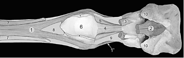
Figure 2-4 (Above) Equine distal forelimb, palmar view. (Digital flexor tendons have been removed except for a small portion of their tendons of insertion. Further exposure of the insertion of the deep digital flexor tendon of insertion was attained by removal of the hoof and much of the digital cushion). 1, Suspensory ligament (interosseous tendon); 1’, extensor branch of suspensory ligament; 2, deep digital flexor tendon (DDFT); 3, superficial digital flexor tendon (SDFT); 4, straight sesamoidean ligament; 5, oblique sesamoidean ligament; 6, location of groove formed by the sesamoid bones and intersesamoidean ligament; 7, splint bones (Mc 2 and 4); 8, Mc 3 (cannon bone); 9, P1 (long pastern bone); 10, collateral cartilages of P3. Both digital flexor tendons lie in a groove (6) formed by the sesamoid bones. The flexor tendons are bound by the palmar annular ligament which has been removed. However, the cut edges of the palmar annular ligament are indicated by asterisks (*). Notice that the suspensory ligament (interosseous tendon) lies on the palmar surface of the cannon bone and splits at the level of the end of the splint bones. Typically, the medial splint bone, Mc2, is slightly longer than Mc4.
- Clinical Note: The sagittal groove may be the point of origin for a longitudinal/sagittal fracture of P1, which is called a screwdriver fracture. The metaphor here pictures the sagittal ridge of Mc3 as the blade of a screwdriver and the sagittal groove of P1/long pastern bone as the groove in the head of a screw (Fig. 7 below). P1 is also a common site for comminuted fractures (i.e., bone is broken into two or more pieces).
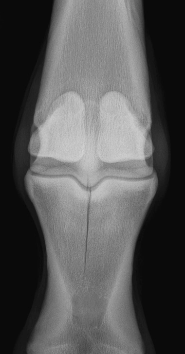
Fig. 7. A dorso-palmar radiographic projection of an incomplete (proximal-distal) sagittal proximal first phalangeal fracture. Essentially all of these sagittal fractures originate in the center of the sagittal groove. Santschi, Elizabeth M. “Articular fetlock injuries in exercising horses.” The Veterinary clinics of North America. Equine practice 24 1 (2008): 117-32 .
- Middle phalanx (aka P2, short pastern bone): P2 is half the length of the long pastern bone.
- P2 has a transverse rounded area on the proximo-palmar border for attachment of the superficial (straight) sesamoidean ligament and the superficial digital flexor tendon (SDFT) (Fig. 2-4/4,3). P2 is also a common site for comminuted fractures (i.e., bone is broken into two or more pieces).
- Distal phalanx (aka P3, coffin bone): named because the hoof (aka ‘foot capsule’) forms a “coffin” that surrounds this bone. It is also called the pedal bone in BOVINE.
- There is a prominent extensor process (Fig. 2-5/e) where the common digital extensor (in the forelimb) or long digital extensor (in the hind limb) tendon inserts.
- The convex dorsal surface of the coffin bone is referred to as the parietal surface because it is adjacent to the hoof wall (‘parietal’ means wall). It may have a small divot or notch at the midline called a crena marginalis (Fig. below).
- The flat “ground” surface of the coffin bone is called the solar surface and is where the deep digital flexor tendon inserts.
- The solar surface has two solar foramina that lead to a solar canal. The solar canal contains an arterial arch formed by anastomoses of the medial and lateral digital aa. Numerous arterial branches radiate from this arterial arch and penetrate the parietal surface to supply deep structures of the hoof.

Fig 1-26: Left Distal Phalanx (P3), solar view: coffin bone, solar foramina, solar canal (from Equine Anatomy Guide: The Forelimb; Mansour, Steiss, Wilhite)
-
- Collateral cartilages are attached to the proximal edges of the “heels” or wings of the coffin bone. These cartilages are usually quite flexible but occasionally become partially ossified to form “sidebones” (Fig. 2-5/s).
- On either side of the articular surface there is a depression of the parietal surface to form facets for attachment of the collateral ligaments of the distal interphalangeal (aka DIP/coffin) joint (Fig. 2-5/C).

Figure 2-5 Equine distal phalanx (P3), parietal (wall) and articular surfaces. A, articular surface; C, facet for distal attachment of the collateral ligament of the distal interphalangeal (DIP)/coffin joint; e, extensor process; s, sidebone; v, one of many vascular foramina where branches from the terminal solar arterial arch emerge.
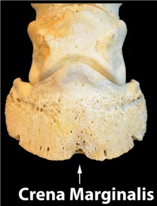
Fig above – crena marginalis, a notch or divot in the coffin bone (P3) that can be seen in radiographs (not clinically a problem).
DIGITS 3 AND 4 OF THE OX (there are a total of four proximal sesamoid bones, and two distal sesamoid bones (aka navicular bones) per limb in the OX).
- Digits 3 and 4 of the OX are quite similar in structure and terminology to EQUINE, except there are two digits instead of one (Fig. 2-3). Hence, there are a total of two proximal phalanges (P1), two middle phalanges (P2), and two distal phalanges (P3), per limb.
- Note: the distal phalanges (P3) are most often referred to as the pedal bones in the bovine animal.
- On each digit, the deep digital flexor tendon attaches on the palmar/solar surface of P3 at the flexor tuberosity. The pressure of this tuberosity likely creates or leads to sole ulcers. (see LAS Bovine Foot Anatomy, Lameness, Approaches)
Objective
A2.2 Identify and describe the joints, joint angles, and joint actions of the (distal) thoracic limb; describe the location of muscle groups that result in flexion/extension of these joints.
Joints of the (Distal) Thoracic Limb
- Clinical Notes: Joint pouches are extensions of the synovial capsule and cavity past joint surface. In more mobile joints these pouches can be more expansive/extensive. When you “tap a joint” (i.e., aspirate) you will not always remove fluid right at the bony union of a joint, but rather near the extension of these synovial cavities. These pouches may also bulge outwards and can visibly be seen due to inflammation. Additionally, anesthesia or medications can be injected in and near the joint capsules. (more Supplemental Clinical in LAS Nerve and Joint Blocks)
- Metacarpophalangeal (aka MCP, fetlock joint) is between Mc3 and P1. On the palmar (or plantar) surface of the joint are the proximal sesamoid bones.
- The fetlock is created by the interlocking of the sagittal ridge of Mc3, the sagittal groove of P1, and the proximal sesamoids. This interlocking prevents sideward movement of the digit, and allows the digit to rotate around the end of the cannon bone for >200 degrees.
- The fetlock has a greater range of motion than most other joints, as it has a relatively large degree of flexion, extension and some hyper-extension. Over hyper-extension is resisted by the suspensory apparatus comprised of the suspensory ligament, proximal sesamoid bones, and the distal sesamoidean ligaments. The joint is stabilized by medial and lateral collateral ligaments of the fetlock.
- The BOVINE has one fetlock joint per foot (Fig. 2-3).
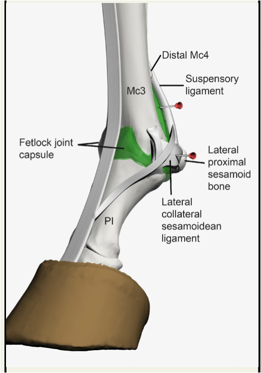
Fig 2-5: Left Forelimb. Lateral View of the Metacarpophalangeal (Fetlock, MCP) Joint (joint capsule/pouch in green). The joint has a dorsal and palmar pouch. The palmar pouch is usually injected using a lateral approach, with the limb planted on the ground (weight bearing). The injection site is between the palpable suspensory ligament, distal part of the lateral splint (button) and the lateral proximal sesamoid bone. Alternatively, injection of the fetlock joint can be performed by penetrating the lateral (or medial) collateral sesamoidean ligament, with the limb flexed. The advantage of this approach is that it causes less hemorrhage in the joint compared to the palmar approach (Equine Joint Injection and Regional Anesthesia by Moyer, Schumacher, Schumacher, 2011; image from Equine Anatomy Guide: The Forelimb; Mansour, Steiss, Wilhite)
- Proximal interphalangeal (aka PIP, pastern joint) is between P1 and P2. The joint is stabilized by medial and lateral collateral ligaments of the pastern joint. It is a relatively low motion joint.
- The BOVINE has two pastern joints per foot (Fig. 2-3).
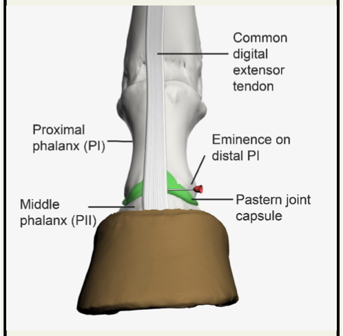
Fig 2-6: Left Forelimb. Dorsal View of the Proximal Interphalangeal (Pastern, PIP) Joint (joint capsule/pouch in green). Dorsal approach to the pastern joint: The joint has a dorsal and palmar pouch. It is usually accessed by injection of the dorsal pouch. The landmarks for the dorsal approach include the eminence on distal P1 (which serves for attachment of the lateral collateral ligament of the pastern joint) and the common digital extensor tendon. The needle is inserted deep to the tendon of the common digital extensor distal to the lateral bony eminence of P1. (Equine Joint Injection and Regional Anesthesia by Moyer, Schumacher, Schumacher, 2011; image from Equine Anatomy Guide: The Forelimb; Mansour, Steiss, Wilhite)
- Distal interphalangeal (aka DIP, coffin/pedal joint) is between P2 and P3 and is a saddle joint. The distal sesamoid tucks between P2 and P3. The joint is stabilized by medial and lateral collateral ligaments of the coffin joint.
- The BOVINE has two coffin joints per foot (Fig. 2-3).
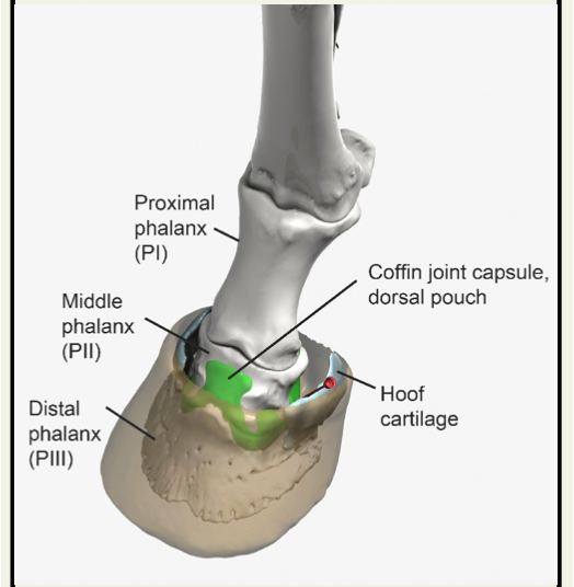
Fig 2-7: Left Forelimb. Dorsolateral View of the Distal Interphalangeal (Coffin, DIP) Joint (joint capsule/pouch in green). Lateral approach to the coffin joint: The coffin joint can be accessed on the lateral side just above the palpable lateral hoof cartilage (collateral cartilage of the foot). The needle is usually angled medially between the medial surface of the cartilage and P2. (Equine Joint Injection and Regional Anesthesia by Moyer, Schumacher, Schumacher, 2011; image from Equine Anatomy Guide: The Forelimb; Mansour, Steiss, Wilhite)
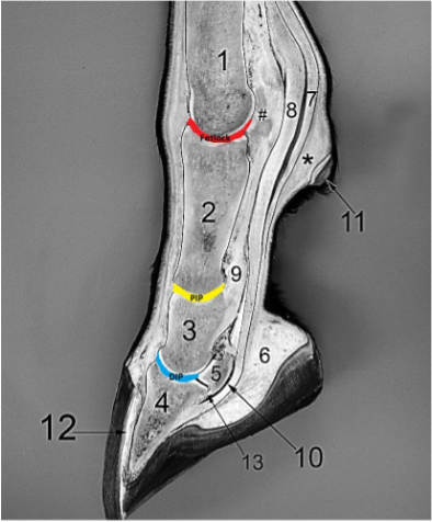
Figure 2-6 Sagittal section equine foot and digit. 1, distal cannon bone (Mc3); 2, P1; 3, P2; 4, P3; 5, navicular bone (distal sesamoid); 6, digital cushion; 7, superficial digital flexor tendon (SDFT); 8, deep digital flexor tendon (DDFT); 9, superficial distal sesamoidean ligament extending from the proximal sesamoids (#) to the fibrocartilage on the proximal palmar aspect of P2; 10, the navicular bursa lying between the navicular bone and the flexor surface of the DDFT; 11, the ergot; *, fibrous pad deep to the horny ergot that is present even when no horny tissue of the ergot is present; 12, horny (insensitive) laminae and adjacent wall forming the so called ‘white line’ that may be observed on the ground surface between the hoof wall and sole; 13, distal sesamoidean impar ligament which connects the navicular bone to P3. Note that the length of P1 is twice that of P2. Fetlock (red): fetlock/metacarpophalangeal/MCP joint; PIP (yellow): proximal interphalangeal/pastern joint; DIP (blue): distal interphalangeal/coffin/pedal joint.
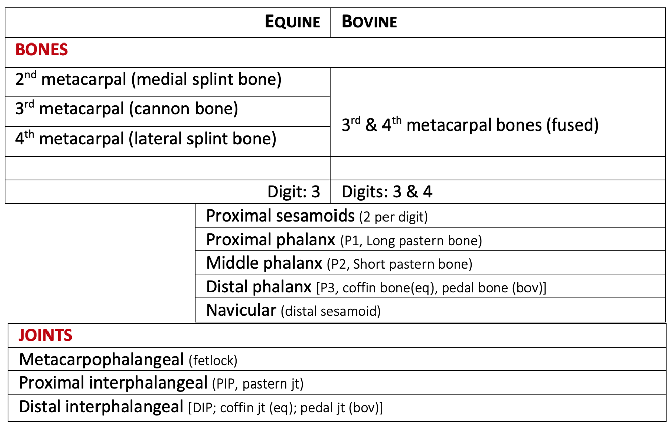
Table for reviewing Distal Bones and Joints in Equine and Bovine
Joint Actions and Muscles (with Innervation) of the (distal) forelimb:
- Extensors of Carpus: extensor carpi radials m. is on the dorsolateral surface of the carpus (innervation – radial n.).
- Flexors of Carpus: flexor carpi ulnaris m., flexor carpi radialis m. (innervation – ulnar, median nn., respectively) are on the palmaromedial surface of the carpus.
- Although innervated by the radial n., the ulnaris lateralis (aka extensor carpi ulnaris) m. is primarily a flexor of the carpus.
- Extensors of Digits: common digital extensor, lateral digital extensor mm. (innervation – radial n.) are on the dorsolateral surface of the carpus and digit(s).
- Flexors of Digits: superficial digital flexor, deep digital flexor mm., (innervation – median, ulnar nn.) are on the palmaromedial surface of the carpus and digit(s).
- Note: The check ligaments (aka accessory ligaments) help prevent hyperextension of the fetlock joint in EQUINE. The check ligaments support the superficial and deep digital flexor m. tendons that run along the palmar surface of the carpus, fetlock, and some of the interphalangeal joints.
- The proximal (radial) check ligament is associated with the superficial digital flexor m. tendon (SDFT). It attaches to the palmar surface of the distal end of the radius and then ties into the SDFT.
- The distal (carpal, inferior) check ligament is associated with the deep digital flexor m. (DDFT). It attaches to the palmar carpal ligament and then ties into the DDFT.
Objective
A2.3 Identify and describe the components of the hoof.
- The “foot” of ungulates is defined as the hoof and its contents. The hoof is sometimes referred to as the capsule of the foot, hence ‘hoof capsule’ is a redundant term.
- Many of the foot structures are best seen on specimens in the anatomy lab.
- Good resource with labeled images: http://vanat.cvm.umn.edu/ungDissect/
Equine Foot (Figs. 3-1 through 3-5 and below)
- The junction of skin and hoof is known as the coronet, from the Latin word corona meaning crown. Some may confuse and interchange the terms coronet and coronary band.
-
- In this transition zone, there is a thin region of epidermis known as the periople. The periople produces a thin waxy layer which protects the hoof wall against drying. It is white, or transparent and easily rubbed off, so it may not be seen. (It is similar to the human cuticle at the skin/nail junction.)
- The 3 main parts of the equine hoof include:
- Wall: most of what can be seen of a hoof in a standing horse is called the wall. The hoof wall is divided into thirds: the toe (front third), quarter (middle third), and heel (back third) (Figs. 5-1, 5-2, 3-2).
- At the heel, the wall is reflected inward and toward the toe as the bars (Fig. 3-1/4).
- The palmar aspects of the heels are referred to as the bulbs (Fig 3-1/3).
- Sole: the semi-flat area on the ground surface of the hoof between the wall and the bars (Figs. 5-5, 3-1/6).
- Clinical Notes: bruises or contusions to the sole can occur. If bruises are between the bar and hoof wall they are often called “corns“. (Supplemental Clinical LAS Corns)
- Frog: is a triangular shaped region on the ground surface of the hoof where the point is called the apex of the frog. There is a single central sulcus of the frog that is rather shallow (Fig. 5-5, 3-1/1,2).
- Two collateral sulci separate the sole and bar from the frog on the ground surface (Fig. 5-5, 3-1/white arrows). This region is usually filled with packed manure and is cleaned out with a hoof pick. There are two collateral sulci per hoof.
- Clinical Notes: Thrush is a degenerative condition of the central sulci and/or collateral sulci of the frog caused by infection from bacteria or fungi. (Supplemental Clinical LAS Thrush)
- Wall: most of what can be seen of a hoof in a standing horse is called the wall. The hoof wall is divided into thirds: the toe (front third), quarter (middle third), and heel (back third) (Figs. 5-1, 5-2, 3-2).
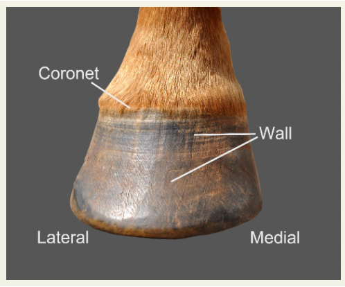
Fig 5-1 Right forefoot, dorsal aspect (from Equine Anatomy Guide: The Forelimb; Mansour, Steiss, Wilhite). Coronet, wall
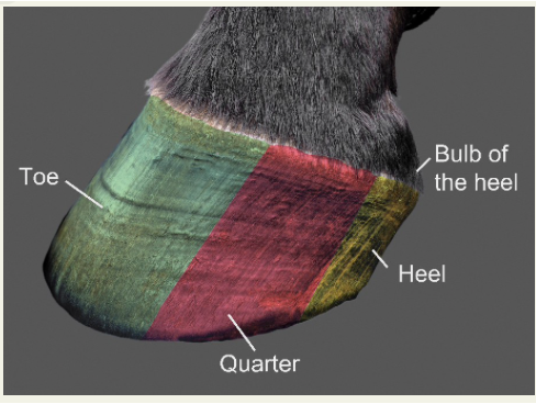
Fig 5-2 Left fore foot, lateral aspect (from Equine Anatomy Guide: The Forelimb; Mansour, Steiss, Wilhite). Hoof wall: toe, quarter, heel, bulb of heel
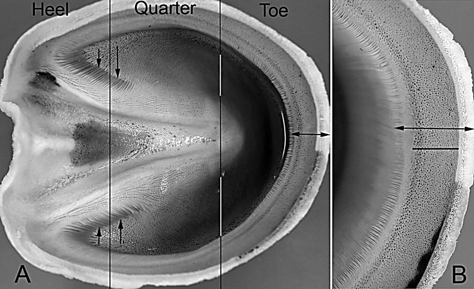
Figure 3-2 A: Proximal view of inner surface of the equine hoof, wet specimen. B: Enlarged view of the wall in the toe region. Double arrow, wall thickness; bar without arrows, extent of coronary groove where small holes of the wall mark the location of soft tissue dermal papillae of the live foot and the horny tubules represent the product of the keratinized stratified squamous epithelium. The horny tubules of the wall are formed by the epithelial covered papillae of the coronary dermis, while the laminae attach the hoof to the dorsal surface of P3 (Fig. 3-3). There are about 600 laminae in the typical hoof. Vertical arrows show that the laminae continue from the inside of the wall on to the bars.
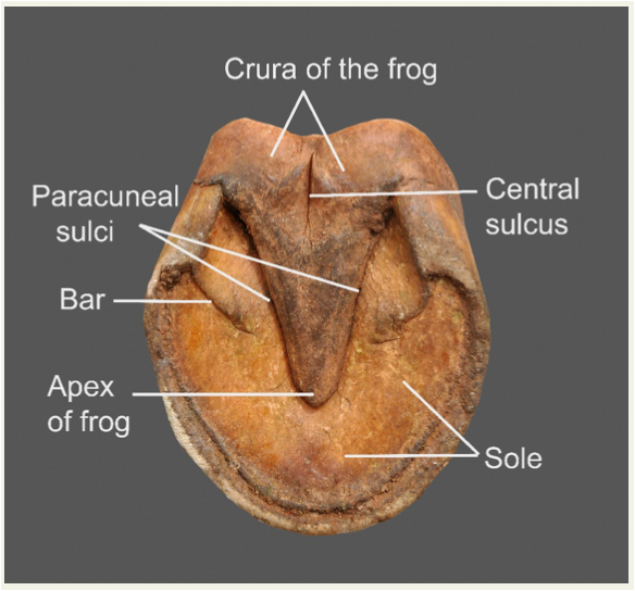
Fig. 5-5 Ground surface aspect of hoof capsule (from Equine Anatomy Guide: The Forelimb; Mansour, Steiss, Wilhite). Frog: apex, central sulcus; sole; bar; collateral sulci (aka paracuneal sulci)
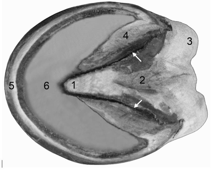 Fig 3-1 Equine foot, ground surface. 1, Apex of the frog; 2, central sulcus of the frog; 3, bulb of heel; 4, bar, which is continuous with 5, wall; 6, sole; arrows, collateral sulci (grooves) that lie between the bars and the frog, and to a lesser extent, between the frog apex and the sole.
- The horny (aka insensitive) laminae are on the inner surface of the wall of the hoof (Figs. 3-2/vertical arrows, 8, 10) and are epidermal. The bars also bear horny laminae.
- The laminar dermis (aka sensitive laminae) extend from the underlying bone (P3) and interdigitate with the horny laminae of the wall of the hoof (Figs. 3-3/LD, 8, 10).
- EQUINE have additional secondary laminae associated with the sensitive laminae which increase surface area, while RUMINANTS do not.
- On the ground surface, the unpigmented laminae (aka ‘white line’) is found at the wall/sole junction (Figs. 3-5/12, 8, 10). The white line represents where the sensitive and insensitive laminae interdigitate.
- Clinical Notes: Hoof abscesses are relatively common and may occur due to a penetrating wound or deep bruise. Horses prone to laminitis may be more prone to abscess due to the insensitive and sensitive laminae boundary already being inflamed. Bacteria and fungi can also enter through the white line causing infection and then degeneration of the hoof wall. (Supplemental Clinical LAS Hoof abscesses)
- The weight of a horse is bearing down on the wall; therefore, there needs to be a very strong connection between the wall, dermis (connective tissue), and P3.
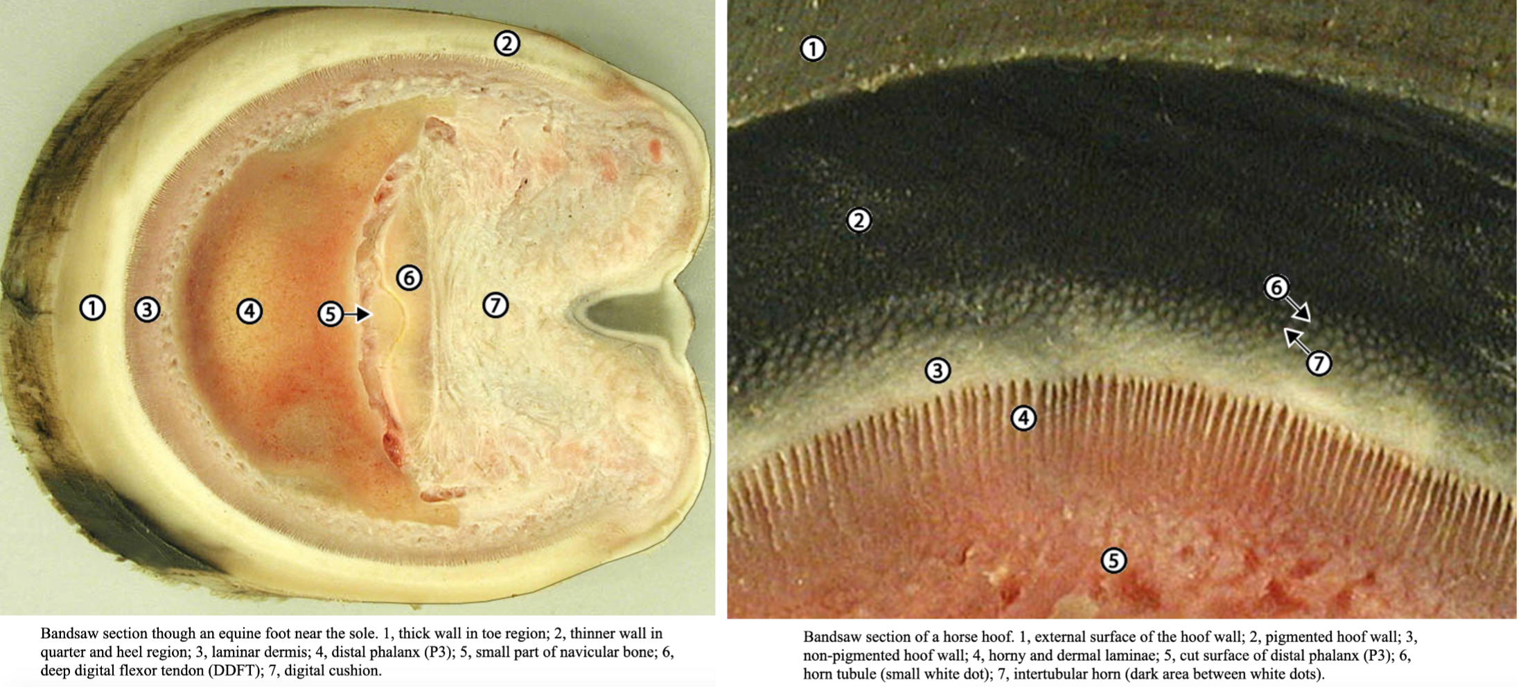
Bandsaw (transverse) section though an equine foot near the sole (LEFT). 1, thick wall in toe region; 2, thinner wall in quarter and heel region; 3, laminar dermis; 4, distal phalanx (P3); 5, small part of navicular bone; 6, deep digital flexor tendon (DDFT); 7, digital cushion. Bandsaw section of a horse hoof (RIGHT). 1, external surface of the hoof wall; 2, pigmented hoof wall; 3, non-pigmented hoof wall “white line“; 4, horny and dermal laminae; 5, cut surface of distal phalanx (P3); 6, horn tubule (small white dot); 7, intertubular horn (dark area between white dots). (images from http://vanat.cvm.umn.edu/ungDissect/Lab04/Lab04.html)
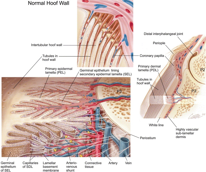
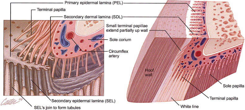
Figs 8, 10. Diagram showing the dermal papillae of the coronet and interdigitating lamellae of the inner hoof wall (TOP); dermal papillae near the sole/ground surface (BOTTOM). The lamellar corium has laminar dermis (aka dermal lamina) that interlock with the horny laminae (aka epidermal lamina) of the inner hoof wall and bars. (https://doi.org/10.1053/j.ctep.2004.07.001)
- The coronary groove is on the inner rim of the hoof (Fig. 5-8 below). Tiny holes in the surface of the groove are filled with dermal papillae from the coronary dermis (aka coronary band/corium) (Figs. 3-3/CD, 8, 10). The coronary dermis/band is simply the dermal tissue deep to the coronary groove of the wall.
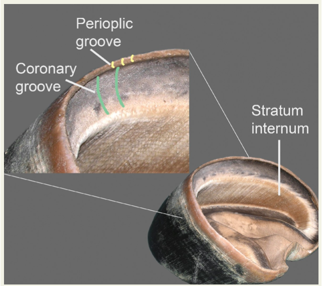
Fig 5-8: Hoof capsule showing internal details: Coronary groove (from Equine Anatomy Guide: The Forelimb; Mansour, Steiss, Wilhite).

Figure 3-3 Plastinated equine foot. LD, laminar dermis (aka sensitive laminae), the region of the dermis that attaches to the insensitive laminae (aka horny laminae) of the wall. These sensitive laminae are what make laminitis so painful because they support the entire weight of the horse. CD, coronary dermis, covered by the wall producing stratified squamous epithelium. The coronary dermis is the true coronary band but this term is often confused with the coronet.
- Observe the collateral cartilages associated with the coffin bone (P3). These cartilages are deep to the skin and above the coronet. (Figs 3-4/C)
- Clinical Notes: on the palmar surface of P3 there are projections above the wall of the hoof. These are the collateral cartilages (medial and lateral) are palpable, and can sometimes ossify. When they ossify they are called “sidebones” and are more common in the thoracic limbs than the pelvic limbs. They are also more commonly seen in heavy/draft breeds of horses. The process of ossification can be a normal part of aging in horses.
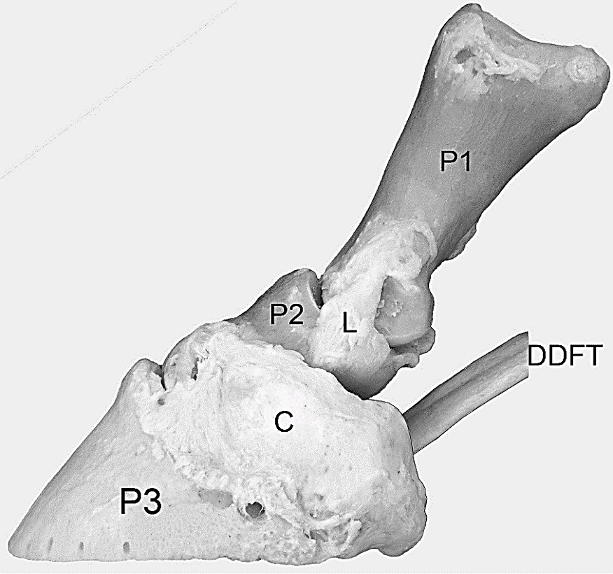
Figure 3-4 Equine digit, lateral view; ligamentous preparation. C, collateral cartilage; L, collateral ligament of the pastern (PIP) joint. The proximal edge of the collateral cartilage can be palpated on the live horse.
- The digital cushion is extensive fibrous fatty tissue between the collateral cartilages, above the frog and parts of the sole, and below the DDFT. It is homologous to the digital pads of the dog, and is a mixture of fat, collagen fibers and small pieces of cartilage. (Fig. 3-5/6)
- The navicular (aka distal sesamoid) bone is located on the palmar (or plantar) surface at the DIP joint (between P2 and P3) (Fig. below, several Figs. above in Objective 2.1, Fig. 24-1).
- The navicular bursa (aka podotrochlear bursa; Figs. 3-5/10 and below) is an important structure between the distal sesamoid (aka navicular) bone and the deep digital flexor tendon.
-
- The deep digital flexor tendon inserts on the solar surface of the coffin bone (P3) (Fig. 3-5).
- The distal sesamoidean impar (i.e., “unpaired”) ligament (aka distal navicular ligament) runs from the distal sesamoid bone to the palmar surface of the coffin bone (Fig. 3-5/13 and below).
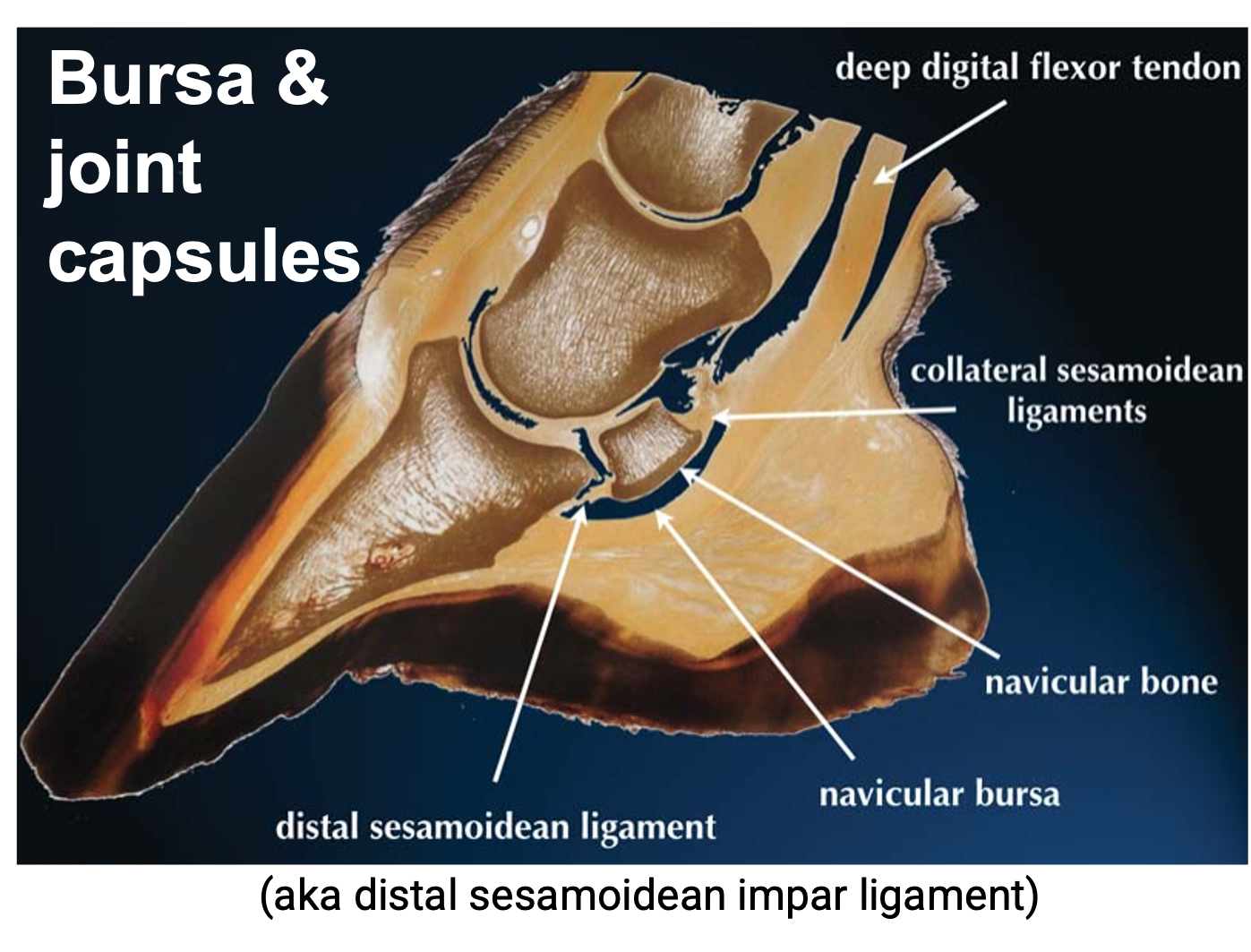
Fig above – sagittal cut through equine hoof: Distal sesamoidean impar ligament, navicular bursa, navicular bone, deep digital flexor tendon.
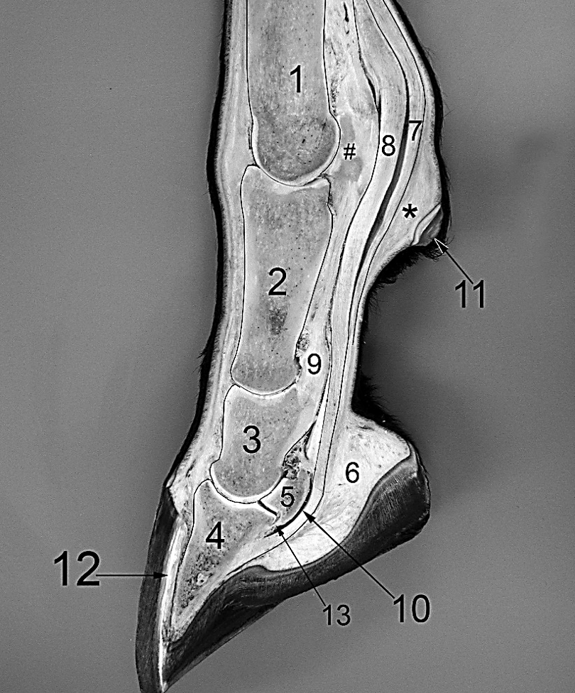
Figure 3-5 Sagittal split equine foot and digit. 1, distal cannon bone (Mc3); 2, P1; 3, P2; 4, P3; 5, navicular bone (distal sesamoid); 6, digital cushion; 7, superficial digital flexor tendon (SDFT); 8, deep digital flexor tendon (DDFT); 9, superficial distal sesamoidean ligament extending from the proximal sesamoids (#) to the fibrocartilage on the proximal palmar aspect of P2; 10, the navicular bursa lying between the navicular bone and the flexor surface of the DDFT; 11, the ergot; *, fibrous pad deep to the horny ergot that is present even when no horny tissue of the ergot is present; 12, horny (insensitive) laminae and adjacent wall forming the so called ‘white line’ that may be observed on the ground surface between the hoof wall and sole; 13, distal sesamoidean impar ligament which connects the navicular bone to P3. Note that the length of P1 is twice that of P2.
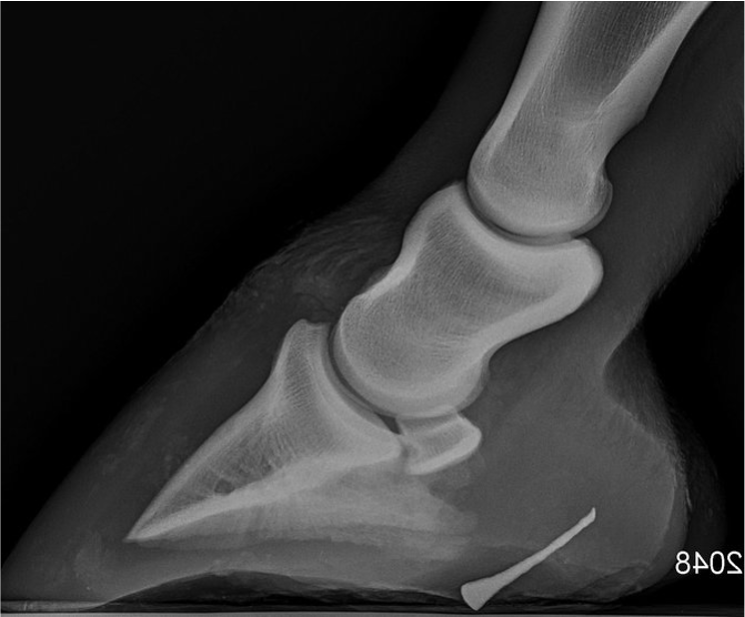
- Clinical Notes: penetration of hoof structures by a nail. Be able to determine and list what structures may be damaged in the hoof due to penetration of the hoof by a foreign body. Depending on which structures are involved, treatment can involve soaking and bandaging or emergency surgery (sometimes called “street nails“).
- In the radiograph below (lateral view of distal limb), you can see a nail has penetrated the more caudal/palmar portion of the hoof. You would need another radiographic view to see exactly where along the midline this nail is reaching.
- From this particular view, the nail could be penetrating: the digital cushion.
Ruminant Foot (Figs. 3-6 and 3-7) – Many of the foot structures are best seen on specimens in the anatomy lab. (see LAS Bovine Foot Anatomy, Lameness, Approaches)
- RUMINANT feet are referred to as digits or hooves. The ruminant foot is similar in some ways to the horse foot, but all of the digital structures and joints are “doubled.”
- There are 4 proximal sesamoids and 2 distal sesamoids (aka navicular bones), and one fetlock, but two pastern, and two coffin joints per foot.
- The coronet is the junction of skin and hoof. In this transition zone, there is a thin region of epidermis known as the periople. (Fig. 3-6) The periople produces a thin waxy layer which protects the hoof wall against drying.
- The coronary groove is on the inner rim of the hoof and represents the area that is filled with dermal papillae from the coronary dermis (aka coronary band/corium).
-
- Digits:
- Each weight-bearing digit contains a proximal (P1/long pastern), middle (P2/short pastern), and distal (P3/coffin/pedal) phalanx.
- There is a proximal interphalangeal (PIP) and distal interphalangeal (DIP/coffin/pedal) joint.
- Digits 3 and 4 are weight bearing.
-
- Digits 2 and 5 are dewclaws in most RUMINANTS (pronghorn antelopes, giraffes, and camelids lack these).
-
- Instead of the digit being symmetrical, each digit has a rounded and thicker abaxial wall (meaning away from the midline or axis) and a flattened and thinner axial wall (meaning towards the midline). The coronet is higher above the ground surface on the abaxial side of the digit than on the axial side. (Figs. below)
- Clinical Notes: the axial wall of the hoof is thinner than the abaxial wall (see figure below). Therefore, the pedal joint aka (coffin joint) is more vulnerable to trauma and infection on the axial side. Overgrowth of the abaxial wall of the hooves of sheep and goats is common and necessitates frequent trimming.
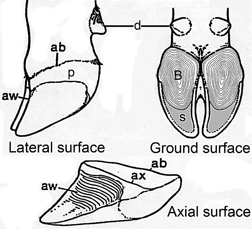
Figure 3-6 Bovine Foot: ab, abaxial coronet; ax, axial coronet; aw, axial wall; B, bulb of heel; d, dewclaw; p, periople; s, sole. The abaxial coronet is higher than the axial coronet.
-
- The interdigital cleft (it continues as the interdigital space) is the separation between the two digits. (Fig. below)
- Two interdigital ligaments join digits 3 and 4. The most significant one is the distal interdigital ligament (Fig. 3-7/10) which crosses from each distal sesamoid bone to the middle phalanx of the adjacent digit. This ligament prevents the digits from splaying too far from the foot axis. This ligament is well developed in CATTLE but poorly developed in SMALL RUMINANTS.
- Ground surface features:
- The digits of RUMINANTS have a large heel portion, known as the bulb (Fig. below, 3-6).
- The sole is the portion of the ground surface of the hoof between the bulb and wall. The distinction between bulb and sole is more defined in SHEEP and GOATS than in CATTLE.
- There is no frog and no collateral cartilages. The sole is reduced in size relative to EQUINE feet.
- The unpigmented laminae aka white line is found at the wall/sole junction (Fig. below), and represents where the sensitive and insensitive laminae interdigitate with each other (similar to EQUINE).
- The horny (aka insensitive) laminae are on the inner surface of the wall of the hoof.
- The laminar dermis (aka sensitive laminae) is more internal to the insensitive laminae of the hoof wall.
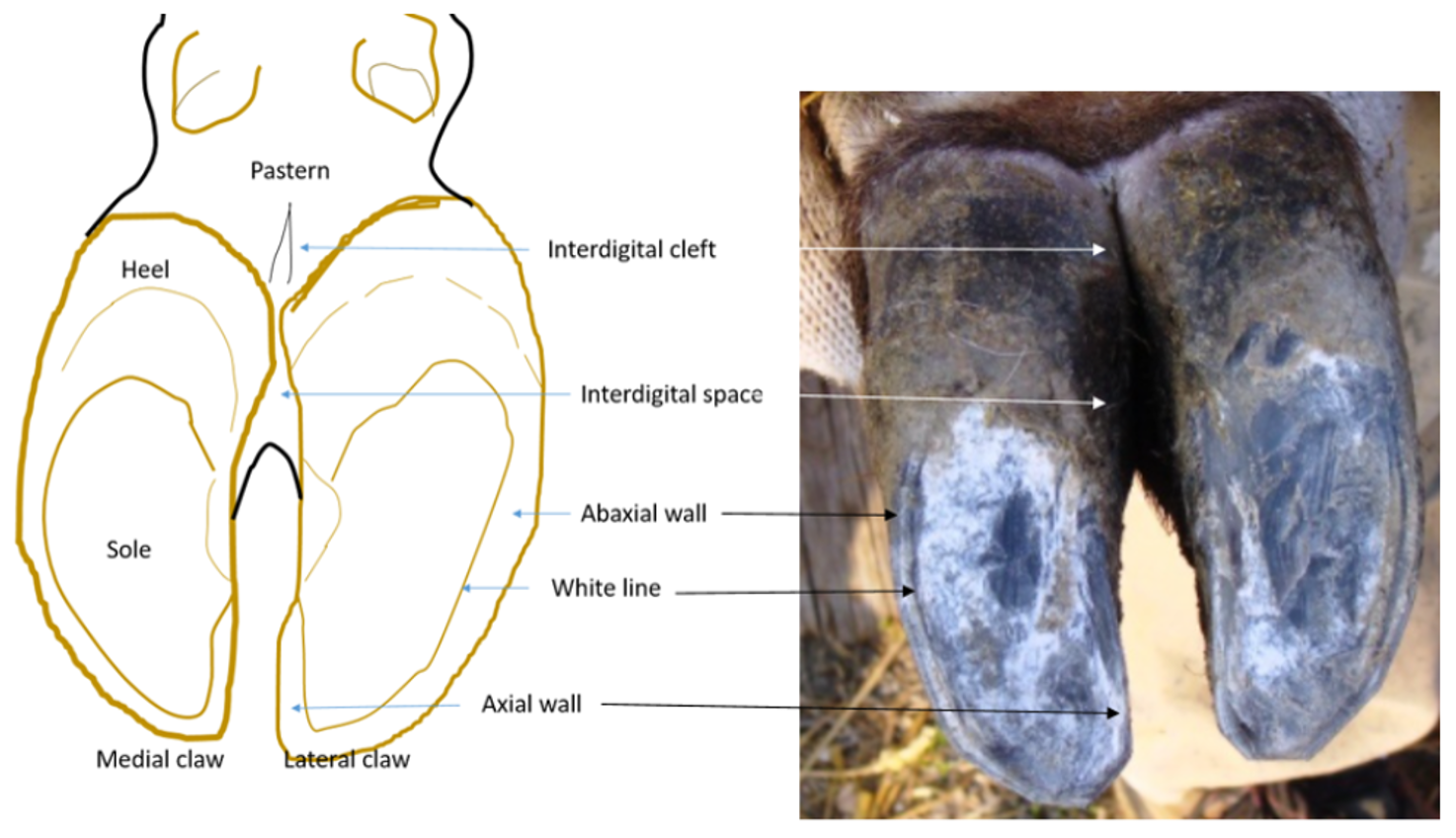
Fig above – Bovine foot, ground surface: medial (#3) and lateral (#4) digits; dewclaws (digits 2 and 5) at top of left image; 2, heel bulb; sole; interdigital space (and cleft); axial wall; abaxial wall; white line. (LAS Bovine Anatomy)
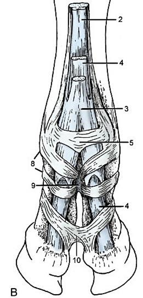
Figure 3-7 B. Bovine foot. Palmar view. 2, Suspensory ligament (interosseous tendon); 3, superficial digital flexor; 4, deep digital flexor; 5, palmar annular ligament; 8, digital annular ligaments; 9, proximal interdigital ligament; 10, distal interdigital ligament. Modified from TVA Figs. 23-29, 30-7
- Note how extensive the digital cushion is in BOVINE, similar to EQUINE (Fig. 30.11/8) and the point of insertion of the deep digital flexor tendon on P3, called the flexor tuberosity. The pressure of this tuberosity likely creates or leads to sole ulcers. (see LAS Bovine Foot Anatomy, Lameness, Approaches)

Fig. 30.11 Sagittal section of the medial digit of the bovine forefoot. 1, Proper (medial) digital extensor tendon; 2, common digital extensor tendon; 3, coronary dermis; 4, laminar dermis; 5, middle phalanx; 6, distal phalanx; 7, sole dermis covered by sole; 8, digital cushion; 9, deep digital flexor tendon; 9′, fibers of deep digital flexor to the middle phalanx and navicular bone; 10, navicular bone; 11, collateral navicular ligament; 12, palmar ligaments of pastern joint; 13, superficial digital flexor tendon. (TVA)
Camelid Foot (Figs 3-8, 3-9)
- The camelid hoof is reduced to a large claw covering the distal phalanx (digits 3 and 4).
- Camelids have padded feet and are classified in the Artiodactyla (even-toed ungulates) suborder Tylopoda (padded foot).
- Small camelids have metatarsal pads that are commonly referred to as chestnuts, but are not part of the foot. These are different from EQUINE chestnuts since they are only in the hindlimb, on the metatarsus (not tarsus), both medial and lateral sides of the metatarsus (larger on lateral), and are flat and soft instead of elevated and horny.
- In the anatomy lab there are dry specimens of camel, llama, and alpaca feet.

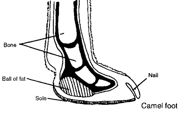
Figure 3-9 Camel foot. The large wide pad of the camel aids in walking on sandy terrain without sinking. Camels have a similar arrangement of digits and phalanges as in alpaca and llama feet.
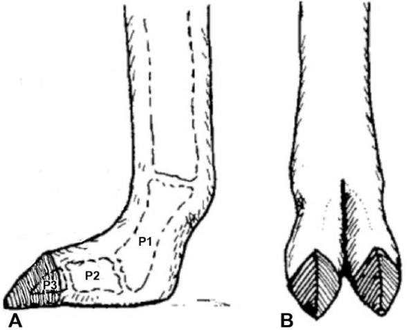
Figure 3-8 Alpaca and Llama feet. Digits 3 and 4. Weight-bearing portion of digits is P2 and P3.
Pig Foot (Figs. 3-10, 3-11)
- In PIGS, digit 4 is larger than digit 3.
- Digits 2 and 5 form the dewclaws, which are elongated and of similar size.
- The PIG foot has a true hoof (similar to that of ruminants), but the bulb is soft like that of padded feet animals.
- The hoof has axial and abaxial walls and a reduced sole.
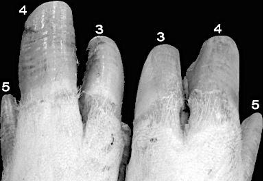
Figure 3-11 Pig feet, dorsal view. The lateral digit (4) is larger than the medial digit (3). The dewclaws are digits 2 and 5 (medial dewclaws are not shown).
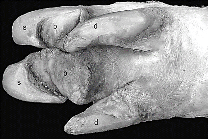
Figure 3-10 Pig foot, palmar view. b, bulb; d, dewclaw; s, sole
Objective
A2.4 Identify and describe the ligaments and tendons of the foot structures in equine and bovine.
EQUINE
- Digital extensors muscle tendons in the EQUINE:
- The common digital extensor m. tendon attaches mainly to the extensor process of P3 (Fig. 23-26/17 below).
- The lateral digital extensor m. tendon attaches to the dorsal aspect of P1.
- Clinical Notes: the digital extensor tendons can be lacerated causing “knuckling over.”
- The primary distal components of the passive forelimb stay apparatus (EQUINE) include the superficial and deep digital flexor tendons and the suspensory apparatus of the fetlock joint.
- Digital flexor muscle tendons:
- The superficial digital flexor tendon (SDFT) is superficial to the deep digital flexor tendon (DDFT) in the metacarpal region. At the level of the fetlock, the SDFT forms a sleeve for the DDFT to pass through. This is called the flexor manica. The SDFT inserts on P1 and P2, near the pastern joint. (Figs. 23-28 and 23-26/18 below).
- Digital flexor muscle tendons:
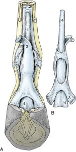
FIG. 23.28 Relations and topography of the superficial and deep flexor tendons. (A) Palmar view, in situ. (B) Dorsal view, isolated. 1, Splint bones; 2, interosseus; 3, superficial digital flexor; 4, deep digital flexor. (TVA)
-
-
- After coursing through the flexor manica, the DDFT continues distally and attaches to the solar surface of P3 (Fig. 23-26/13 below).
- The distal (carpal, inferior) check ligament attaches to the palmar surface of the carpus (palmar carpal ligament) and proximal end of the cannon bone and then ties into the DDFT.
- Clinical Note: understanding the relative position and shape of the ligaments and tendons of the palmar surface of the distal forelimb will be important for palpation and ultrasonography.
- The distal (carpal, inferior) check ligament attaches to the palmar surface of the carpus (palmar carpal ligament) and proximal end of the cannon bone and then ties into the DDFT.
- After coursing through the flexor manica, the DDFT continues distally and attaches to the solar surface of P3 (Fig. 23-26/13 below).
-
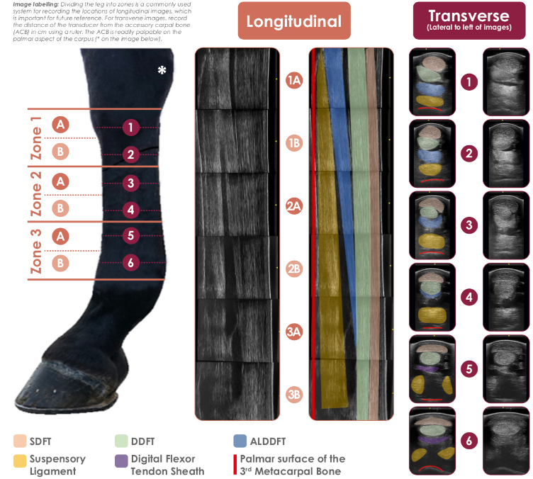
Figure above – This guide shows the normal ultrasonographic anatomy of the metacarpal soft tissues. The superficial (SDFT) and deep digital flexor tendons (DDFT), accessory ligament of the DDFT (ALDDFT) and suspensory ligament are evaluated primarily from the palmar aspect, in longitudinal and transverse sections. (from www.IMV-imaging.com)
-
-
- Clinical Notes: club foot is a type of flexural deformity of the coffin (DIP) joint and is a common problem in young, growing horses. The contracture of the deep digital flexor tendon causes coffin joint flexion. Characteristics of a club foot are a prominent or bulging coronary band, a very upright hoof wall angle, and a long heel (if the condition is severe the heel may not touch the ground). (Supplemental Clinical LAS Flexural limb deformities) (Supplemental Clinical LAS Inferior check desmotomy)
-
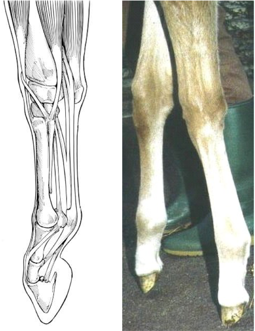
Fig. above – Type 2 club foot: coffin joint, deep digital flexor muscle tendon
-
-
- The digital tendon sheath surrounds the superficial and deep digital flexor tendons from the distal one-fourth of the metacarpal region to the middle of P2. (Fig. 23-26/14, Fig.3-26 below). This sheath can become inflamed and appear as a swelling on the palmar aspect of the proximal sesamoids (sometimes called “wind puffs” or “wind galls”). Sheaths and tendons can also be inflamed or infected (tenosynovitis, tendonitis) due to ascending foot infections (in both EQUINE and BOVINE). (Supplemental Clinical LAS Tendonitis and bowed tendons)
-
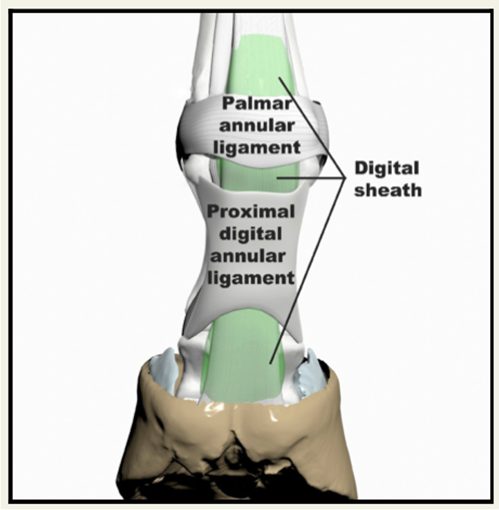
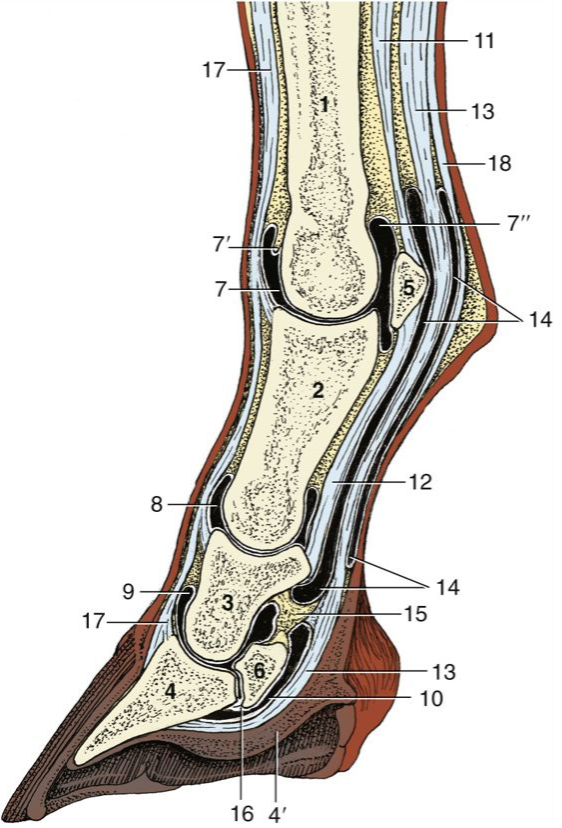
Figure 23-26 Sagittal section of equine digit (semi-schematic). 1, metacarpal bone/cannon; 2, proximal phalanx; 3, middle phalanx; 4, distal phalanx; 4ʹ, digital cushion; 5, proximal sesamoid bone; 6, distal sesamoid (navicular) bone; 7, dorsal pouch of fetlock joint; 7ʹ, capsular fold; 7ʺ, palmar pouch of fetlock joint; 8, 9, dorsal pouch of pastern and coffin joints; 10, navicular bursa; 11, suspensory ligament (interosseous tendon); 12, straight sesamoidean ligament; 13, deep flexor tendon; 14, digital sheath; 15, connective tissue bridge; 16, distal navicular ligament; 17, common digital extensor tendon; 18, superficial flexor tendon. (TVA)
-
- The suspensory apparatus of the fetlock joint supports the fetlock by preventing hyperextension. The components of the suspensory apparatus include the proximal sesamoid bones, the suspensory ligament (interosseous tendon), and the distal sesamoidean ligaments (of the proximal sesamoids). These structures form a “sling” that supports the fetlock joint.
-
-
- The suspensory ligament (aka interosseous tendon) originates from the distal row of carpal bones and proximal Mc3 and runs along the palmar aspect of the cannon bone. At the level of the metacarpus, the suspensory ligament is deep to the digital flexor tendons (Figs. 23-26, 3-24). Proximal to the fetlock, the suspensory ligament divides into two thick branches (medial and lateral), which insert on the abaxial surface of the medial and lateral proximal sesamoid bones.
- Each thick branch of the suspensory ligament gives rise to a thinner extensor branch, which courses dorsally and joins the common digital extensor m. tendon on the dorsal surface of the foot (near P2) (Figure. 2-5 Left Forelimb). Tension in the extensor branches helps counteract flexion of the coffin joint caused by pull of the DDFT.
- The distal sesamoidean ligaments of the proximal sesamoids (Fig. 2-4/4,3, 3-24):
- The superficial (aka straight) sesamoidean ligament courses from the base of the proximal sesamoid bones to the proximal end of P2.
- The middle (aka oblique) sesamoidean ligament extends from the distal outer edges of the proximal sesamoid bones to the triangular roughened area on the palmar surface of P1.
- The suspensory ligament (aka interosseous tendon) originates from the distal row of carpal bones and proximal Mc3 and runs along the palmar aspect of the cannon bone. At the level of the metacarpus, the suspensory ligament is deep to the digital flexor tendons (Figs. 23-26, 3-24). Proximal to the fetlock, the suspensory ligament divides into two thick branches (medial and lateral), which insert on the abaxial surface of the medial and lateral proximal sesamoid bones.
-

- The collateral ligaments between phalanges (e.g., joints: fetlock/MCP, pastern/PIP, coffin/DIP) provide support medially and laterally on hinge joints (see image below; EQUINE shown – bovine also have these but are not discussed).
- Medial and lateral collateral ligaments of the fetlock/MCP: C in image below, between cannon/Mc3 and P1.
- Medial and lateral collateral ligaments of the pastern joint: between P1 and P2.
- Medial and lateral collateral ligaments of the coffin joint: A in image below, between P2 and P3.
- Medial and lateral collateral ligaments of the navicular bone (aka sesamoidean, aka suspensory ligaments of the navicular bone): B in image below, between P1 and navicular bone.
- The proximal sesamoids also have collateral ligaments, between Mc3 and each sesamoid (D in image below).

- Annular ligaments (EQUINE):
- Palmar annular ligament: holds superficial and deep digital flexor tendons in place on the palmar surface of the fetlock. (Fig. 23.29/5).
- Proximal digital annular ligament: superficial to the digital flexor tendons on the palmar aspect of P1, makes an “X” shape in EQUINE (Fig. 23.29/6).
- Distal digital annular ligament: superficial to the deep digital flexor tendons on the palmar aspect of P2 and P3, makes a “U” shape in EQUINE (Fig. 23.29/7).
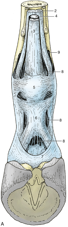
FIG. 23.29 (A) Annular ligaments of the digit. 1, Splint bones; 2, suspensory ligament (interosseous tendon); 3, superficial digital flexor; 4, deep digital flexor; 5, palmar annular ligament; 6, proximal digital annular ligament; 7, distal digital annular ligament; 8, digital sheath; 9, palmar pouch of fetlock joint. (TVA)
BOVINE ligamentous attachments distally:
- Digital flexor muscle tendons (one tendon for each digit):
- In the metacarpal region, the SDFT is superficially positioned. At the level of the fetlock, the SDFT forms a sleeve for the DDFT to pass through. This is called the flexor manica. The SDFT inserts on P2 of digits 3 and 4 (Fig. 30.6/12, Fig. 3-7/3 below).
- The DDFT is deep on the palmar surface proximal to the fetlock, then becomes superficial distally and attaches to the solar surface of P3 of digits 3 and 4 (Fig. 30.6/11, Fig. 3-7/4 below).
- The digital tendon sheath travels along the outside of the digital flexor tendons from the around the proximal fetlock to the mid pastern (Fig. 30.6/15 below). In bovine, the tendon sheath can become inflamed or infected due to ascending infections from the foot. (e.g., Supplemental Clinical LAS Foot Rot)
- There is a palmar annular ligament and digital annular ligaments, as well as the distal (and proximal) interdigital ligaments (Fig. 3-7/5,8,10 below).
Figure 3-7 B. Bovine foot. Palmar view. 2, Suspensory ligament (interosseous tendon); 3, superficial digital flexor; 4, deep digital flexor; 5, palmar annular ligament; 8, digital annular ligaments; 9, proximal interdigital ligament; 10, distal interdigital ligament. Modified from TVA Figs. 23-29, 30-7
- Bovine also have a suspensory ligament (aka interosseous tendon) on the palmar aspect of Mc3/Mc4 that extends distally and splits into four branches which insert on the proximal sesamoid bones. It has more muscle fibers than in the EQUINE (Fig. 30.6/16 below; Fig. 3-7/2 above).
- The extensor branches of the suspensory ligament reach the common digital extensor m. tendon and the lateral digital extensor m. tendon on the dorsal surface of the foot (near P2) (Fig. 30.9/4,5 below).
- There are also distal sesamoidean ligaments for each digit between the proximal sesamoids and P1 (Fig. 30.6/13 below).
- Digital extensors muscle tendons:
- The common digital extensor m. tendons attach to digits 3 and 4 (Fig. 30.6/6, 30.9 below).
- The lateral digital extensor m. tendon attaches to digit 4 (Fig. 30.6/1 below).
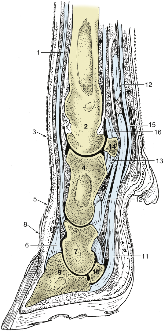
FIG. 30.6 Sagittal section of the bovine foot, splitting the lateral digit. 1, Lateral digital extensor; 2, metacarpal bone (Mc3,4); 3, fetlock (MCP) joint; 4, proximal phalanx (P1); 5, pastern (PIP) joint; 6, common digital extensor; 7, middle phalanx (P2); 8, coffin (DIP/pedal) joint; 9, distal phalanx (P3/coffin/pedal); 10, navicular (distal sesamoid) bone; 11, deep digital flexor tendon; 12, superficial digital flexor tendon; 13, distal sesamoidean ligaments; 14, proximal sesamoid bone; 15, digital sheath; 16, suspensory ligament (interosseous tendon). (TVA)
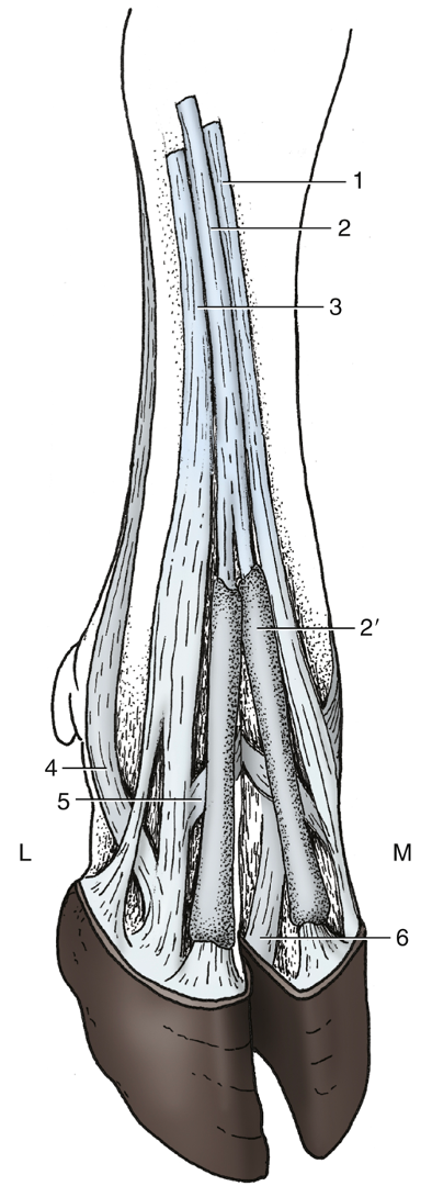
FIG. 30.9 Dorsal view of the bovine right forefoot. L, Lateral; M, medial; 1, medial tendon of common digital extensor to the medial digit; 2 and 2′, common digital extensor and its sheaths, respectively; 3, lateral digital extensor; 4 and 5, abaxial and axial extensor branches, respectively, of the interosseous to the lateral digital extensor; 6, common axial collateral ligament. (TVA)
Objective
A2.5 Describe the main vessels and nerves supplying the foot and digit(s).
Vessels
- Arteries in EQUINE: The main artery in the antebrachium is the median a. The median a. continues to the level of the carpus and then becomes the medial palmar a. (Fig. 23.39)
- The medial palmar a. courses in the metacarpal region with the medial palmar nerve and vein.
- Clinical Notes: The medial palmar a. is the main blood supply to the digit. A severe laceration or cut to the palmar cannon, fetlock, or pastern region can reduce all blood flow to the foot.
- Just proximal to the fetlock the medial palmar a. splits into the medial and lateral palmar digital aa. (Fig. 23.39).
- The medial palmar a. courses in the metacarpal region with the medial palmar nerve and vein.
- Veins in EQUINE: The medial and lateral palmar digital veins drain blood from the foot primarily into the medial palmar v. which mainly drains into the cephalic v. The cephalic v. drains into the external jugular v.
- The dorsal aspect of the distal forelimb is drained by the accessory cephalic v., which drains into the cephalic v.
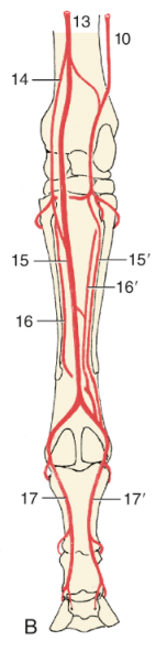
FIG. 23.39 The major arteries (aa.) of the right equine forelimb: (B) palmar view. 10, collateral ulnar a.; 13, median a.; 14, radial a.; 15 and 15′, medial and lateral palmar aa., respectively; 16 and 16′, medial and lateral palmar metacarpal aa., respectively; 17 and 17′, medial and lateral palmar digital aa., respectively. (TVA)

FIG. 30.13 The principal arteries on the bovine right forelimb; medial view. 1, Axillary artery (a.); 2, subscapular a.; 3, deep brachial a.; 4, brachial a.; 5, collateral ulnar a.; 6, common interosseous a.; 7, median a.; 8, radial a.; 9, palmar common digital a. III; 10, dorsal common digital a. III. (TVA)
- Arteries in BOVINE: The median a. changes name and is continued as the palmar common digital a. III. This name change occurs just before/just proximal to the fetlock (i.e., at the level of the fetlock). Between the carpus and fetlock, the main blood supply to the bovine digits is still called the median a. At the fetlock, the name change occurs – so the main blood supply from the fetlock to the digits comes from the palmar common digital a. III. (Step #16 in Distal Thoracic Limb of the LAA Dissection eBook)
- Veins in BOVINE:
- To numb the foot for diagnostics or treatment, IV regional anesthesia (IVRA) is often performed. (Supplemental Clinical LAS Bovine Foot Anatomy, Lameness, Approaches)
-
- Distal veins commonly used for clinical procedures in bovine include:
- The dorsal common digital v. is located on the dorsal surface of the distal metacarpus and fetlock (Fig. 30.14 and below), and it drains into the accessory cephalic v. (Step #23 in Distal Thoracic Limb of the LAA Dissection eBook)
- The medial (aka axial) or lateral (aka abaxial) palmar (on forelimbs) or plantar (on hind limbs) digital vv. drain to more proximal veins in the forelimb or hind limb.
- Distal veins commonly used for clinical procedures in bovine include:
Figure 3. A 19 ga butterfly needle placement for RIVP in the dorsal common digital v. of the bovine distal limb. Photo courtesy Dr. John Gilliam, Oklahoma State University.
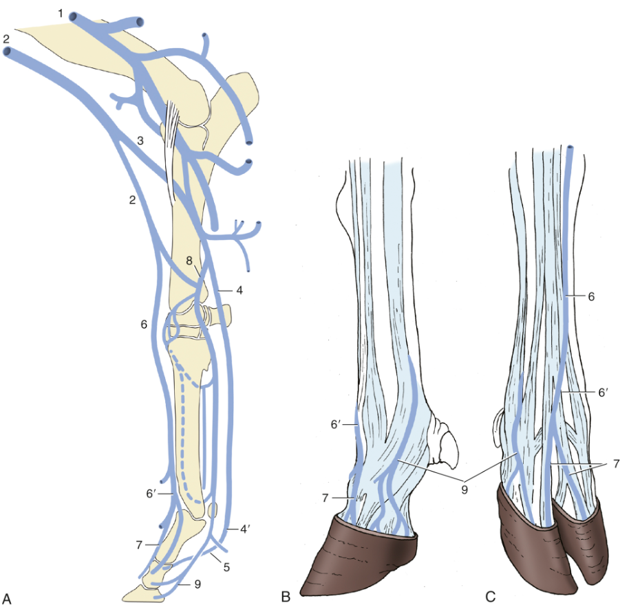
Figure 30.14 The principal veins (vv.) of the bovine forelimb. (A) Right limb, medial view. (B) Left foot, lateral view. (C) Right foot, dorsal view. 1, Brachial vein (v.); 2, cephalic v.; 3, median cubital v.; 4, median v.; 4′, palmar common digital v. III; 5, axial palmar digital vv.; 6, accessory cephalic v.; 6′, dorsal common digital v. III; 7, dorsal digital vv.; 8, radial v.; 9, abaxial palmar digital vv. (TVA)
Nerves (Supplemental Clinical in LAS Nerve and Joint Blocks)
- EQUINE: There are two main nerves found distally in the thoracic limb: median and ulnar nn. They provide motor innervation to the flexors of the carpus and digits (e.g., flexor carpi radialis, superficial digital flexor, deep digital flexor, flexor carpi ulnaris mm.).
- The median and ulnar nn. course distally along the medial and caudal aspects of the antebrachium, respectively.
- Proximal to the carpus, the median n. branches into the medial and lateral palmar nn. The ulnar n. sends a branch to join the lateral palmar n. The medial palmar n. runs within the carpal canal.
- Just distal to the carpus, the lateral palmar n. gives rise to a deep branch which innervates the interosseous tendon and gives rise to medial and lateral palmar metacarpal nn. These nerves innervate the deep structures of the metacarpus and fetlock.
- In the metacarpal region, the medial and lateral palmar nn. course between the interosseous tendon and the flexor tendons on the medial and lateral sides of the limb. Near the mid-cannon region, the medial palmar n. gives off a communicating branch of the palmar nn., which runs over the superficial digital flexor tendon to join the lateral palmar n.
-
Clinical Notes: The location of the communicating branch needs to be considered when performing some of the nerve blocks of the foot because sensory fibers cross from the lateral to the medial side. This could cause confusion in lower palmar blocks if the block is performed below the level of the communicating branch on one side and above the location of the communicating branch on the other side.
-
- Just proximal to the digit (near the MCP/fetlock joint), the names of the palmar nn. change to medial and lateral palmar digital nn. (often shortened to PDN).
- The palmar digital nn. give off dorsal branches on the abaxial sides of the proximal sesamoids; these dorsal branches innervate the dorsal aspect of the fetlock and pastern joint capsules. If these branches are disturbed, the horse may not be able to sense the placement of its hoof.
- Note: in the most distal extent of the thoracic limbs, the nerves carry sensory information, as there are no muscle bellies in the distal thoracic limb. Most of the sensory information from the medial side of foot, and some of the lateral, is carried within the median n. Most of the lateral sensory information is carried within the ulnar n.
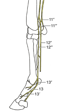
FIG. 23.43 Distribution of the nerves in the right forelimb, medial view. 11, ulnar n.; 11′, caudal cutaneous antebrachial n.; 11″, palmar branch; 11′″, dorsal branch; 12, median n.; 12′, muscular branches; 12″, lateral palmar n.; 12′″, medial palmar n.; 13, medial palmar digital n.; 13′ and 13″, dorsal branches. (TVA)
- BOVINE: the median n. gives rise to a communicating branch and several digital nerves run along the medial and lateral aspects of each digit (as well as dorsal branches from the radial and ulnar nn.).
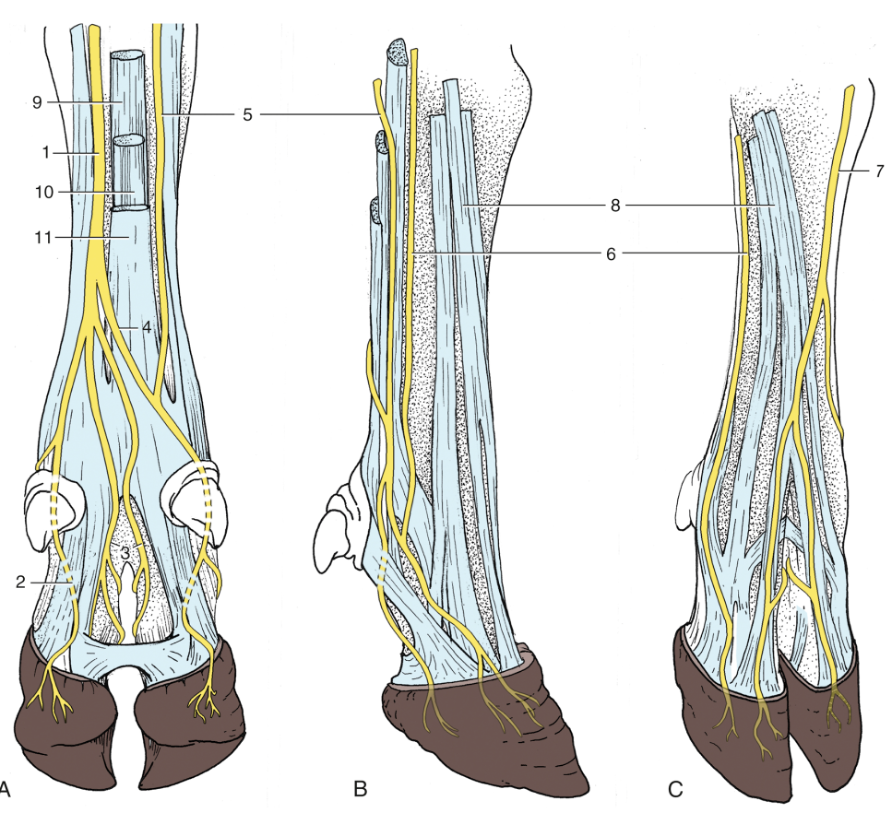
Figure 30.16 The principal nerves of the bovine right forefoot in (A) palmar, (B) lateral, and (C) dorsal views. 1, Median nerve (n.); 2, palmar abaxial digital n.; 3, palmar axial digital nerves; 4, communicating branch; 5, palmar branch of ulnar n.; 6, dorsal branch of ulnar n.; 7, superficial branch of radial n.; 8, digital extensor tendons; 9, suspensory ligament (interosseous tendon); 10, deep flexor tendon; 11, superficial flexor tendon. (TVA)
Objective
A2.6 Describe laminitis and navicular syndrome in equine and common foot disorders in bovine; identify the related anatomical structures.
- Laminitis aka “Founder” is inflammation of the laminae (Supplemental Clinical LAS Laminitis):
- Many different mechanisms can cause laminitis, but lead to the same breakdown or inability of the laminae to hold P3 (aka coffin bone) in place. Over time and depending on severity, there can be separation of P3 partially or completely from the hoof wall.
- P3 tends to rotate downward due to gravity and the pull of the deep digital flexor tendon (DDFT), as the DDFT is attached to the palmar surface of P3.
- Possible treatments are based on the underlying cause(s) and may include: pain relievers (NSAIDs), changing diet, limit walking, reduce swelling, shoeing, and sometimes surgery.
- Chronic and severe cases: Cutting the distal (aka inferior) check ligament may be sufficient in a few cases. In many cases, the deep digital flexor tendon is transected in the pastern region or in the mid-cannon bone region. This temporarily relieves the pull on the coffin bone. The tendon will heal so the surgery must be accompanied by appropriate farrier work to ensure it heals with the desired conformation. (Supplemental Clinical LAS Inferior check desmotomy)

Fig. above – Sagittal section of a horse foot with severe laminitis pathology. 1, proximal phalanx = P1; 2, middle phalanx = P2; 3, distal phalanx rotated away from the hoof wall due to laminar breakdown and the pull of the DDFT on P3; 4, distal sesamoid (navicular) bone; 5, proximal interphalangeal (PIP) joint; 6, distal interphalangeal (DIP) joint; 7, deep digital flexor tendon (DDFT); 8, navicular (podotrochlear) bursa; 9, distal sesamoidean impar ligament; 10, superficial (straight) distal sesamoidean ligament; 11, digital cushion; 12, widened laminar area due to breakdown of laminae. Inset image shows a white fibrocartilaginous portion of the DDFT between the arrows. (http://vanat.cvm.umn.edu/ungDissect/Lab04/Img4-12.html)
- Navicular syndrome or disease is a complex chronic degenerative condition of the navicular apparatus that is a common cause of forelimb lameness in horses. The navicular apparatus involves the navicular bone, including nearby tendons, bursae, joints, and ligaments. It is often bilateral and commonly found in Quarter horses. (Supplemental Clinical LAS Navicular syndrome)
- Relationship of navicular bone to nearby structures (aka the navicular apparatus; see Fig. below and section A2.1 above for text and images).
- The navicular (aka distal sesamoid) bone is located on the palmar (or plantar) surface at the DIP joint (between P2 and P3) and articulates with these bones.
- The navicular bone is deep to the frog, so hoof testers would show sensitivity over the frog.
- The navicular bursa is an important structure between the navicular bone and the deep digital flexor tendon. The DDFT inserts on the solar surface of the coffin bone (P3).
- Clinical Notes: The structures of the “heel” are compact and close together, so it can be hard to separate them to perform diagnostic exams and to see them on imaging. Inflammation and infection that impact one structure often impacts others. For example, bursitis can lead to navicular bone and tendon damage, as well as adhesions. Additionally, if an anesthetic is used to block the bursa, the palmar digital nn. are also blocked.
- The distal sesamoidean impar ligament (aka distal navicular ligament) runs from the distal sesamoid bone to the palmar surface of the coffin bone. There are also medial and lateral collateral ligaments of the navicular bone.
- Synovial invaginations (synovial fossae, see Fig. 24-1/A distal surface) into the navicular bone (sometimes they look like lollipops in radiographs of the navicular bone) are clinically important as they allow for medications injected into the coffin joint to perfuse the navicular bone. Unfortunately, this also means inflammatory mediators in the joint can have access into the bone and if the joint is under higher pressure, the bone is too.
- Relationship of navicular bone to nearby structures (aka the navicular apparatus; see Fig. below and section A2.1 above for text and images).
FIG. 24-1 The navicular bone. A, view of the distal surface with synovial fossae (small holes). 1, The small articular surface with the distal phalanx (P3); 2, the projection off the distal border where the impar ligament attaches. B, view of the proximal surface. 1, The articular surface with the middle phalanx (P2); 2, the proximal border itself where the suspensory navicular ligament attaches; 3, the central eminence. The view in B is analogous to that obtained in a palmaroproximal-palmarodistal radiograph. (https://veteriankey.com/the-equine-navicular-bone/)
-
- Contrast dye can be injected into the navicular bursa and joint spaces (Positive contrast in DIP and navicular bursa) to show how extensive the synovial membranes and spaces are beyond the initial joint and articular surfaces.
Fig above – sagittal cut through equine hoof: Distal sesamoidean impar ligament, navicular bursa, navicular bone, deep digital flexor tendon.
-
- Palmar digital neurectomy is one option to relieve pain from navicular disease. (Supplemental Clinical LAS Palmar digital neurectomy) The palmar digital n. (PDN) is transected both medially and laterally in the pastern region (see images below). The nerve is typically on the axial aspect of the neurovascular bundle and is closely associated (fused with) the palmar digital a. The ligament of the ergot crosses over the nerve and is shiny (see images below of superficial and deep layers of palmar surfaces of pastern).
- The ligament of the ergot can be mistaken for the palmar digital n. when performing PD neurectomies.
- Palmar digital neurectomy is one option to relieve pain from navicular disease. (Supplemental Clinical LAS Palmar digital neurectomy) The palmar digital n. (PDN) is transected both medially and laterally in the pastern region (see images below). The nerve is typically on the axial aspect of the neurovascular bundle and is closely associated (fused with) the palmar digital a. The ligament of the ergot crosses over the nerve and is shiny (see images below of superficial and deep layers of palmar surfaces of pastern).
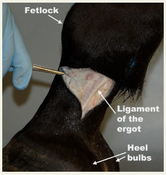
Fig 3-28 Left equine forelimb. Lateral view of pastern region. The ligament of the ergot appears through a skin incision. The digital vein, artery and nerve are deep to the ligament of the ergot. The ligament of the ergot on the medial side would appear identical. The ergot is located on the palmar midline, hidden by the tuft of hairs at the fetlock. (from Equine Anatomy Guide: The Forelimb; Mansour, Steiss, Wilhite)
Fig. above – Palmar view of distal equine forelimb. Medial and lateral palmar digital nn.; medial and lateral palmar digital aa.
- Notes on Foot Disorders in Bovine (most of the text is from Supplemental Clinical LAS Bovine Foot Anatomy, Lameness, Approaches) – The vast majority (85%) of bovine lameness originates in the foot. The medial fore digit and lateral hind digit are the most common sites.
- Nerve blocks of the foot can include performing a “ring block” of the nerves near the mid-cannon region (e.g., branches of the median, ulnar, radial nn.) or using a tourniquet for IV regional anesthesia (IVRA).
- Digital dermatitis: an infectious and contagious bacterial infection (Treponema) of the skin, commonly seen in the interdigital cleft of wet, traumatized feet. It leads to lameness and foot erosions or growths. Treatment typically consists of applying topical tetracycline based antibiotics to active lesions using a wrap or a paste. Foot baths are useful in preventing bacterial infections. (Supplemental Clinical LAS Digital dermatitis)
-
- Foot rot: a sporadic infection (opportunistic infection of wet traumatized feet by gram negative anaerobes) of the soft tissues of the foot in dairy and beef cattle and can range from mild to severe. There is usually a sudden onset of lameness accompanied by the symmetrical swelling of the lower leg above the digit and a foul smell. Swelling above the coronary band that affects both digits and includes the interdigital space is almost pathognomonic for foot rot. Systemic antibiotics are used for treatment. Ascending infection can develop and lead to coffin joint or flexor tendon sheath infections. Foot baths are useful to prevent bacterial infections. (Supplemental Clinical LAS Foot Rot)
- Sole ulcer: develop as the flexor tuberosity of P3 (aka pedal bone) pushes on the sole from inside the foot. Ulcers occur most commonly in the outside/lateral/4th digit in rear legs and are associated with varying degrees of changes in weight bearing due to loss of the supporting structures in the foot. Treatment includes removing loose horn and placing a block on the opposite digit. Prevention is key. Cows should have comfortable areas in which to lie down, be on a regular trimming schedule, and be on a good nutritional plan.
(Supplemental Clinical LAS Sole ulcers) - White line disease: encompasses a range of lesions that typically occur in the abaxial white line region towards the heel or outside digit of the rear foot. White line lesions are compromises of the laminar junction. Bacteria and debris can enter this space, and lesions can range from hemorrhages to separations and abscesses or cause further damage. White line disease is associated with cattle moving too fast or slipping while walking. Treatment involves opening up the infected area and removing loose horn. A block is placed on the opposite digit to permit the area to heal. (Supplemental Clinical LAS White line disease)
-
- Foot sepsis: several structures in the digit can become infected, typically due to ascending infections from foot rot or foot abscesses that develop from sole ulcers or white line disease. Interdigital trauma can also be a source of infection. The pedal bone (P3), the navicular bone, the coffin joint and the deep digital flexor tendon can be impacted, often in various combinations. Antibiotics and/or joint lavage are rarely successful in treating these conditions; digit amputation is a means of rapid removal of the problem but does limit lifespan of the animal due to breakdown of the opposite digit; ankylosis involves debridement and fusion of the infected joint and can lead to a full lifespan in larger or valuable animals – it does take more time to resolve the pain. (Supplemental Clinical LAS Foot sepsis)
