4 Human Anatomy and Physiology
Paul Iaizzo
It is critical for medical devices developers to:
- Learn terminologies and anatomic, physiologic and pathophysiologic principles.
- Analyze images (multi-modal and comparative) and procedures from clinical cases.
- Study anatomical specimens: human when possible, but those from large mammalian models are also useful.
- Consult with clinical and anatomical experts.
- Develop 3D models of relevant anatomies and devices: i.e., devices in varied anatomies.
- Use computer simulations and study them with mixed reality approaches if possible.
Learning Anatomical, Physiological and Pathophysiological Terminologies
There are many anatomical terms that the medical device developers should be familiar with as they start the process of device innovation. For example, the developer/designer must be able to identify the standard human anatomic positions and anatomic planes to describe whole body or specific body part locations (see figure.)
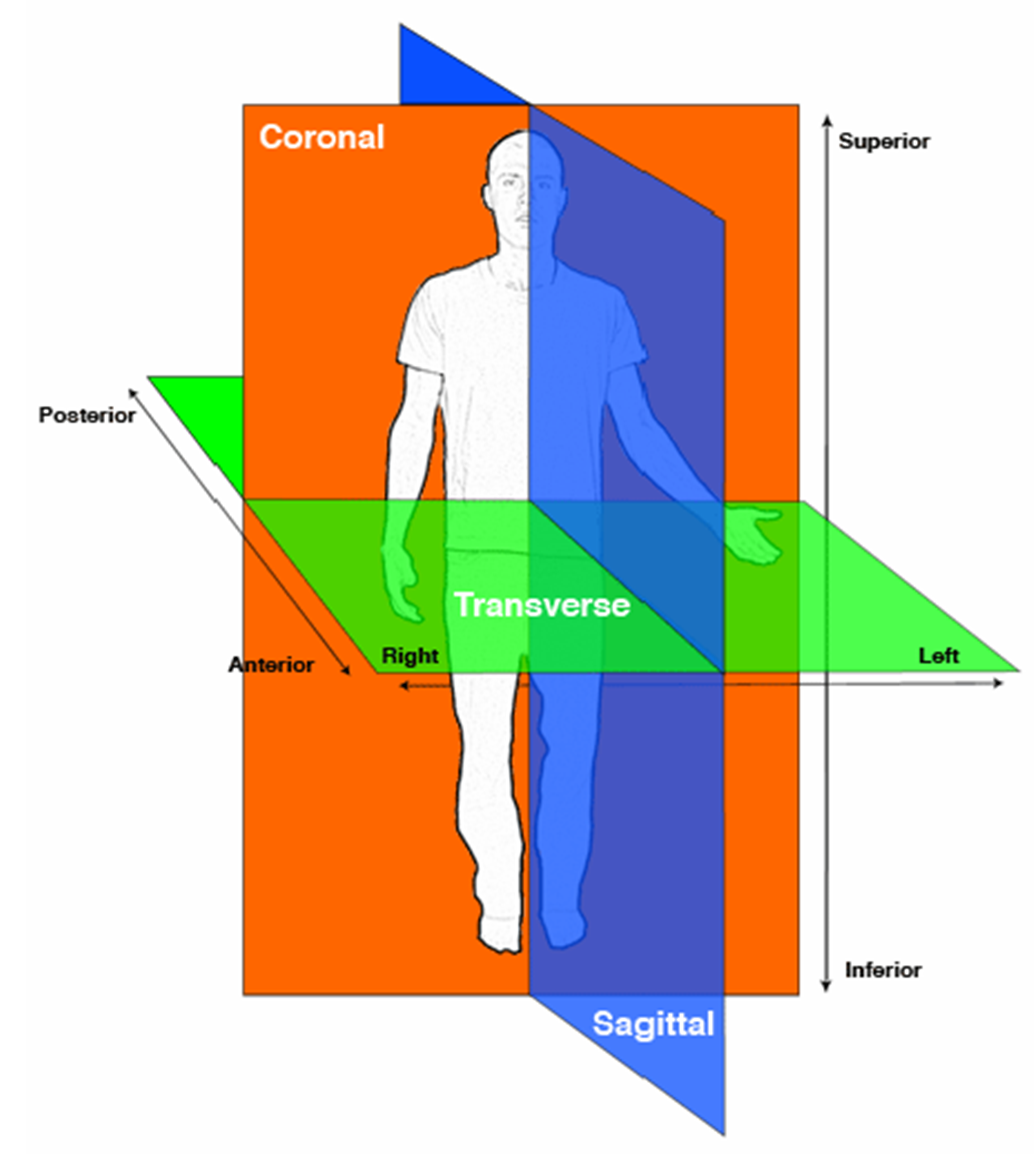
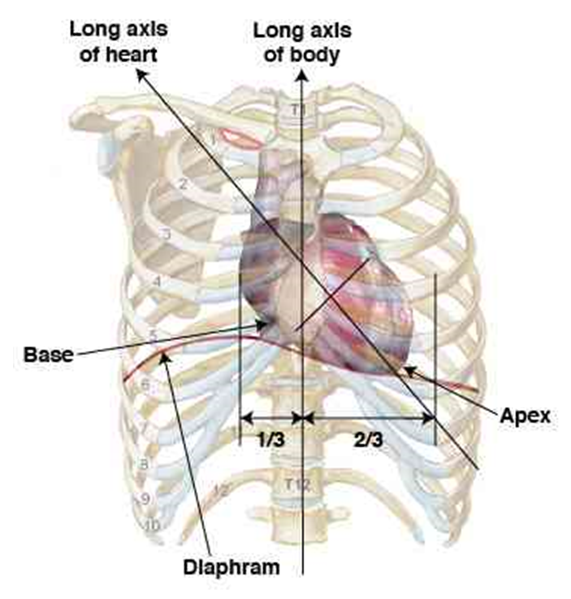
Regardless of the position of the body or organ upon examination, the anatomy of an organ or the whole system should be described as if observed from the vantage point of the standard anatomic position. The standard human anatomic position is divided by three orthogonal planes: (1) the sagittal plane, which divides the body into right and left portions; (2) the coronal plane, which divides the body into anterior and posterior portions; and (3) the transverse plane, which divides the body into superior and inferior portions. Another example of top-level anatomy is the relative location of the human heart within the ribcage as shown in this figure. For more details on anatomy see the tutorials on the “Atlas of Human Cardiac Anatomy” free access website: e.g., http://www.vhlab.umn.edu/atlas/anatomy-tutorial/anatomic-position.shtml
Learning Anatomy
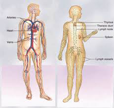
There are numerous avenues for studying human anatomy and gaining expertise relevant to your area of technology. University courses, websites and textbooks (some dating back hundreds of years that can be fun to explore) are just some of those avenues. The classic Netter textbooks with their excellent anatomical drawings are an excellent place to start. One example of a university-based resource is the advanced cardiac anatomy and physiology week-long short course held every January at the University of Minnesota. If you are working at a large company you should seek out in-house expertise. Noteworthy, human anatomy is highly variable and can be further modified by disease and by various treatments. Thus, one must study the full range of anatomies that might be seen by the proposed device both pre- and post-treatment. For example, if you need to understand the relative association of the cardiovascular system relative to lymphatic system, there are many such good depictions such as the one shown in this figure.
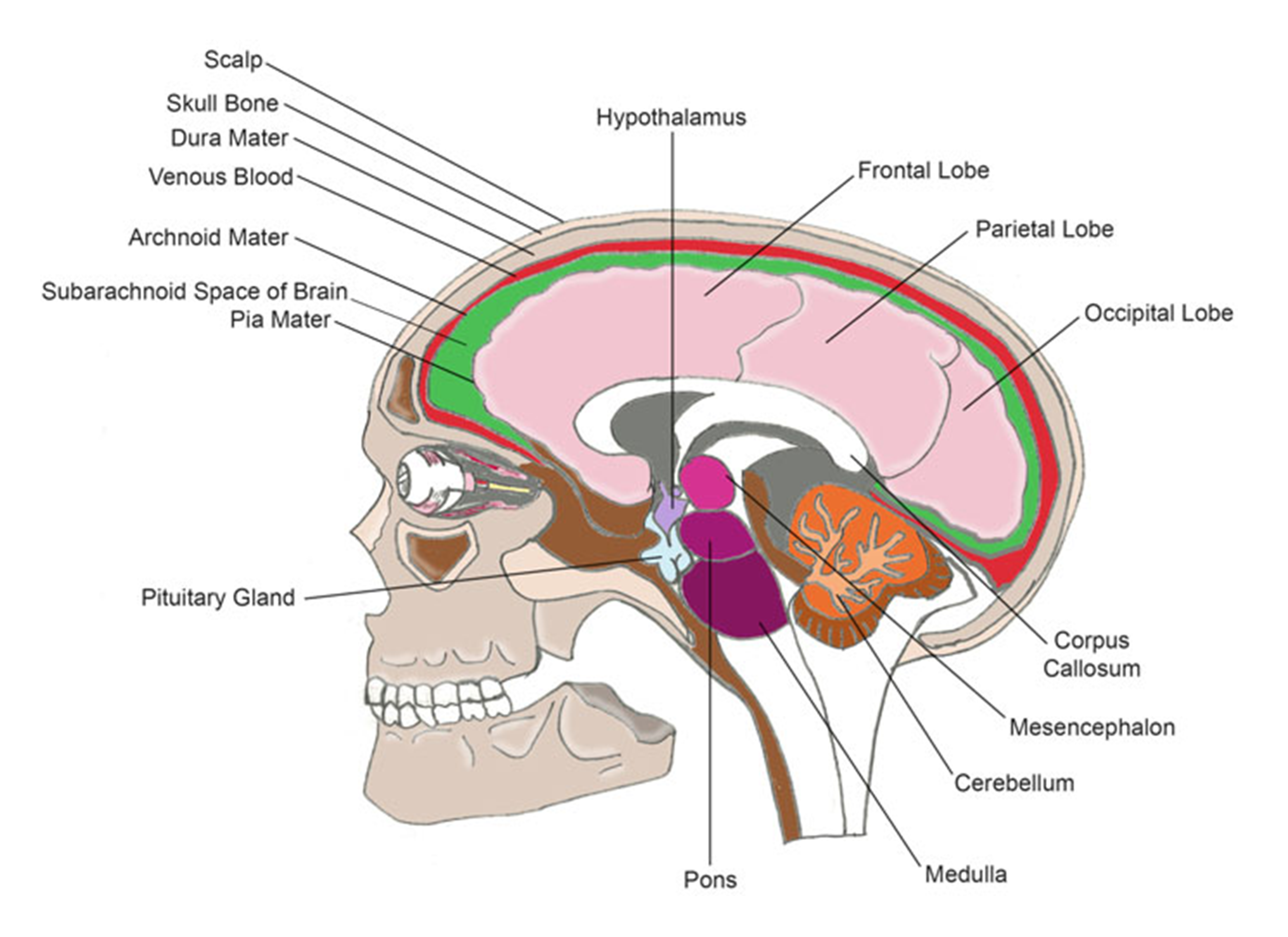
Or, if developing a device that works on the brain, the “Handbook of Neural Engineering” is a good starting point for learning detailed anatomy. Examples from this book include this figure, which shows general brain structure,
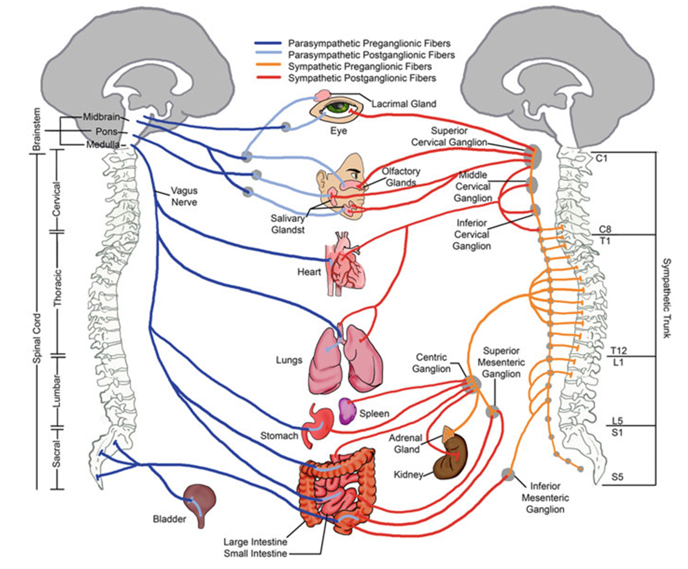
and this figure, which shows the anatomical and physiological differences within the autonomic nervous system.
One should study an array of human anatomical specimens associated with the proposed therapy; including both pre- and post-therapy specimens. Yet, this can be difficult to accomplish and/or can entail major expense. Instead, a good place to start is to dissect and study appropriate animal models. For example, one can learn much about human cardiac anatomy by dissecting swine hearts, which can be obtained from a local butcher shop at low cost.
Computational and Physical Models
Computational modeling is a useful tool for clinicians and device designers to better understand associated anatomies that may lead to optimization of therapeutic treatments. In addition, physical models derived from medical imaging can be valuable design tools. For example, the Visible Heart® Laboratories at the University of Minnesota has developed 3D printed models for education that have been presented at scientific meetings around the world and have been given to physicians to aid in patient education. The group also uses 3D printing to create scaled-up models of specific structures to better understand complex anatomies. Prototype cardiac devices can be placed and tested in a soft printed silicon heart with varying anatomy and pathologies as an analog to human specimens.
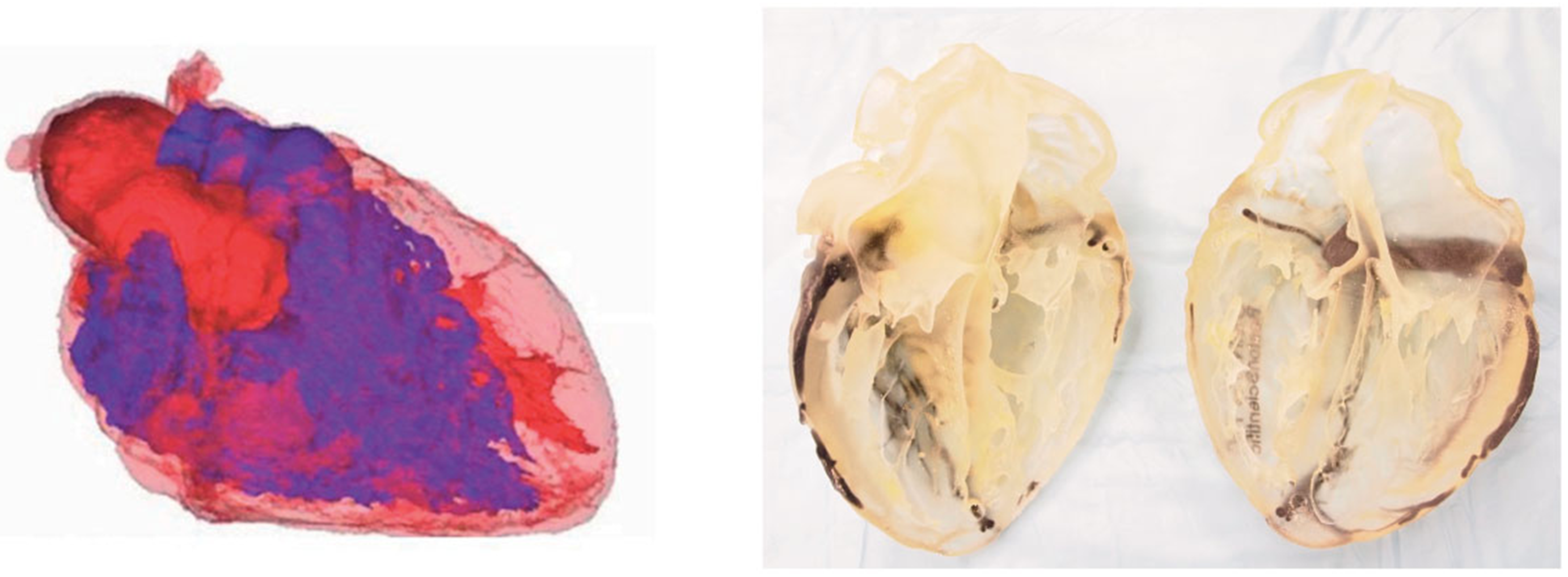
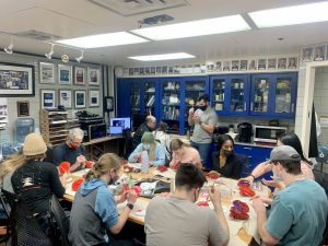
Another example of how 3D printed models can be used to learn anatomy is shown below where lab students are hand-painting anatomical models. This is the 3D analog of the popular “Anatomy Coloring Book” and is a way to understand detailed cardiac structures and how varied they can be.
Summary
If you are planning to develop medical devices or associated therapies, it is critical that you learn all appropriate associated terminologies as well thouroghly understand the underlying anatomic, physiologic and pathophysiologic properties. You should as quickly become an engaged student of these topics, utilizing all available internal and external resources possible. Try to gain access to associated anatomical specimens, which may be accomplished via specified University courses, and try to gain access to images from clinical cases (today, much is readily available online). Also, try yourself to dissect the appropriate animal tissues for the associated anatomies, for example by obtaining specimens from a butcher shop. Throughout your educational journey, consult with clinical and anatomical experts. With recent advances in 3D anatomical modeling and computer simulations, much value can be gained by virtual prototyping of the implantation of your medical device or application of your therapy in a variety of human anatomies. Throughout your medical device career, to remain successful, you should be a vigilant student of human anatomy and physiology.

