94 Sponges and Cnidarians
By the end of this section, you will be able to:
- Describe the organizational features of the simplest animals
- Describe the organizational features of cnidarians
The kingdom of animals is informally divided into invertebrate animals, those without a backbone, and vertebrate animals, those with a backbone. Although in general we are most familiar with vertebrate animals, the vast majority of animal species, about 95 percent, are invertebrates. Invertebrates include a huge diversity of animals, millions of species in about 32 phyla, which we can just begin to touch on here.
The sponges and the cnidarians represent the simplest of animals. Sponges appear to represent an early stage of multicellularity in the animal clade. Although they have specialized cells for particular functions, they lack true tissues in which specialized cells are organized into functional groups. Sponges are similar to what might have been the ancestor of animals: colonial, flagellated protists. The cnidarians, or the jellyfish and their kin, are the simplest animal group that displays true tissues, although they possess only two tissue layers.
Sponges
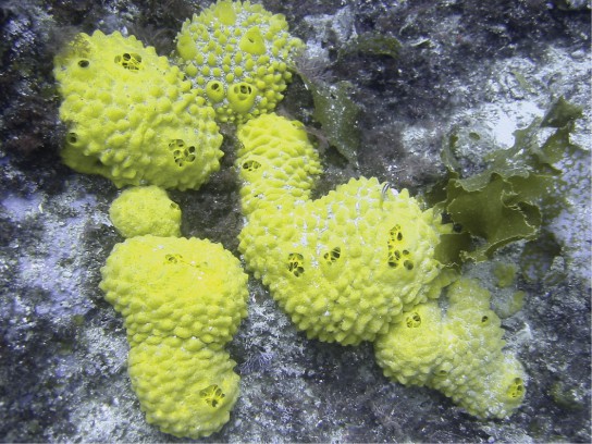
Animals in subkingdom Parazoa represent the simplest animals and include the sponges, or phylum Porifera (Figure 1). All sponges are aquatic and the majority of species are marine. Sponges live in intimate contact with water, which plays a role in their feeding, gas exchange, and excretion. Much of the body structure of the sponge is dedicated to moving water through the body so it can filter out food, absorb dissolved oxygen, and eliminate wastes.
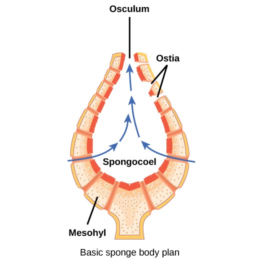
The body of the simplest sponges takes the shape of a cylinder with a large central cavity, the spongocoel. Water enters the spongocoel from numerous pores in the body wall. Water flows out through a large opening called the osculum (Figure 2). However, sponges exhibit a diversity of body forms, which vary in the size and branching of the spongocoel, the number of osculi, and where the cells that filter food from the water are located.
Sponges consist of an outer layer of flattened cells and an inner layer of cells called choanocytes separated by a jelly-like substance called mesohyl. The mesohyl contains embedded amoeboid cells that secrete tiny needles called spicules or protein fibers that help give the sponge its structural strength. The cell body of the choanocyte is embedded in mesohyl but protruding into the spongocoel is a mesh-like collar surrounding a single flagellum. The beating of flagella from all choanocytes moves water through the sponge. Food particles are trapped in mucus produced by the sieve-like collar of the choanocytes and are ingested by phagocytosis. This process is called intracellular digestion. Amoebocytes take up nutrients repackaged in food vacuoles of the choanocytes and deliver them to other cells within the sponge.
Physiological Processes in Sponges
Despite their lack of complexity, sponges are clearly successful organisms, having persisted on Earth for more than half a billion years. Lacking a true digestive system, sponges depend on the intracellular digestive processes of their choanocytes for their energy intake. The limit of this type of digestion is that food particles must be smaller than individual cells. Gas exchange, circulation, and excretion occur by diffusion between cells and the water.
Sponges reproduce both sexually and asexually. Asexual reproduction is either by fragmentation (in which a piece of the sponge breaks off and develops into a new individual), or budding (an outgrowth from the parent that eventually detaches). A type of asexual reproduction found only in freshwater sponges occurs through the formation of gemmules, clusters of cells surrounded by a tough outer layer. Gemmules survive hostile environments and can attach to a substrate and grow into a new sponge.
Sponges are monoecious (or hermaphroditic), meaning one individual can produce both eggs and sperm. Sponges may be sequentially hermaphroditic, producing eggs first and sperm later. Eggs arise from amoebocytes and are retained within the spongocoel, whereas sperm arise from choanocytes and are ejected through the osculum. Sperm carried by water currents fertilize the eggs of other sponges. Early larval development occurs within the sponge, and free-swimming larvae are then released through the osculum. This is the only time that sponges exhibit mobility. Sponges are sessile as adults and spend their lives attached to a fixed substrate.
Cnidarians
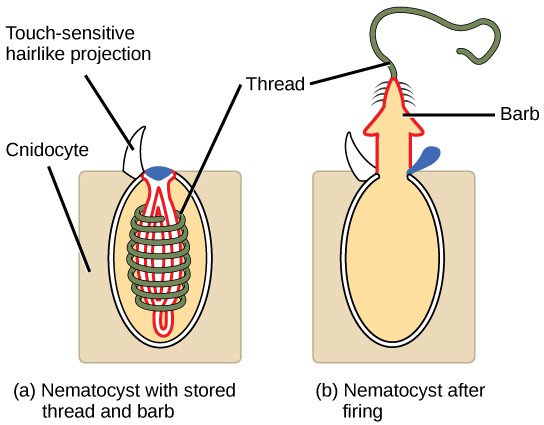
The phylum Cnidaria includes animals that show radial or biradial symmetry and are diploblastic. Nearly all (about 99 percent) cnidarians are marine species. Cnidarians have specialized cells known as cnidocytes (“stinging cells”) containing organelles called nematocysts. These cells are concentrated around the mouth and tentacles of the animal and can immobilize prey with toxins. Nematocysts contain coiled threads that may bear barbs. The outer wall of the cell has a hairlike projection that is sensitive to touch. When touched, the cells fire the toxin-containing coiled threads that can penetrate and stun the predator or prey (see Figure 3).
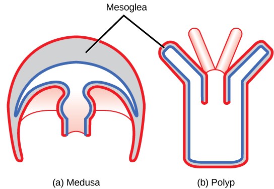
Cnidarians display two distinct body plans: polyp or “stalk” and medusa or “bell” (Figure 4). Examples of the polyp form are freshwater species of the genus Hydra; perhaps the best-known medusoid animals are the jellies (jellyfish). Polyps are sessile as adults, with a single opening to the digestive system (the mouth) facing up with tentacles surrounding it. Medusae are motile, with the mouth and tentacles hanging from the bell-shaped body. In other cnidarians, both a polyp and medusa form exist, and the life cycle alternates between these forms.
Physiological Processes of Cnidarians
All cnidarians have two tissue layers. The outer layer is called the epidermis, whereas the inner layer is called the gastrodermis and lines the digestive cavity. Between these two layers is a non-living, jelly-like mesoglea. There are differentiated cell types in each tissue layer, such as nerve cells, enzyme-secreting cells, and nutrient-absorbing cells, as well as intercellular connections between the cells. However, organs and organ systems are not present in this phylum.
The nervous system is primitive, with nerve cells scattered across the body in a network. The function of the nerve cells is to carry signals from sensory cells and to contractile cells. Groups of cells in the nerve net form nerve cords that may be essential for more rapid transmission. Cnidarians perform extracellular digestion, with digestion completed by intracellular digestive processes. Food is taken into the gastrovascular cavity, enzymes are secreted into the cavity, and the cells lining the cavity absorb the nutrient products of the extracellular digestive process. The gastrovascular cavity has only one opening that serves as both a mouth and an anus (an incomplete digestive system). Like the sponges, Cnidarian cells exchange oxygen, carbon dioxide, and nitrogenous wastes by diffusion between cells in the epidermis and gastrodermis with water.
Cnidarian Diversity
The phylum Cnidaria contains about 10,000 described species divided into four classes: Anthozoa, Scyphozoa, Cubozoa, and Hydrozoa.
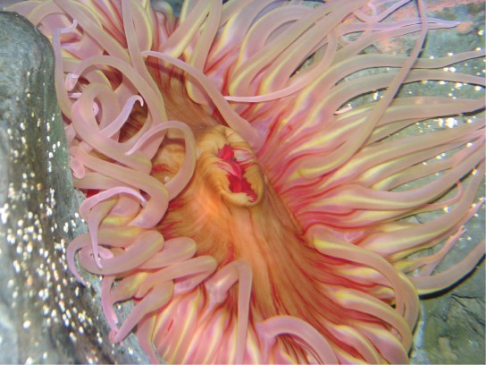
The class Anthozoa includes all cnidarians that exhibit a sessile polyp body plan only; in other words, there is no medusa stage within their life cycle. Examples include sea anemones, sea pens, and corals, with an estimated number of 6,100 described species. Sea anemones are usually brightly colored and can attain a size of 1.8 to 10 cm in diameter. These animals are usually cylindrical in shape and are attached to a substrate. A mouth opening is surrounded by tentacles bearing cnidocytes (Figure 5).
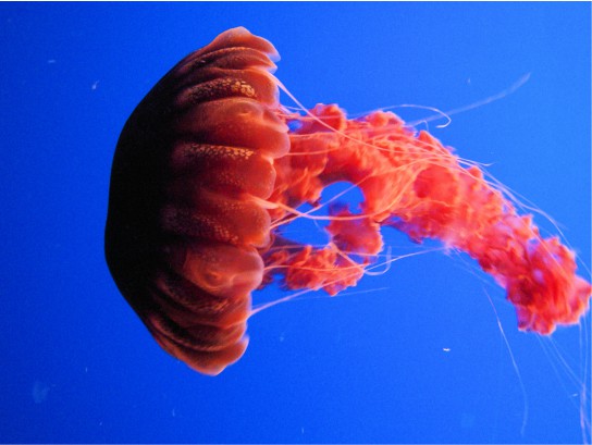
Scyphozoans include all the jellies and are motile and exclusively marine with about 200 described species. The medusa is the dominant stage in the life cycle, although there is also a polyp stage. Species range from 2 cm in length to the largest scyphozoan species, Cyanea capillata, at 2 m across. Jellies display a characteristic bell-like body shape (Figure 6).
The class Cubozoa includes jellies that are square in cross-section and so are known as “box jellyfish.” These species may achieve sizes of 15–25 cm. Cubozoans are anatomically similar to the jellyfish. A prominent difference between the two classes is the arrangement of tentacles. Cubozoans have muscular pads called pedalia at the corners of the square bell canopy, with one or more tentacles attached to each pedalium. In some cases, the digestive system may extend into the pedalia. Cubozoans typically exist in a polyp form that develops from a larva. The polyps may bud to form more polyps and then transform into the medusoid forms.
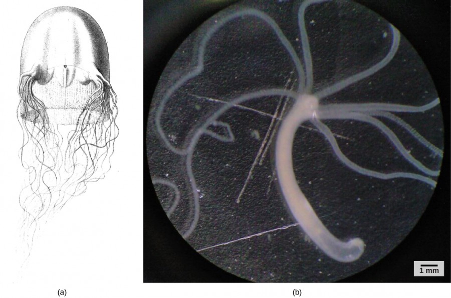
Hydrozoa includes nearly 3,500 species,1 most of which are marine. Most species in this class have both polyp and medusa forms in their life cycle. Many hydrozoans form colonies composed of branches of specialized polyps that share a gastrovascular cavity. Colonies may also be free-floating and contain both medusa and polyp individuals in the colony, as in the Portuguese Man O’War (Physalia) or By-the-Wind Sailor (Velella). Other species are solitary polyps or solitary medusae. The characteristic shared by all of these species is that their gonads are derived from epidermal tissue, whereas in all other cnidarians, they are derived from gastrodermal tissue (Figure 7ab).
Footnotes
- “The Hydrozoa Directory,” Peter Schuchert, Muséum Genève, last updated November 2012, http://www.ville-ge.ch/mhng/hydrozoa/hydrozoa-directory.htm.
- amoebocyte
- an amoeba-like cell of sponges whose functions include distribution of nutrients to other cells in the sponge
- budding
- a form of asexual reproduction that occurs through the growth of a new organism as a branch on an adult organism that breaks off and becomes independent; found in plants, sponges, cnidarians, and some other invertebrates
- choanocyte
- a cell type unique to sponges with a flagellum surrounded by a collar used to maintain water flow through the sponge, and capture and digest food particles
- Cnidaria
- a phylum of animals that are diploblastic and have radial symmetry and stinging cells
- cnidocyte
- a specialized stinging cell found in Cnidaria
- epidermis
- the layer of cells that lines the outer surface of an animal
- extracellular digestion
- a form of digestion, the breakdown of food, which occurs outside of cells with the aid of enzymes released by cells
- fragmentation
- a form of asexual reproduction in which a portion of the body of an organism breaks off and develops into a living independent organism; found in plants, sponges, and some other invertebrates
- gastrodermis
- the layer of cells that lines the gastrovascular cavity of cnidarians
- gastrovascular cavity
- the central cavity bounded by the gastrodermis in cnidarians
- gemmule
- a structure produced by asexual reproduction in freshwater sponges that is able to survive harsh conditions
- intracellular digestion
- the digestion of matter brought into a cell by phagocytosis
- medusa
- a free-floating cnidarian body plan with a mouth on the underside and tentacles hanging down from a bell
- mesoglea
- the non-living, gel-like matrix present in between ectoderm and endoderm in cnidarians
- mesohyl
- the collagen-like gel containing suspended cells that perform various functions in sponges
- monoecious
- having both sexes in one body, hermaphroditic
- nematocyst
- the harpoon-like organelle within a cnidocyte with a pointed projectile and poison to stun and entangle prey
- osculum
- the large opening in a sponge body through which water leaves
- polyp
- the stalk-like, sessile life form of a cnidarians with mouth and tentacles facing upward, usually sessile but may be able to glide along a surface
- Porifera
- a phylum of animals with no true tissues, but a porous body with a rudimentary endoskeleton
- spicule
- a short sliver or spike-like structure, in sponges, they are formed of silicon dioxide, calcium carbonate, or protein, and are found in the mesohyl
- spongocoel
- the central cavity within the body of some sponges

