
3 Part 2: Ventral Head Dissection
IMPORTANT NOTE: For this part of the chapter, the Guide directions will refer to either “ALL specimens” (meaning dissect pony and calf specimens in the same way) or to one species in particular, i.e., “ALL (PONY) specimens” or “ALL (CALF) specimens.” This added instructional note is needed because the dissection may differ slightly according to species.
ventral midline dissection
- ALL specimens: Follow the steps below to perform a midline dissection of the surgical approach to the larynx (used for ventriculectomy in horses).
- You will perform the midline dissection that mimics the surgical approach to the larynx via the thyroid notch for a ventriculectomy (used in cases of “roaring” in horses).
-
- Dissection Note: This dissection applies more to the pony specimens but should also be completed on calf specimens for comparison.
-
- You will perform the midline dissection that mimics the surgical approach to the larynx via the thyroid notch for a ventriculectomy (used in cases of “roaring” in horses).
- ALL (PONY) specimens: Identify the mandibular lymph nodes.
- On the ventral surface of the pony head, identify the large V-shaped mass of mandibular lymph nodes, which may be involved in strangles infections in horses.
- ALL (CALF) specimens: Identify the mandibular salivary glands.
- On the ventral surface of the calf head, identify the large mass of mandibular salivary glands.
-
- Comparative Note: Note that these are NOT the mandibular lymph nodes which were seen in this location in the pony. The lymph nodes would be located deep to these salivary glands in the calf.
-
- On the ventral surface of the calf head, identify the large mass of mandibular salivary glands.
- ALL specimens: Identify the sternohyoideus mm. inserting on the basihyoid bone.
- Locate the insertions of the sternohyoideus mm. on the basihyoid bone (deep to the mandibular lymph nodes in the pony).
-
- Dissection Note: Note that you will not clearly see the basihyoid bone until your head has been split in half. So be sure to return to these terms once you have your head split!
-
- Separate the sternohyoideus muscles along the midline seam between left and right sides and retract them from the midline to expose a triangular depression on the ventral aspect of the larynx, associated with the thyroid cartilage, with its apex pointing rostrally (see Fig. 7-5). This is the thyroid notch (“God’s gift” to equine surgeons).
- Locate the insertions of the sternohyoideus mm. on the basihyoid bone (deep to the mandibular lymph nodes in the pony).
- ALL specimens: Identify the thyroid notch and the cricothyroid ligament.
- As mentioned above, the thyroid notch is a triangular depression of the larynx that is associated with the thyroid cartilage. Identify this notch on your specimen.
- The thyroid notch is covered over by the cricothyroid ligament – a sheet of connective tissue extending from the rostral border of cricoid cartilage, over the thyroid notch and attaching to the thyroid cartilage. Identify this ligament on your specimen.
-
- After identifying the cricothyroid ligament, incise it on the ventral midline to enter the laryngeal cavity.
-
- ALL (PONY) specimens: After incising the cricothyroid ligament, try to pass your a finger through the thyroid notch and locate the laryngeal ventricle within the larynx. (You can pass your index finger into the ventricle of a large horse, but possibly not in a pony, so, try your pinkie finger. If your fingers won’t fit, carefully explore the laryngeal ventricle with a probe.)
- Comparative Note: Note that the laryngeal ventricles are not present in the calf.
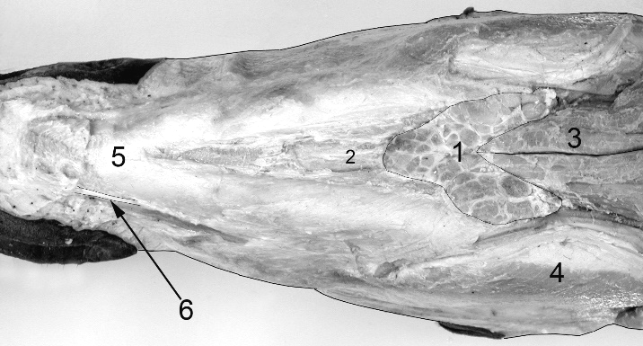
Figure 7-4. Equine head dissection, ventral view. 1, mandibular lymph nodes; 2,geniohyoideus m.; 3, sternohyoideus m.; 4, masseter m.; 5, mandibular symphysis; 6, depresser labii inferioris m. tendon.
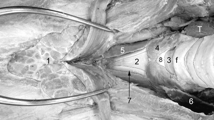
Figure 7-5. Equine head dissection, ventral view, close-up view of larynx. Surgical spreaders are reflecting the sternohyoideus and omohyoideus mm. off of the larynx. 1, mandibular lymph nodes; 2, cricothyroid ligament; 3, cricotracheal ligament; 4, cricothyroideus m.; 5, thyrohyoideus m.; 6, sternothyroideus m.; 7, edge of thyroid cartilage adjacent to the thyroid notch; 8, cricoid cartilage; f, first tracheal ring; T, thyroid gland
splitting the head
7. ALL specimens: After removing the skin, identifying the levator labii superioris m. (midline tendon) from Part 1, and performing the ventral midline dissection described here in Part 2, you are ready to have your specimen head split in half.
8. ALL specimens: Bring the skinned specimen head to an instructor to be split in half using a band saw.
Dissection Videos for this Section of Material
Ventral Midline Dissection
- Pony (1:52-3:20): https://youtu.be/zg4Lq5xThOo
- Calf (2:35-3:52): https://youtu.be/aaIeUwHXTOs
