
2 Part 1: Superficial Structures, Skinning and External Head Dissection
IMPORTANT NOTE: For this part of the chapter, the Guide directions will refer to either “ALL specimens” (meaning dissect pony and calf specimens in the same way) or to one species in particular, i.e., “ALL (PONY) specimens” or “ALL (CALF) specimens.” This added instructional note is needed because the dissection may differ slightly according to species.
Superficial structures
- ALL specimens: Prior to skinning, note the differences in the upper lips of calf and pony specimens.
- Before removing the skin of the head, note that the equine upper lip is very mobile, while the bovine is a rather rigid hairless region called the nasolabial plate.
-
- ALL (CALF) specimens: Identify the nasolabial plate.
- Comparative Species Notes: A similar nasolabial plate is seen in some members of the deer family (cervids) such as white tail deer, elk and caribou, but not in the moose. Goats and sheep lack a nasolabial plate and their hairy upper lip is split, which enables them to graze closer than cattle.
-
- Before removing the skin of the head, note that the equine upper lip is very mobile, while the bovine is a rather rigid hairless region called the nasolabial plate.
- ALL specimens: Remove the petroleum jelly from the nostrils and oral cavity. Attempt to identify the orifice of the nasolacrimal duct.
- Before you begin to examine the nose and mouth of the specimen, you will need to remove as much of the petroleum jelly from the nostrils and oral cavity as possible (the petroleum jelly helps to prevent drying during storage).
- Examine the nostril and attempt to find the small, round orifice of the nasolacrimal duct in the floor of the nostril (Fig. 7-1A). (This may be very difficult, especially in the calf specimens.)
- ALL (PONY) specimens: Identify the nasal diverticulum (aka false nostril).
- In the pony specimen, probe the well-developed nasal diverticulum (aka false nostril) with your pinky finger. (This diverticulum is located laterodorsal to the main orifice of the nostril).
- On the upper edge of the nostril, palpate the wide part of the alar cartilage. This cartilage is an aid in flaring the nostrils (Fig. 7-1B) to bring in more air during exertion; the nasal diverticulum collapses at the same time, increasing the airway diameter in the rostral part of the nasal cavity. This may be the reason for existence of the nasal diverticulum.
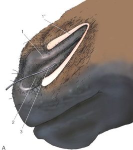
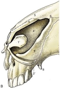
Figure 7-1 A. Left nostril opened laterally to expose equine nasal diverticulum. B. Equine nasal cartilages.
1, Alar fold, supported by the lamina (1’) of the alar cartilage; dorsal to the alar fold is the nasal diverticulum (1’’); 2, floor of nostril supported by the cornu of the alar cartilage (2’) – the floor leads into the nasal cavity; 3, cannula in nasolacrimal duct; 4, nasal septum; 5, nasal bone; 6, nasoincisive notch; 7, incisive bone; 8, canine tooth. (Duplication of TVA Figure 18-3)
Skinning the head
4. ALL specimens: Remove the skin from the head.
-
- Remove as much of the hairy skin as possible from the entire head of your pony and calf specimens.
- Leave a small rim of skin around the eyes, nostrils, and lips. Skin only to the base of the ears.
- In the pony, be especially careful to leave the skin around the edges of the nostrils to avoid damage to the nasal diverticulum.
- Once the skin is removed, the head specimen should sit upright on a table so that both sides can be dissected simultaneously.
External head dissection
5. ALL specimens: Identify the levator labii superioris m.
-
- ALL (PONY) specimens: We do not have time to do justice to the muscles of facial expression, but the levator labii superioris m. of the horse (Fig. 7-2A and 7-3A) deserves attention due to its importance in causing the characteristic “lip curl” associated with the flehmen response, an important facet of equine behavior.
-
- Find the common tendon of insertion of this muscle into the upper lip (labii superioris).
- Trace the well-defined tendons caudally to the two conical muscle bellies lying under thinner superficial facial muscles on the maxilla.
- Remove the overlying superficial facial muscles to expose and identify the levator labii superioris m. on both sides.
-
- ALL (CALF) specimens: We do not have time to do justice to the muscles of facial expression, but you should identify the levator labii superioris m. (Fig. 7-2B) to compare with the same structure on the pony.
-
- Find the common tendon of insertion of this muscle into the upper lip (labii superioris).
- Trace the tendons caudally to the two muscle bellies lying under thinner superficial facial muscles.
- Remove the overlying superficial facial muscles to expose and identify the levator labii superioris m. on both sides.
-
- Dissection Note: Note that other muscles have common midline tendons which meet on a midline raphe (seam) but the levator labii superioris is the only one with a well-defined midline tendon (especially prominent in the horse).
- ALL (PONY) specimens: We do not have time to do justice to the muscles of facial expression, but the levator labii superioris m. of the horse (Fig. 7-2A and 7-3A) deserves attention due to its importance in causing the characteristic “lip curl” associated with the flehmen response, an important facet of equine behavior.
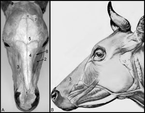
Figure 7-2 A. Equine head dissection, dorsal view. B. Bovine head dissection, left lateral view.
1, common tendon of levator labii superioris m.; 2, levator labii superioris m.; 3, levator nasolabialis covering levator labii superioris m.; 4, nasal bone; 5, frontal bone; 6,parietal bone; 7,temporalis m.; 8, maxillary bone
6. ALL specimens: Identify the infraorbital n.
-
- Elevate the fleshy part of the levator labii superioris m. to find the infraorbital n. (Fig. 7-3) emerging from the infraorbital foramen on both left and right sides.
-
- The infraorbital nerve is the main continuation of the maxillary nerve and is the main sensory nerve of the muzzle region; the infraorbital n. also supplies the upper incisors and cheek teeth.
-
- Elevate the fleshy part of the levator labii superioris m. to find the infraorbital n. (Fig. 7-3) emerging from the infraorbital foramen on both left and right sides.
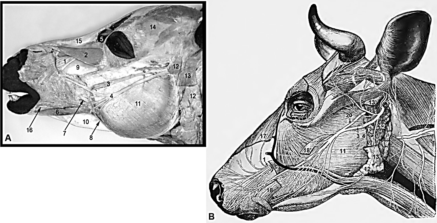
Figure 7-3 A. Equine head dissection, left lateral view; B. Bovine head dissection, left lateral view.
1, infraorbital n.; 2, levator labii superioris m.; 3, 4, dorsal and ventral buccal branches of the facial n.; 5, conchofrontal sinus; 6, depressor labii inferioris m.; 7, parotid duct; 8, facial a.; 9, maxilla; 10, mandible; 11, masseter m.; 12, parotid salivary gland (partially removed in bovine); 13, stump of the parotidoauricularis m.; 14, postorbital fat pad; 15, nasal bone; 16, mental nerve; 17, levator nasolabialis m.; 18, zygomaticus; 19, 19′ mandibular salivary gland (extensive in bovine); 20, mandibular n. branch.
Dissection Videos for this Section of Material
Superficial Structures, Skinning and External Head Dissection
- Pony
- External Head Dissection (0:00-1:52):https://youtu.be/zg4Lq5xThOo
- Calf
- External Head Dissection (0:00-2:35): https://youtu.be/aaIeUwHXTOs
