
Part 2: Pelvic Inlet
Abby Brown
IMPORTANT NOTE: For this part of the chapter, the Guide directions will refer to either “ALL specimens” (meaning dissect pony and calf specimens in the same way) or to one species in particular, i.e., “ALL (PONY) specimens” or “ALL (CALF) specimens.” This added instructional note is needed because the dissection may differ slightly according to species. The dissection will also vary based on the sex of the animal.
- ALL MALE specimens: In the pelvic inlet, note the region of the vaginal ring; identify the ductus deferens and testicular artery and vein, as well as noting the location of the deep inguinal ring.
- In the male specimens, on the peritoneal side of the pelvic inlet (inguinal region) visualize the region of the vaginal ring and identify the ductus deferens entering the vaginal ring.
-
- Dissection Notes:
-
- Recall that there are two vaginal rings, one on the left side and one on the right side of the body, so you should identify a ductus deferens on the left and another on the right.
-
- The vaginal ring is the opening formed by the parietal peritoneum as it leaves the abdomen and enters the inguinal canal to form the vaginal tunic/process. An accumulation of some amount of fat usually surrounds the vaginal ring region.
-
- The vaginal ring marks the position of the deep inguinal ring, which is formed by the reflection of transversalis fascia outside the vaginal ring. The deep inguinal ring represents the entrance to the inguinal canal.
- Recall that there are two vaginal rings, one on the left side and one on the right side of the body, so you should identify a ductus deferens on the left and another on the right.
-
- Dissection Notes:
-
- Identify the testicular artery and testicular vein also entering the inguinal canal near the ductus deferens on each side.
- In the male specimens, on the peritoneal side of the pelvic inlet (inguinal region) visualize the region of the vaginal ring and identify the ductus deferens entering the vaginal ring.
- ALL FEMALE specimens: On the peritoneal side of the pelvic inlet (inguinal region), identify the round ligament of the uterus.
- Trace the round ligament of the uterus to the area where it enters the region of the vaginal ring, but note that the vaginal ring is sealed in all female ungulates.
-
- Dissection Note: Recall that there are two vaginal rings, one on the left side and one on the right side of the body, so you should identify a round ligament of the uterus on the left and another on the right.
-
- Trace the round ligament of the uterus to the area where it enters the region of the vaginal ring, but note that the vaginal ring is sealed in all female ungulates.
- ALL specimens: Carefully remove some of the peritoneum that lines the pelvic inlet; observe/identify any remaining portions of the rectus abdominis mm. and the internal abdominal oblique mm. Calf specimens: Also identify any remaining/visible portion of the transversus abdominis m. (note that it may not be present).
- ALL specimens: Identify the prepubic tendon. (Fig. 6-1, TVA 547).
- The prepubic tendon is a complex structure of interwoven collagenous fibers that attaches to the cranial aspect of the pubis (extending between contralateral iliopubic eminences), and fuses with the tendons of the rectus abdominis muscles to form a tough sling that supports the abdominal viscera.
-
- In the equine, recall from Chapter 2 that the accessory ligament of the femoral head unites with the prepubic tendon (see Chapter 5 and TVA 547).
-
- Important Note: You should also be able to identify the prepubic tendon on the dried/ligamentous equine pelvic specimen available in lab (from the museum collection).
- The prepubic tendon is a complex structure of interwoven collagenous fibers that attaches to the cranial aspect of the pubis (extending between contralateral iliopubic eminences), and fuses with the tendons of the rectus abdominis muscles to form a tough sling that supports the abdominal viscera.
- ALL specimens: On one side of the specimen, identify the aponeurosis of the external abdominal oblique m. and look for the superficial inguinal ring in the inguinal region of the aponeurosis.
- The superficial inguinal ring is a slit in the aponeurosis of the external abdominal oblique muscle that allows passage of the external pudendal artery and vein (and the spermatic cord in males), and marks the external opening of the inguinal canal.
- Recall that the inguinal canal (TVA 549) is merely a potential space between the oblique muscles, through which the spermatic cord (male) and external pudendal artery and vein (male and female) pass.
-
- The inguinal canal can also be defined as a ‘channel’, ‘slit’, or short ‘passageway’ through the abdominal wall, extending from the deep inguinal ring (inside the abdomen) to the superficial inguinal ring (outside the abdominal wall), and allows passage of the various structures described. (Keep in mind that the inguinal canal is not a visually ‘distinct’ structure, but is a concept that is important to understand.)
-
- ALL specimens: Trace the inguinal canal as it travels between the oblique muscle and identify the region of the deep inguinal ring (inside the abdominal cavity) The boundaries of the deep inguinal ring are as follows:
-
- The medial free edge of rectus abdominis m.
- The cranial free edge of internal abdominal oblique m. (i.e., caudal edge of this muscle)
- The caudal edge of external abdominal oblique m. (also referred to as the inguinal ligament)
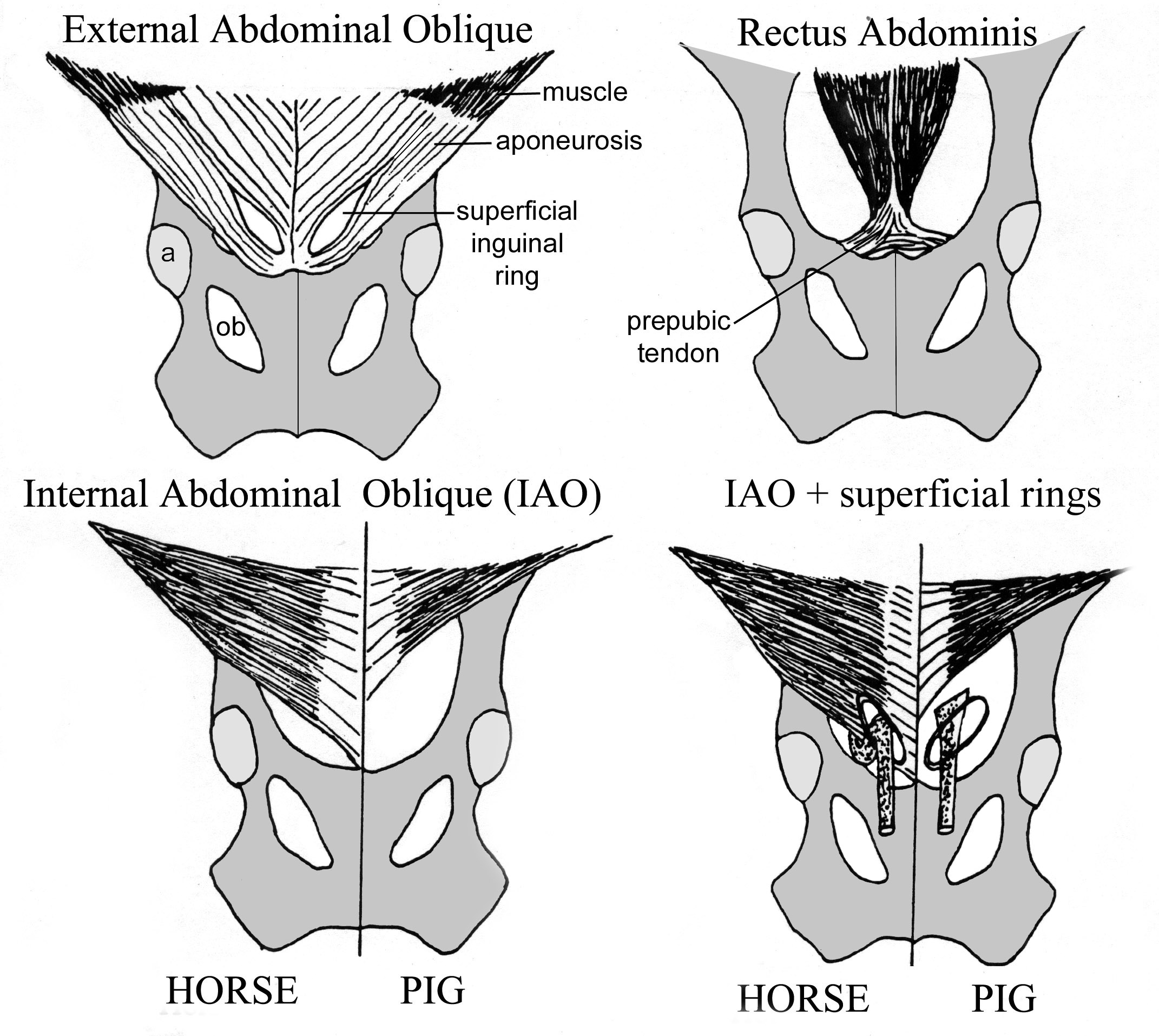
Figure 6-1. Ventral views of the layers that form the inguinal canal. a, acetabulum; ob, obturator foramen.
7. ALL MALE specimens: Identify the cremaster m., spermatic cord, vaginal tunics and trace the previously identified testicular a. & v.
-
- In the male specimens, caudolaterally, identify the cremaster m. which is a strip of muscle originating from the caudal edge of the internal abdominal oblique muscle.
-
- Trace the cremaster m. onto the surface of the spermatic cord, which it accompanies.
-
- Identify the spermatic cord and note its associated vaginal tunics (parietal and visceral).
-
- Note that the vaginal tunics originated from peritoneum. With testicular descent (from abdominal cavity to scrotum) the testis and associated structures become enveloped with serous membrane that is then termed ‘vaginal tunics’ (parietal and viscera). Terminology Note: The word ‘vagina’ = sheath.
-
- Re-identify the testicular artery and testicular vein. Trace these vessels alongside the spermatic cord as it travels through the inguinal canal.
- In the male specimens, caudolaterally, identify the cremaster m. which is a strip of muscle originating from the caudal edge of the internal abdominal oblique muscle.
OTHER INTERNAL PELVIC STRUCTURES
8. ALL (PONY) specimens: Identify the internal obturator muscle. Follow the tendon of this muscle from medial to lateral, as it passes out through the lesser sciatic foramen.
9. ALL (CALF) specimens: On the medial/inner surface of the sacroscicatic ligament, find the depression that corresponds to the lesser sciatic foramen. Note that in the calf there is no true internal obturator muscle. The external obturator m. has an internal part (intrapelvic part) that is continuous with the main (external) part through the obturator foramen. Therefore, no tendon passes through the lesser sciatic foramen as it does in the horse.
Dissection Videos for this Section of Material
Pelvic Inlet
- Pony
- Male Pelvic Inlet & Perineum: https://youtu.be/sLf20VYjmXU
- Female Pelvic Inlet & Perineum: https://youtu.be/KUBAoRBIp1Q
- Calf
- Male Pelvic Inlet & Perineum: https://youtu.be/FggEeCp4qIc
