
Vision Loss and V1
102 Columns and Hypercolumns in V1
Learning Objectives
Know how the V1 neurons are organized according to eye of origin.
Know how the V1 neurons are organized according to their orientation preferences (depth vs surface).
Know what V1 hypercolumns are.
In 1958, Hubel and Wiesel discovered the organization of the visual cortex by cortical columns through a landmark experiment. In this study, they exposed a cat to a variety of visual orientations of light and recorded the activations from the visual cortex. They found that certain parts of the visual cortex responded to unique orientations of light, establishing the idea of the cortical column.
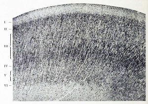
A cortical column is a cluster of several thousand neurons, spanning the ~3 mm cortical depth, that respond to one unique visual orientation. Columnar organization is an important property of the visual cortex (V1) across two directions: depth vs surface.
- If you sample neural responses as you move through the depth, they stay approximately the same. This means that as you move down through cortex, you find neurons with the same orientation preferences.
- If you sample neural responses as you move across the surface, they change. For V1, this means that as you move across the cortex, you find neurons with different orientation preferences. Orientation preference changes systematically as you move, creating a pattern of orientation preference that people refer to as pinwheels.
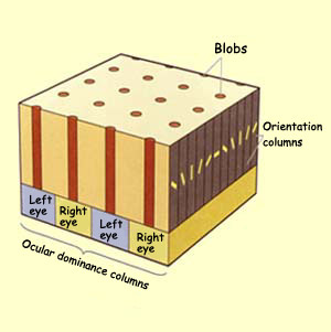
V1 neurons can also either be color selective or non-selective, and color selectivity is organized into a pattern of its own. Color-selective clusters of neurons are referred to as blobs because of their appearance when tissue is stained for cytochrome oxidase (an enzyme found in greater concentrations in tissue that has higher oxygen demand). Blobs tend to be found at the center of orientation pinwheels, which in turn are centered on ocular dominance columns.
Ocular dominance columns (ODC) are bands of tissue that respond more strongly to inputs from either the left or the right eye. These are organized into stripes in V1. The highest segregation in responsiveness between the left and right eye is located in Layer 4 of the cortex, where V1 receives input from the thalamus. While neurons in superficial and deep layers typically show a preference for one eye or the other (i.e., ODCs are stripes that run across the cortical surface and through the cortex depth), neurons outside of Layer 4 (input layer) typically receive inputs from both eyes and thus are binocularly driven.
Hypercolumns are ~1 mm blocks in V1 that contain a full set of receptive fields for a given region in space: an ocular dominance column for each eye, and a full set of orientation pinwheels (usually one in each ODC, but the organization is not perfect).
For more in depth explanations of the topics, check this link out
Cheryl Olman PSY 3031 Detailed Outline
Provided by: University of Minnesota
Download for free at http://vision.psych.umn.edu/users/caolman/courses/PSY3031/
License of original source: CC Attribution 4.0Wikipedia, Cortical column
URL: https://en.wikipedia.org/wiki/Cortical_column
License: CC BY-SA 3.0
