
Basics: neuroscience and psychophysics
7 Synapses
Learning Objectives
Know that a synapse is a gap between two neurons where communication happens chemically
Understand that the effect of a given neurotransmitter is determined by the post-synaptic receptors
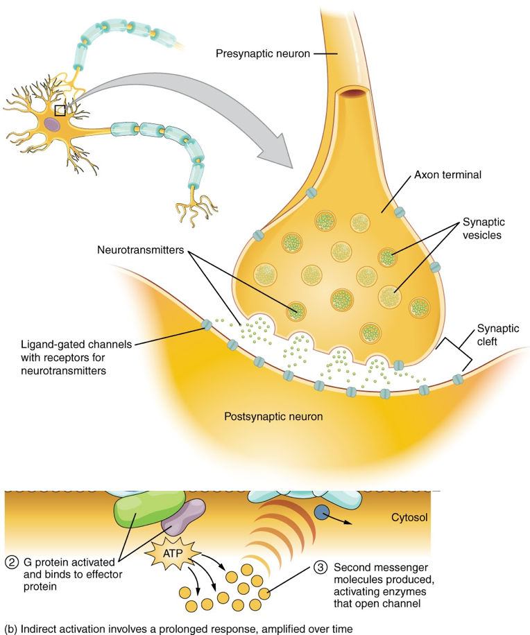
The synapse or “gap” is the place where information is transmitted from one neuron to another. Synapses usually form between axon terminals and dendritic spines, but this is not universally true. There are also axon-to-axon, dendrite-to-dendrite, and axon-to-cell body synapses. The neuron transmitting the signal is called the presynaptic neuron, and the neuron receiving the signal is called the postsynaptic neuron. Note that these designations are relative to a particular synapse—most neurons are both presynaptic and postsynaptic. There are two types of synapses: chemical and electrical.
When an action potential reaches the axon terminals, voltage-gated Ca2+ channels in the membrane of the synaptic end bulb open. The concentration of Ca2+ increases inside the end bulb, and the Ca2+ ion associates with proteins in the outer surface of neurotransmitter vesicles. The Ca2+ facilitates the merging of the vesicle with the presynaptic membrane so that the neurotransmitter is released (a process called exocytosis) into the small gap between the cells, known as the synaptic cleft.
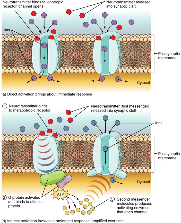
Once in the synaptic cleft, the neurotransmitter diffuses the short distance to the postsynaptic membrane and can interact with neurotransmitter receptors. Receptors are specific for the neurotransmitter, and the two fit together like a key and lock. One neurotransmitter binds to its receptor and will not bind to receptors for other neurotransmitters, making the binding a specific chemical event.
The effect of a neurotransmitter on the postsynaptic element is entirely dependent on the receptor protein. First, if there is no receptor protein in the membrane of the postsynaptic element, then the neurotransmitter has no effect. The depolarizing or hyperpolarizing effect is also dependent on the receptor. For example, when acetylcholine binds to the nicotinic receptor, the postsynaptic cell is depolarized. This is because the receptor is a cation channel and positively charged Na+ will rush into the cell. However, when acetylcholine binds to the muscarinic receptor, of which there are several variants, it might cause depolarization or hyperpolarization of the target cell.
The amino acid neurotransmitters, glutamate, glycine, and GABA, are almost exclusively associated with just one effect. Glutamate is considered an excitatory amino acid, but only because Glu receptors in the adult cause depolarization of the postsynaptic cell. Glycine and GABA are considered inhibitory amino acids, again because their receptors cause hyperpolarization.
Glutamate is interesting because it can cause excitation by activating ionotropic receptors, but it can also activate metabotropic receptors that act on the post-synaptic cell in ways other than immediately changing the membrane potential. Ionotropic receptors are fast-acting and have (relatively) short-lived consequences. Metabotropic receptors, on the other hand, can start intracellular processes with effects as far-reaching as changing the regulation of genes. Glutamate’s ionotropic receptors are NMDA, AMPA and kainate (named after the other molecules that also activate them). Glutamate’s three metabotropic receptors are called mGluRs and can either increase or decrease the excitability of the post-synaptic cell.
CC LICENSED CONTENT, SHARED PREVIOUSLY
OpenStax, Anatomy and Physiology Section 12.5 Communication Between Neurons
Provided by: Rice University.
Access for free at https://openstax.org/books/anatomy-and-physiology/pages/1-introduction
License: CC-BY 4.0
Adapted by: Cheryl Olman
Libretexts, 7.3 Synapses
Provided by: Libretexts
Access for free at:
https://med.libretexts.org/Bookshelves/Veterinary_Medicine/Book%3A_Introductory_Animal_Physiology_(Hinic-Frlog)/7%3A_Electrical_Signals/7.3%3A_Synapses
License: CC BY-NC-SA 3.0.
