
Light and Eyeballs
82 Eyeball Anatomy
Learning Objectives
Being able to describe different parts of a human eyeball.
Know which parts of the eyeball are involved in accommodation.
Know what a blind spot on the retina is, and where it’s located.
Structure of the Eye
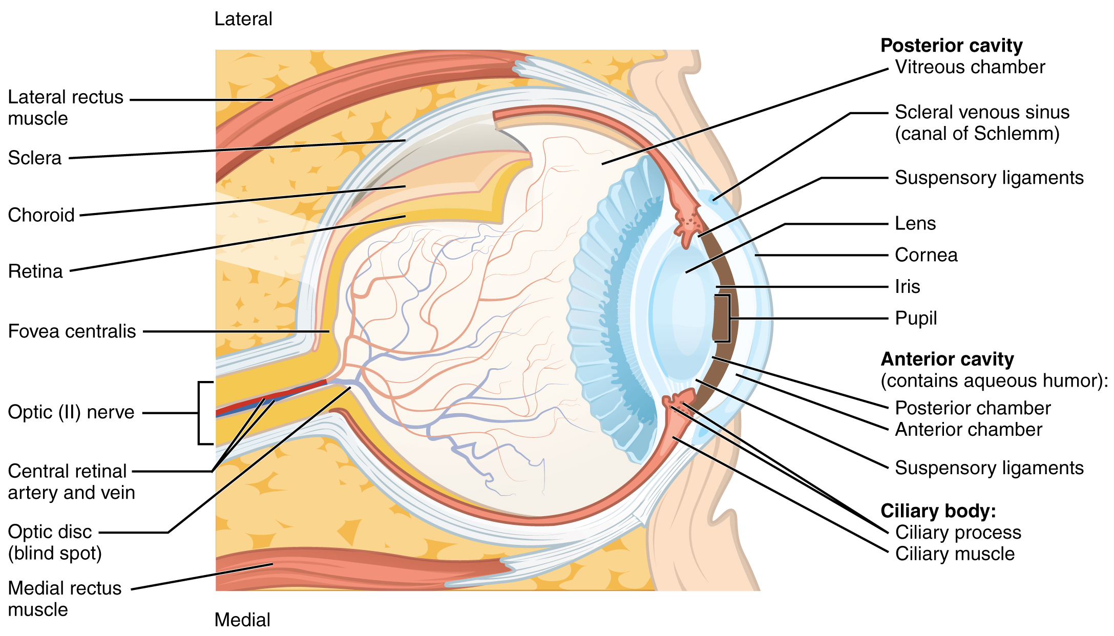
The eye is made up of various components which function together to allow us to see. Each component has an important role, and works together to allow us to see. Many of these components are used to focus incoming light in a specific way. Light enters at the most anterior part of the eye and first goes through the cornea. The cornea is the transparent but alive, curved structure at the front of the eye, with embedded nerve endings for pain, touch and thermal sensation. It has 80% of the focusing power of the eye but is not flexible. Around the cornea is the sclera which is the white, hard outside of the eyeball. Light then passes through the aqueous humor, a low viscosity fluid behind the cornea, and then through the pupil. The pupil is the hole through which light enters the eye and is shaped by the iris. The iris is the muscular, colored tissue that gives each individual their eye color. The iris is also able to open and close in response to different lighting conditions, resulting in a change to pupil size and the amount of light let into the eye. Light then passes through the lens which is a flexible, clear substance that provides 20% of the focusing power of the eye. The lens is able to change its shape (curvature) through the contraction and relaxation of ciliary (or lens) muscles, the muscles attached to the lens. Light then enters the posterior cavity of the eye which is filled with highly viscous fluid called the vitreous humor. Finally, light reaches the retina, a sheet of neurons at the back of the eye.
The retina contains photoreceptors (cones and rods) which are able to convert light into signals that are then sent to the brain through the optic nerve. Located at a specific point on the retina is the fovea where light from the center of gaze lands on the retina. Visual acuity, or the sharpness of vision, is the greatest at the fovea with a decrease in acuity in either direction from the fovea. The macula, retinal area surrounding the fovea, has slightly worse acuity, and the peripheral retina has the least acuity resulting in blurry edges. Additionally there is a blind spot located on the retina where there are no photoreceptors (because axons heading for the optic nerve occupy that space) known as the optic disc. Your brain fills in the hole with a copy of what’s around the blind spot. The axons pass through the retina where the optic nerve begins and where there are no photoreceptors at the very back of the eye. This is what creates a “blind spot” in the retina and a corresponding blind spot in our visual field.
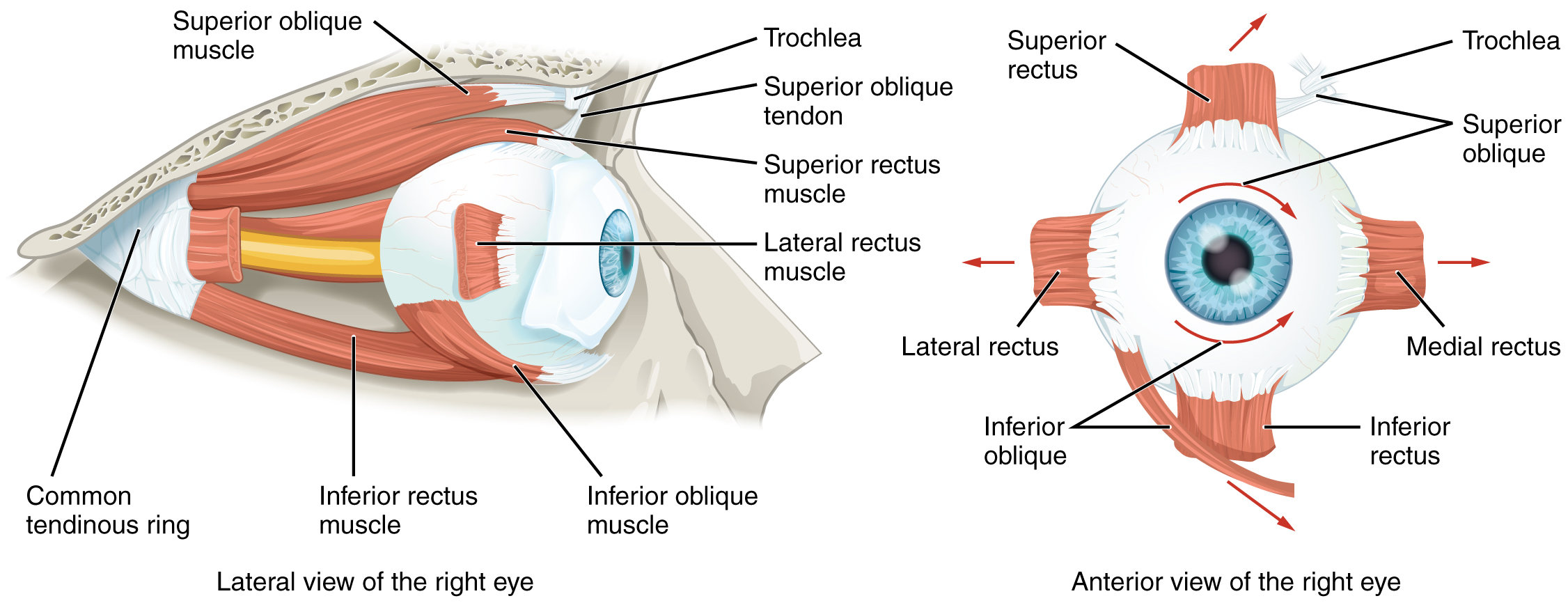
Additionally, there are components of the eye that are important for maintaining function and mobility. At the back of the retina lies a single layer of cells called the pigment epithelium which is the black layer behind the retina where visual pigments are replenished. Additionally, the choroid, which is a layer of highly vascularized connective tissue between the retina and sclera, provides a blood supply to the eyeball. There are also a variety of muscles that are responsible for eye movement. There are four main muscles arranged at the cardinal points around the eye called superior rectus, medial rectus, inferior rectus, and lateral rectus. Contraction of one of these muscles results in the eye moving toward the contracting muscle. There are also two muscles called superior oblique and inferior oblique which are responsible for the rotation of the eye due to the eyes not perfectly aligning on the sagittal plane.
Each one of these components work together to give us vision. Dysfunction of any part of the eye can lead to various degrees of vision loss or even blindness. A common example of dysfunction is with the lens. 20% of the focusing power of the eye is in the lens. Although it is only 20% of the focusing power, it is important because it is flexible and gives us the ability to accommodate. Accommodation is the ability to focus on things that are near or far away. Presbyopia is when the lens gets hard and can’t be squished by the ciliary muscles to focus on things that are near. Eventually, your arms won’t be long enough to hold reading material far enough away for your eyes to focus on it. Cataracts are the crystallization of the lens that scatters light, making it hard to perceive detail.
Exercises
CC LICENSED CONTENT, SHARED PREVIOUSLY
OpenStax, Anatomy and Physiology Chapter 14.1 Sensory Perception
Provided by: Rice University.
Access for free at https://openstax.org/books/anatomy-and-physiology/pages/14-1-sensory-perception
License: CC-BY 4.0
Adapted by: Ran Rice
Cheryl Olman PSY 3031 Detailed Outline
Provided by: University of Minnesota
Download for free at http://vision.psych.umn.edu/users/caolman/courses/PSY3031/
License of original source: CC Attribution 4.0
Adapted by: Ran Rice and Jacob Powers
