
Light and Eyeballs
84 Presbyopia
Learning Objectives
Know what the cause of presbyopia is.
Know what the current treatment options for presbyopia are.
Be able to describe another common eye problem that comes with age.
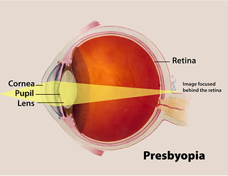
Presbyopia is a condition associated with the aging of the eye that results in progressively worsening ability to focus clearly on close objects. Symptoms include difficulty reading small print, having to hold reading material farther away, headaches, and eyestrain, and the severity of symptoms differs by person. In addition, other types of refractive errors may exist at the same time as presbyopia. Presbyopia is a normal part of the aging process; it occurs due to hardening of the lens of the eye, causing the eye to focus light behind rather than on the retina when looking at close objects. It is a type of refractive error along with nearsightedness, farsightedness, and astigmatism. Diagnosis is by an eye examination, and treatment is typically with eyeglasses that have a higher focusing power in the lower portion of the lens. Off-the-shelf reading glasses may be sufficient for some.[1] Contact lenses may also occasionally be used.[2]
While Presbyopia is a normal part of the aging process, a more severe disorder that may also come with aging is Age-Related Macular Degeneration (AMD). AMD is the leading cause of worldwide blindness in the elderly, is a bilateral ocular condition that affects the central area of the retina known as the macula. Clinically, AMD is classified into the nonexudative “dry” or atrophic form and the exudative “wet” or neovascular form. More severe vision loss is typically associated with the “wet” form that occurs in about 15% of all patients with AMD, but up to 20% of legal blindness from AMD is due to the “dry” form [3]. The typical clinical sign of “dry” AMD is pigment disruption and drusen (small yellowish deposits) in the retina. Drusen may be small “hard” (small with discrete margins) or “soft” (larger with indistinct edges). They lie between the RPE and an adjacent basement membrane complex known as Bruch’s membrane (BM). The designation of exudative or “wet” AMD implies that fluid, exudates, and/or blood are present in the extracellular space between the neural retina and the RPE (the subretinal space), and/or between the RPE and Bruch’s membrane (the sub-RPE space) in the case of RPE detachments. Additionally, while Presbyopia may be treated with something as simple as eyeglasses, AMD is much more difficult to treat, with there being no current treatments for its early stages. However, once it reaches the intermediate-late stages, treatments such as vitamins and minerals, or a number of procedures may help to stop vision loss.
Here is a quick refresher video on how the eye works.
CC LICENSED CONTENT, SHARED PREVIOUSLY
Wikipedia, Presbyopia
URL: https://en.wikipedia.org/wiki/Presbyopia
License: CC BY SA 3.0
Webvision: The Organization of the Retina and Visual System, Age-related macular degeneration (AMD)
Authored by: Gregory S. Hageman, Karen Gaehrs, Lincoln V. Johnson and Don Anderson
URL: https://webvision.med.utah.edu/book/part-xii-cell-biology-of-retinal-degenerations/age-related-macular-degeneration-amd/
License: CC BY-NC
Adapted by: Kori Skrypek and Kayla Besse
References
- Facts About Presbyopia”. NEI. October 2010. Archived from the original on 4 October 2016. Retrieved 11 September 2016.
- Pérez-Prados, Roque; Piñero, David P; Pérez-Cambrodí, Rafael J; Madrid-Costa, David (March 2017). “Soft multifocal simultaneous image contact lenses: a review”. Clinical and Experimental Optometry. 100 (2): 107–127. doi:10.1111/cxo.12488. PMID 27800638.
- Sunness JS. The natural history of geographic atrophy, the advanced atrophic form of age-related macular degeneration. Mol Vis. 1999;5:25. [PubMed]
