
Vision Loss and V1
93 Macular Degeneration
Learning Objectives
Be able to describe the cause of macular degeneration.
Know what the two main types of age-related macular degeneration are.
Age-related macular degeneration (AMD), the leading cause of worldwide blindness in the elderly, is a bilateral ocular condition that affects the central area of the retina known as the macula. Although the macula represents only the central 4% of the retinal area, it accounts for the vast majority of useful (detailed) photopic (daytime) vision. Thus, lesions developing in this region can have a major impact on visual function. The two main types of age-related macular degeneration are dry and wet.
Wet AMD is when abnormal blood vessels behind the retina start to grow under the macula, ultimately leading to blood and fluid leakage. Bleeding, leaking, and scarring from these blood vessels cause damage and lead to rapid central vision loss.
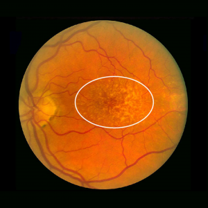
Dry AMD is when the macula thins over time as part of the aging process, gradually blurring central vision. As less of the macula functions over time, central vision is gradually lost in the affected eye. An early warning sign of dry AMD is the presence of excessive drusen. Drusen are deposits of fats and proteins (cellular debris) that are white or yellow in appearance. The presence of a few small drusen in eyes over 60 is normal; large amounts of drusen are associated with AMD.
AMD doesn’t cause complete blindness, but losing your central vision makes it harder to see faces, drive, or do close-up work like cooking or fixing things around the house. AMD happens very slowly in some people. Even if you have early AMD, you may not experience vision loss for a long time. For other people, AMD progresses faster and can lead to central vision loss in one eye or both eyes. As AMD progresses, many people see a blurry area near the center of their vision. Over time, this blurry area may get bigger or you may see blank spots. Things may also seem less bright than before. Some people may also notice that straight lines start to look wavy. This can be a warning sign for late AMD, and folks can check for this warning sign using an Amsler grid.
Age-related macular degeneration is generally thought to progress along a continuum from dry AMD to wet AMD. Dry AMD is progressive, with gradual loss of visual function that may span over many years. The typical clinical sign of dry AMD is the pigment disruption and drusen (small yellowish deposits in Fig.9.3.1) in the retina. Approximately 10-15% of all AMD patients will eventually develop the wet form. Wet AMD implies that fluid, exudates, and/or blood are present in the extracellular space between the neural retina and the retinal pigment epithelium (RPE, i.e., the subretinal space) and/or, as in the case of RPE detachments, between the RPE and Bruch’s membrane (i.e. the sub-RPE space). Wet AMD can advance to severe central vision loss.
Exercises
Webvision: The Organization of the Retina and Visual System, Age-related macular degeneration (AMD)
Authored by: Gregory S. Hageman, Karen Gaehrs, Lincoln V. Johnson and Don Anderson
URL:https://webvision.med.utah.edu/book/part-xii-cell-biology-of-retinal-degenerations/age-related-macular-degeneration-amd/
License: CC BY-NCQUIZ by Holley Jensen
