
Vision Loss and V1
98 Magnocellular and Parvocellular pathways
Learning Objectives
Know the differences between the magnocellular and parvocellular pathways.
Know how the M and P pathway are organized in the LGN.
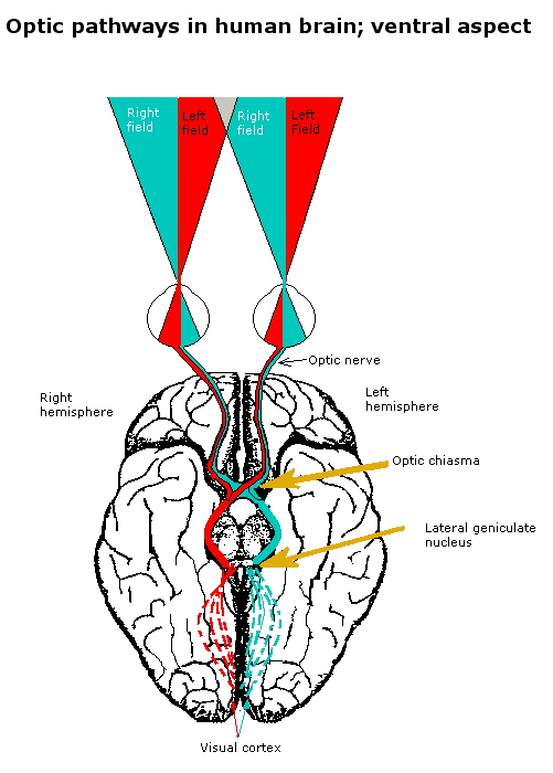
There are two visual nuclei in the thalamus: the lateral geniculate nucleus (LGN) and the pulvinar, which we won’t talk about. Neurons in the LGN have receptive fields that are center/surround like the retinal ganglion cells. The LGN has 6 layers, segregating inputs/outputs according to the eye of origin, which eye the information is coming from, on- or off- receptive field, whether the center is excitatory or inhibitory, and magnocellular or parvocellular pathway (more details below). The names of the pathways come from the fact that the retinal ganglion cells have large (magno) or small (parvo) cell bodies. It’s useful to think of two streams of information coming from the eyes to the brain.
The magnocellular pathway carries information about large, fast things (low spatial frequency, high temporal frequency) and is colorblind. The parvocellular pathway carries information about small, slow, colorful things (high spatial frequency, low temporal frequency).
Table 9.7.1. Characteristics of LGN neurons. In humans the LGN is normally described as having six distinctive layers. The inner two layers (1 and 2) are magnocellular layers, while the outer four layers (3, 4, 5, 6), are parvocellular layers. An additional set of neurons, known as the koniocellular layers, are found ventral to each of the magnocellular and parvocellular layers. (Credit: Wikipedia, https://en.wikipedia.org/wiki/Lateral_geniculate_nucleus, CC-BY-SA 3.0).
| Type | Size* | RGCSource | Type of Information | Location | Response | |
| M: Magnocellular cells | Large | Parasol cells | perception of movement, depth, and small differences in brightness | Layers 1 and 2 | rapid and transient | |
| P: Parvocellular cells (or “parvicellular”) | Small | Midget cells | perception of color and form (fine details) | Layers 3, 4, 5 and 6 | slow and sustained | |
| K: Koniocellular cells (or “interlaminar”) | Very small cell bodies | Bistratified cells | Between each of the M and P layers |
Disruptions in the magnocellular and parvocellular pathways can have a major effect on the complicated function of visual processing. A prominent instance concerns individuals suffering from a disorder called “motion blindness,” wherein impairments to the magnocellular pathway prevent them from perceiving motion. These people can see clearly in static, but they are not able to notice fluid movement, which makes daily chores like pouring a drink or crossing the street difficult. The importance of the magnocellular route in processing dynamic elements of the visual scene is demonstrated by this scenario (Zeki, 2015) On the other hand, abnormalities in the parvocellular pathway may result in problems with color and fine detail recognition. The parvocellular pathway is essential for processing high spatial frequency information and color, as demonstrated by the difficulties patients with such disturbances may have with tasks requiring high spatial resolution, such as reading or recognizing faces. These real-world instances highlight the intricate nature of visual processing in the brain and highlight the different but complementary roles played by the parvocellular and magnocellular pathways in human visual perception.

Cheryl Olman PSY 3031 Detailed Outline
Provided by: University of Minnesota
Download for free at http://vision.psych.umn.edu/users/caolman/courses/PSY3031/
License of original source: CC Attribution 4.0
Adapted by: Katya Tomlin
