
Light and Eyeballs
85 The Retinal Network
Learning Objectives
Be able to describe the different layers and cell types in a retina.
Know what the differences are between rods and cones.
Understand the impact of synaptic convergence on acuity and sensitivity.
The retina is composed of several layers and contains specialized cells for the initial processing of visual stimuli. The photoreceptors (rods and cones) change their membrane potential when stimulated by light energy. The change in membrane potential alters the amount of neurotransmitter that the photoreceptor cells release onto bipolar cells in the outer synaptic layer. It is the bipolar cell in the retina that connects a photoreceptor to a retinal ganglion cell (RGC) in the inner synaptic layer. There, amacrine cells additionally contribute to retinal processing before an action potential is produced by the RGC. The axons of RGCs, which lie at the innermost layer of the retina, collect at the optic disc and leave the eye as the optic nerve (Fig.8.5.1). Because these axons pass through the retina, there are no photoreceptors at the very back of the eye, where the optic nerve begins. This creates a “blind spot” in the retina and a corresponding blind spot in our visual field.
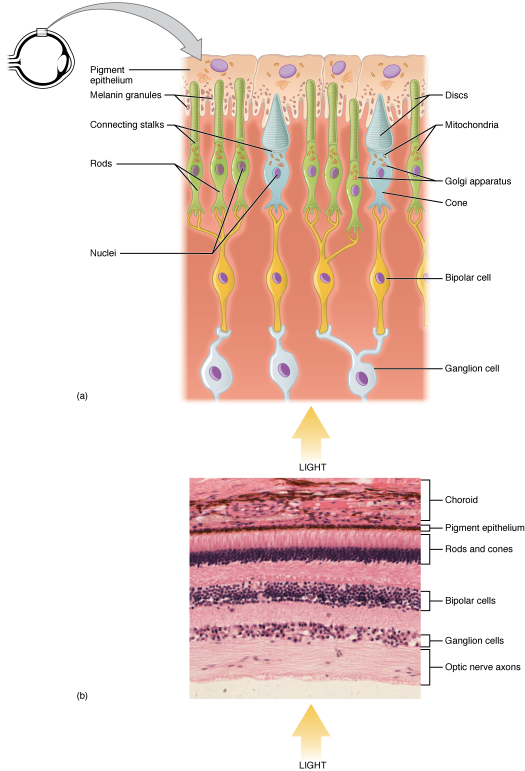
The retinal surface includes two types of photoreceptors called rods and cones. Rods are very sensitive to light and serve as the basis for vision in the dark. Since rods are monochromatic (i.e., respond to only one color), very limited color perception is possible in the dark. There are three types of cones activated during daylight, each enabling perception of color on limited sections of the visible part of the electromagnetic spectrum: those responding to short (bluish), medium (greenish), and long (reddish) wavelengths.
The outer segments of the rods and cones are mixed together to tessellate the back half of the eyeball and catch light from everywhere, with two exceptions. In the fovea, there are only cones. In the blind spot, there are no photoreceptors because all of the axons are leaving the eye.
Synaptic Convergence of Photoreceptors
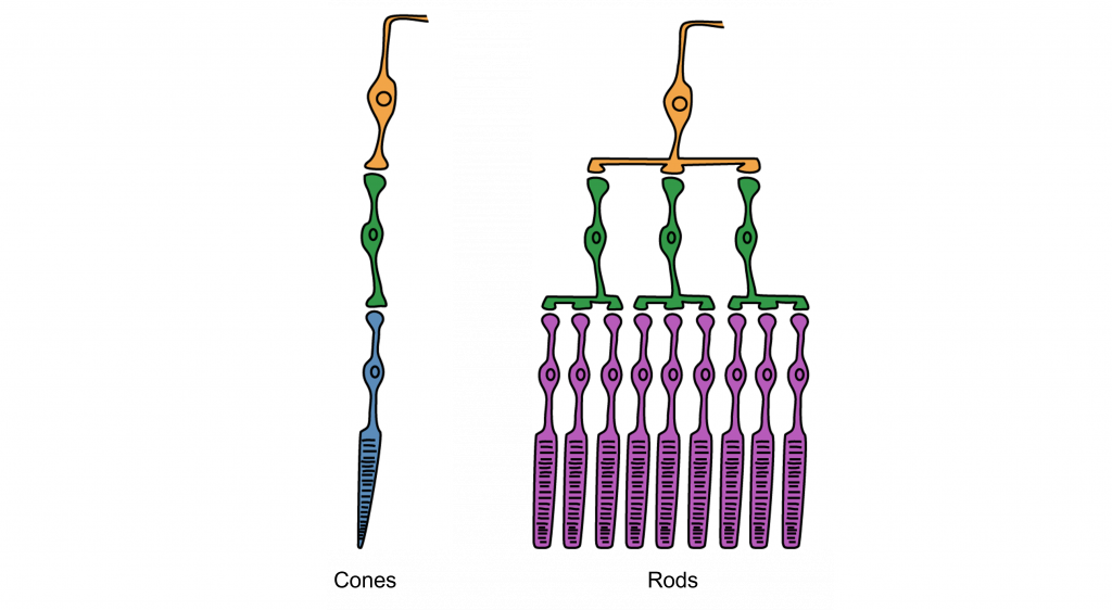
Rod cells are organized to have high synaptic convergence, where several rod cells (up to 30) feed into a single downstream route of communication (the bipolar cells, to be specific). An advantage of a high-convergence network is the ability to add many small signals together to create a seemingly larger signal. Consider stargazing at night, for example. Each rod is able to detect low levels of light, but signals from multiple rod cells, when summed together, allows you to recognize faint light sources such as a star. A disadvantage of this type of organization is that it is difficult to identify exactly which photoreceptor is activated by the incoming light, which is why accuracy is poor when seeing stimuli in our peripheral vision. This is one of the reasons that we cannot actually read text in our peripheral vision or see the distinct edges of a star. Rod photoreceptors are maximally active in low-light conditions.
Unlike rod cells, cone cells have very low synaptic convergence. In fact, at the point of highest visual acuity, a single cone photoreceptor communicates with a single pathway to the brain. The signaling from low-convergence networks is not additive, so they are less effective at low light conditions. However, because of this low-convergence organization, cone cells are highly effective at precisely identifying the location of incoming light.
OpenStax, Anatomy and Physiology Chapter 14.1 Sensory Perception
Provided by: Rice University.
Access for free at https://openstax.org/books/anatomy-and-physiology/pages/14-1-sensory-perception
License: CC-BY 4.0
Adapted by: Kathryn TaterkaPsychology by Jeffrey C. Levy, VisionProvided by: B.C. Faculty Pressbooks
URL: https://pressbooks.bccampus.ca/thescienceofhumanpotential/chapter/vision/
License: CC BY 4.0
Adapted by: Kathryn TaterkaConvergence is adapted from Introduction to Neuroscience Copyright © 2022 by Valerie Hedges is licensed under a Creative Commons Attribution-NonCommercial-ShareAlike 4.0 International License.
