
Light and Eyeballs
87 Eccentricity
Learning Objectives
Know the idea of eccentricity in degrees, and how big our fovea and macula are.
Be able to describe the density of cones and rods from the fovea to the periphery.
Know the visual acuity in our foveal vision and peripheral vision.
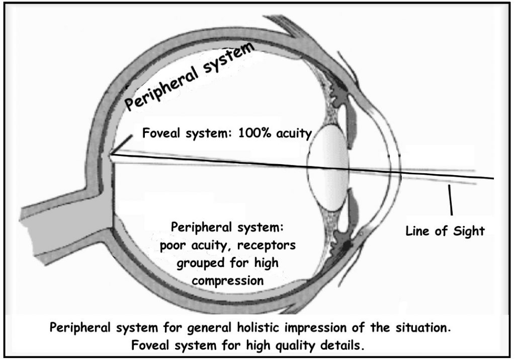
In a normal-sighted individual, the lens will focus images perfectly on a small indentation in the back of the eye known as the fovea, which is part of the retina, the light-sensitive lining of the eye. The fovea contains densely packed cones, which are the family of photoreceptors that express three different kinds of pigment to enable color vision and have relatively low levels of synaptic convergence onto the retinal ganglion cells (output from the retina) to enable high visual acuity. Thus, the fovea provides tremendous spatial resolution and color vision (but only in high light levels).
The fovea lies in a region of the retina called the macula. Although the macula comprises only 4% of the retinal area and 10% of the entire visual field, it is responsible for the majority of useful photopic vision. The fovea lies at the center of the macula and is approximately 2mm in diameter. The fovea represents the central 1 degree of the visual field; the macula is the central 5 degrees of visual angle.
Degrees of visual angle are calculated by drawing an imaginary wedge of light, where the point of the wedge is at the pupil, and the base of the wedge is out in the world, where the light is coming from. A full circle has 360 degrees. Obviously, light from behind your head can’t enter your eye directly; light from an arc that is about 180, or maybe 200 degrees can enter our eyes. So a 5 degree arc (the macula) is a tiny portion of the world, but that’s where the vast majority of our visual information comes from.
While cones are concentrated in the fovea where images tend to be focused, rods (another type of photoreceptor) are located throughout the remainder of the retina. Rods are specialized photoreceptors that work well in low light conditions, and while they lack the spatial resolution and color function of the cones, they are involved in our vision in dimly lit environments as well as in our perception of movement on the periphery of our visual field.
The peripheral retina is dominated by rods, but also contains cones. We have low acuity in the periphery because there is strong convergence: many rods map to a single output ganglion cell (since rods are more sensitive to light than cones). Convergence, in which each ganglion cell pools responses from multiple photoreceptors, increases sensitivity but decreases acuity. There is lots of convergence for rods and much less convergence for cones.
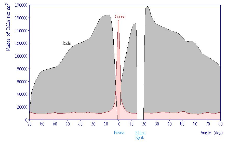 Figure 8.. Graph depicting the number of cells in (mm2) across the retina. Cones are concentrated at the center of the retina (0 degrees) in the fovea. There are many more rods than there are cones. The rods are located in the periphery, but not at the center of the retina. There are neither cones nor rods at the blind spot, which is where the retinal ganglion cells exit the retina. Credit: Distribution_of_Cones_and_Rods_on_Human_Retina © Jochen Burghardt is licensed under a CC BY-NC-SA (Attribution NonCommercial ShareAlike) license
CC LICENSED CONTENT, SHARED PREVIOUSLY
Webvision: The Organization of the Retina and Visual System, Age-related macular degeneration (AMD)
Authored by: Gregory S. Hageman, Karen Gaehrs, Lincoln V. Johnson and Don Anderson
URL: https://webvision.med.utah.edu/book/part-xii-cell-biology-of-retinal-degenerations/age-related-macular-degeneration-amd/
License: CC BY-NC
Adapted by: Kori Skrypek
OpenStax, Psychology Chapter 5.3 Vision
Provided by: Rice University.
Download for free at http://cnx.org/contents/4abf04bf-93a0-45c3-9cbc-2cefd46e68cc@5.103.
License: CC-BY 4.0
Adapted by: Kori Skrypek
Figure and legend on photoreceptor density are adapted from Introduction to Neuroscience Copyright © 2022 by Valerie Hedges is licensed under a Creative Commons Attribution-NonCommercial-ShareAlike 4.0 International License,
