
Vision Loss and V1
99 Retinotopic Organization of V1
Learning Objectives
Know where our primary visual cortex (V1) is located.
Know how the V1 neurons are organized according to their receptive fields in visual space (retinotopic organization).
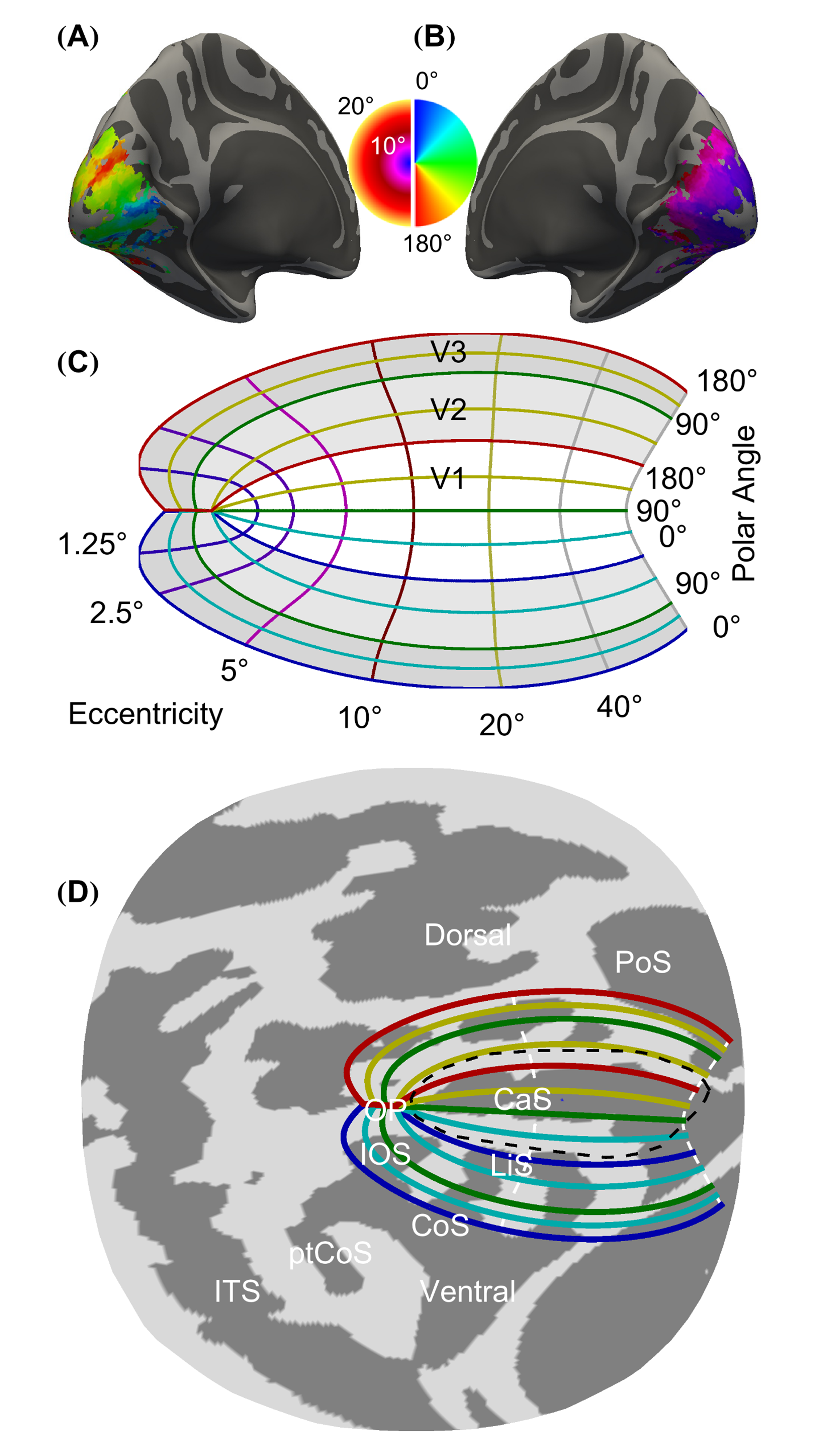
The primary visual cortex (V1) is located in the calcarine sulcus and has retinotopic organization and cortical magnification. The calcarine sulcus is in the occipital cortex, on the medial aspect. If you dissect away the white matter, the gray matter can be laid out like a flat sheet. When you do this to V1, it is roughly U-shaped, and we use this visualization a lot because it helps us to identify how the V1 cortex is organized.
Retinotopic organization means that neurons with receptive fields close together in visual space have cell bodies close together in the cortex. Below is an interactive visualization of how the visual world is mapped to V1. What it shows is how signals from each retina are sent to V1 on the opposite side of the brain. Essentially, the image is flipped in our brain as signals from our left eye are processed and mapped by the right side of V1 and vice versa. Furthermore, the neurons that help map the image are spatially located in corresponding regions in V1. For example, a stimulus in the center of one’s right hand visual field will be mapped by the center neuron in the V1 region on the left side.
Area V1 has retinotopic organization, meaning that it contains a complete map of the visual field (visual map) covered by the two eyes. In most species, V1 is considered to have a single map of the visual field, but in cats there’s two of them: one for area 17 and one for area 18.
Neurons in area V1 are classically divided into two types: simple and complex, based on the structure of their receptive field. In simple cells, receptive fields have separate ON and OFF subregions. ON and OFF subregions differ in their responses to the onset of stimuli on a gray background: ON subregions respond to white bars, and OFF subregions respond to black bars. In complex cells, ON and OFF regions are superimposed, i.e. every location in the receptive field responds both to white and black bars.
Scholarpedia, “Area V1” by Matteo Carandini, University College London, London UK
URL: http://www.scholarpedia.org/article/Area_V1
License: CC BY-NC-SA 3.0
Adapted by: Frank Wiest-PuchalaCheryl Olman PSY 3031 Detailed Outline
Provided by: University of Minnesota
Download for free at http://vision.psych.umn.edu/users/caolman/courses/PSY3031/
License of original source: CC Attribution 4.0
Adapted by: Frank Wiest-Puchala
