
Visual Development and Object Recognition
119 Neuroimaging
Learning Objectives
Know what the four main neuroimaging methods are (PET, EEG, MEG, fMRI).
Know how these four methods work.
Know what the pros and cons are of using different methods.
Neuroimaging exists as a way for scientists to monitor brain activity. There are four major neuroimaging methods that are commonly used by scientists: positron emission tomography (PET), electroencephalography (EEG), functional magnetic imaging (fMRI) and magnetoencephalogram (MEG).
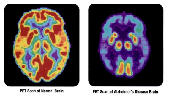
Positron emission tomography (PET) scans create pictures of the living, active brain. An individual receiving a PET scan drinks or is injected with a mildly radioactive substance called a tracer. Once in the bloodstream, the amount of tracer in any given region of the brain can be monitored. As brain areas become more active, more blood flows to that area. A computer monitors the movement of the tracer and creates a rough map of active and inactive areas of the brain during a given behavior (Fig.11.3.1).
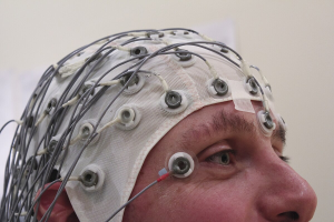
In some situations, it is helpful to gain an understanding of the overall activity of a person’s brain, without needing information on the actual location of the activity. Electroencephalography (EEG) serves this purpose by providing a measure of a brain’s electrical activity. An array of electrodes is placed around a person’s head. The signals received by the electrodes result in a printout of the electrical activity of his or her brain, or brainwaves, showing both the frequency (number of waves per second) and amplitude (height) of the recorded brainwaves, accurate within milliseconds. Such information is especially helpful to researchers studying sleep patterns among individuals with sleep disorders.
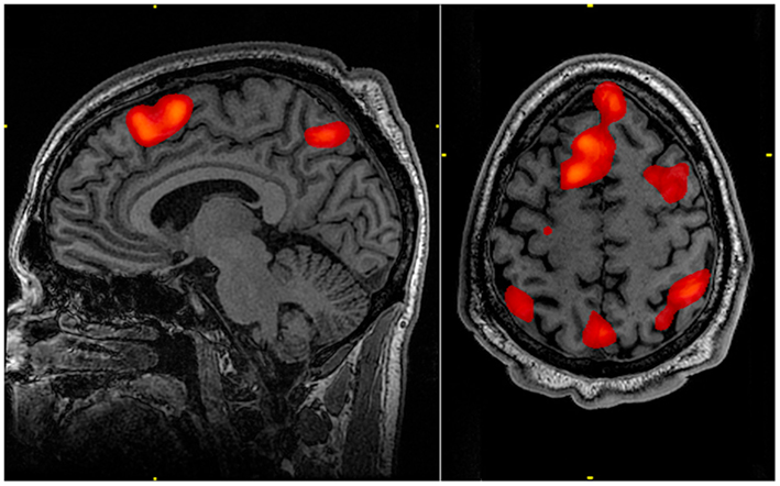
Functional magnetic resonance imaging (fMRI) operates on the same principles but it shows changes in brain activity over time by tracking blood flow and oxygen levels (Fig.11.3.1).
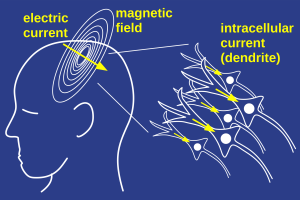
Magnetoencephalography (MEG) is another technique for non-invasively measuring neural activity. The flow of electrical charge (the current) associated with neural activity produces very weak magnetic fields that can be detected by sensors placed near the participant’s scalp (Fig.11.3.1). The number of sensors used varies from a few to several hundred. Due to the fact that the magnetic fields of interest are so small, special rooms that are shielded from magnetic fields in the environment are needed in order to avoid contamination of the signal being measured.
There are many pros and cons to using each method. The fMRI provides more detailed images of the brain’s structure, as well as better accuracy in time, than is possible in PET scans. One major advantage of EEG is its temporal resolution. Data can be recorded thousands of times per second, allowing researchers to document events that happen in less than a millisecond. MEG analytic strategies are nearly identical to those used in EEG, but the MEG recording apparatus is much more expensive than EEG, so MEG is much less widely available.
| Neuroimaging Method | Pros | Cons |
| Positron emission tomography (PET) |
|
|
| Electroencephalography (EEG) |
|
|
| Functional Magnetic Resonance Imaging (fMRI) |
|
|
| Magnetoencephalography (MEG) |
|
|
CC LICENSED CONTENT, SHARED PREVIOUSLY
OpenStax, Psychology Chapter 3.4 The Brain and Spinal Cord
Provided by: Rice University.
Download for free at http://cnx.org/contents/4abf04bf-93a0-45c3-9cbc-2cefd46e68cc@12.2.
License: Creative Commons Attribution 4.0
NOBA, Psychophysiological Methods in Neuroscience
Provided by: University of Delaware & University of California, Los Angeles
URL: https://nobaproject.com/textbooks/introduction-to-psychology-the-full-noba-collection/modules/psychophysiological-methods-in-neuroscience
License: CC BY-NC-SA 4.0
