Basics: neuroscience and psychophysics
4 Neuroscience Review
Learning Objectives
Make sure that you’re familiar with each of these concepts—this introductory section recaps background vocabulary to support other learning goals.
This Neuroscience section is composed of excerpts from Chapter 12 of the OpenStax Anatomy and Physiology textbook to highlight key concepts that are most relevant to a study of sensation and perception.
Nervous tissue is composed of two types of cells, neurons and glial cells. Neurons are the primary cells associated with the nervous system. They are responsible for the computation and communication that occurs within the nervous system. They are electrically active and release chemical signals to target cells. Glial cells, or glia, are known to play a supporting role for nervous tissue. Ongoing research pursues an expanded role that glial cells might play in signaling, but neurons are still considered the basis of this function. Neurons are important, but without glial support, they would not be able to perform their functions.
Parts of a Neuron
The main part of a neuron is the cell body, or the soma. The cell body contains the nucleus and most of the major organelles. Neurons have many extensions of their cell membranes, which are generally referred to as processes. Neurons are usually described as having one axon—a fiber that emerges from the cell body and projects to target cells. That single axon can branch repeatedly to communicate with multiple target cells. It is the axon that propagates the nerve impulse, which is then communicated to one or more cells.
The other processes of the neuron are dendrites, which receive information from other neurons at specialized areas of contact called synapses. The dendrites are usually highly branched processes, providing locations for other neurons to communicate with the soma. Information flows through a neuron from the dendrites, across the cell body, and down the axon. This gives the neuron a polarity, meaning that information flows in this one direction.
Types of Neurons
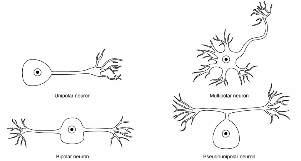
There are many different types of neurons, and they can be classified using many different criteria. The first way to classify neurons is by the number of processes attached to the cell body. Using the standard model of neurons, one of these processes is the axon, and the rest are dendrites. Because information flows through the neuron from dendrites → cell body → axon, these names are based on the neuron’s polarity.

Unipolar cells have only one process emerging from the cell. True unipolar cells are only found in invertebrate animals, so the unipolar cells in humans are more appropriately called “pseudo-unipolar” cells. Invertebrate unipolar cells do not have dendrites. Human unipolar cells have an axon that emerges from the cell body, but it splits so that the axon can extend along a very long distance. At one end of the axon are dendrites, and at the other end, the axon forms synaptic connections with a target. Unipolar cells are exclusively sensory neurons and have two unique characteristics. First, their dendrites receive sensory information- sometimes directly from the stimulus itself. Secondly, the cell bodies of unipolar neurons are always found in ganglia. Sensory reception is a peripheral function, so the cell body is in the periphery- though closer to the CNS in a ganglion. The axon projects from the dendrite endings, past the cell body in a ganglion, and into the central nervous system.
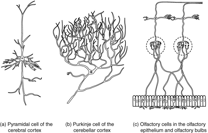
Bipolar cells have two processes, which extend from each end of the cell body, opposite to each other. One is the axon and one the dendrite. Bipolar cells are not very common. They are found mainly in the olfactory epithelium (where smell stimuli are sensed), and as part of the retina.
Multipolar neurons have one axon and two or more dendrites. With the exception of the unipolar sensory ganglion cells, and the two specific bipolar cells mentioned above, all other neurons are multipolar. Some research suggests that certain neurons in the CNS do not conform to the standard model of “one, and only one” axon.
Some sources describe a fourth type of neuron, called an anaxonic neuron. The name suggests that it has no axon, but this is not accurate. Anaxonic neurons are very small, and if you look through a microscope, you will not be able to distinguish any process specifically as an axon or a dendrite. Any of those processes can function as an axon depending on the conditions at any given time. Nevertheless, there is not one specific process that is definitively the axon. These neurons have multiple processes and are therefore multipolar.
Neurons can also be classified on the basis of where they are found, who found them, what they do, or even what chemicals they use to communicate with each other. Some neurons referred to in this section are named on the basis of those sorts of classifications. For example, a multipolar neuron that has a very important role to play in a part of the brain called the cerebellum is known as a Purkinje (pronounced per-KIN-gee) cell. It is named after the anatomist who discovered it (Jan Evangilista Purkinje, 1787–1869).

CC LICENSED CONTENT, SHARED PREVIOUSLY
OpenStax, Anatomy and Physiology Section 12.2 Nervous Tissue
Provided by: Rice University.
Access for free at https://openstax.org/books/anatomy-and-physiology/pages/1-introduction
License: CC-BY 4.0
Adapted by: Cheryl Olman

