
Part 1: Skinning Your Cadaver
Abby Brown
Superficial Structures
- Before you begin to skin out your cadaver, identify the superficial structures found on the distal limbs, as described for each species. (Figure 1-1)
- Pony Specimen: Identify the chestnut, ergot and coronet.
- In the horse, identify the chestnut on the medial antebrachium, located significantly above the carpus.
- On the palmar surface of the fetlock, identify the ergot. (Normally, in a non-clipped animal, there is an area of long hair, referred to as the feather that usually obscures the ergot. However, clipping the hair off the fetlock during preparation of the specimen should have exposed this area sufficiently to see the ergot.) Note that the ergot correlates to the metacarpal or metatarsal pad of the dog.
- Observe the skin to hoof transition; this is called the coronet, referring to the ‘crown’ of the hoof. Deep, and distal, to the coronet is a band of dermal tissue (coronary band) which will be discussed later.
- Calf Specimen: Identify the dewclaws and the coronet.
- Identify the dewclaws on the palmar surface of your calf limbs.
- Observe the skin to claw transition; this is called the coronet.
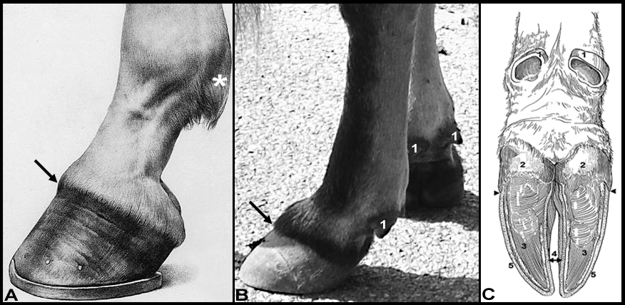 Figure 1-1. A. Horse front foot, lateral view; B. Bovine front foot, lateral view; C. Bovine foot, palmar/plantar view. *Region of ergot (horse); arrows, coronet (horse and bovine); bovine: 1, Dewclaws (digits 2 and 5); Claw (digits 3 and 4): 2, bulb of claws; 3, sole of claws; 4, wall (axial surfaces); 5, wall (abaxial surfaces); arrowhead, junction of wall and bulb. (Modified from Anatomy and Physiology of Farm Animals; Frandson, Wilke, Fails)
- Pony Specimen: Identify the chestnut, ergot and coronet.
- As you begin the proximal thoracic limb dissection, you should note that the forelimb is bound to the neck and chest by at least seven extrinsic muscles. (Recall from Anatomy I that extrinsic muscles have one attachment on the body/trunk and one attachment on the limb itself.) The following dissections will be done on both sides of the pony and calf hanging specimens to expose these extrinsic muscles.
Skinning Your Cadaver
3. Make skin incisions as directed for each species and reflect the skin off of the neck and proximal forelimbs. Identify the area of the nuchal fatty crest in the horse. As you reflect the skin, note that you will identify the cutaneous trunci m. (and omobrachialis portion over the shoulder) and you will identify the superficial thoracic vein (spur vein) in the horse.
-
- Calf skin reflection: Begin by making a single dorsal midline incision over the neck region.
- Pony skin reflection: Incise the skin along the ventral edge of the mane (on both sides). Deep to the mane is a tough mass of fibrous fatty tissue called the nuchal fatty crest (Fig. 1-2, N); it will be studied in more detail when we dissect the neck.
- In both species, continue the incisions caudally along the dorsal midline to the level of the mid-thorax.
- At each end of the dorsal incision (cranially and caudally) make vertical incisions; ventrally you will join the left and right vertical incisions.
- For the cranial incision:
-
- in the pony, make the vertical cut just caudal to the ear
- in the calf, make the vertical cut just caudal to the halter
-
- For the cranial incision:
- Make a midline incision down the ventral aspect of the neck from the head to the sternum. From that ventral midline cut, extend an incision down the medial side of each forelimb to a point just below the elbow. Encircle each forelimb with an incision about 5-10 cm distal to the point of the elbow (Fig. 1-2, upper).
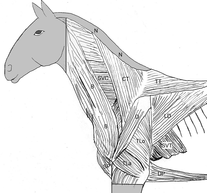
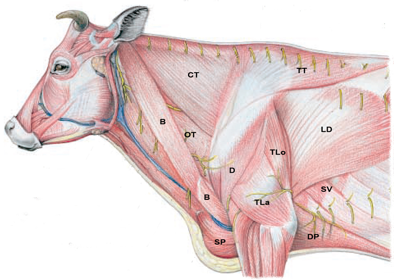
Figure 1-2 Extrinsic and intrinsic muscles of the horse (upper) and ox (lower). (N) Indicates the location of the nuchal fatty crest present only in the horse. Extrinsic muscles: CT, Cervical trapezius; TT, thoracic trapezius; B, brachiocephalicus;SP, superficial pectorals; DP, deep pectorals; LD, latissimus dorsi;SVC, serratus ventralis cervicis; SVT, serratus ventralis thoracis. Labeled intrinsic forelimb muscles: D, Deltoideus; TLo, long head of the triceps brachii; TLa, lateral head of the triceps brachii. (Not shown is the cutaneous trunci m. which may come off when the skin is reflected. If present, it will cover the upper forelimb and structures caudal to it.) (Modified from Budras and Habel, 1st ed.)
4. Reflect the skin from the neck and thorax as one large piece on each side. (You may choose to either remove the reflected skin or keep it attached along the ventral midline of the abdomen to use for wrapping your cadaver.)
-
- As you reflect the skin in the pony and calf, you should notice a large, flat muscle on the cadaver (or it may be adhered to the inner surface of the reflected skin) extending caudally over the thorax; this is the cutaneous trunci muscle which you should identify. It is responsible for twitching the skin in the live animal. It may be difficult to separate the cutaneous muscles from the skin and is not essential to do so; cutaneous mm. may be reflected with the skin if needed.
- An additional (thin) cutaneous muscle can be found covering the proximal, lateral part of the forelimb/shoulder region in both species; this is the omobrachialis m.
5. Reflect the cutaneous mm. distally toward the ventral midline.
-
- The omobrachialis m. should first be reflected caudally off the shoulder toward the cutaneous trunci m. and then reflected ventrally with it.
- The insertion of the cutaneous trunci m. should be transected just caudal to the forelimb as it is reflected ventrally.
6. Pony Specimen: As you reflect the cutaneous trunci m. in the pony, observe the superficial thoracic vein (aka spur vein) just caudal to the elbow region. It will be seen as you reflect the cutaneous trunci m. distally and will need to be transected to fully reflect the cutaneous trunci m. This vein runs along the dorsal edge of the ascending deep pectoral muscle.
7. Proceed with the reflection/removal of the cutaneous muscles; however, be careful to leave the underlying latissimus dorsi m. and the ventrally located deep pectoral muscles intact on the specimen.
-
- Demonstration Specimen Variance: The cutaneous trunci muscle will be separated out and left attached on one side of the demonstration specimen. (If your specimen is well dissected your group may choose to leave the cutaneous trunci m. attached on one side as well.)
Dissection Videos for this Section of Material
Superficial Structures and Skinning Your Cadaver
- Pony: watch 0:00-3:25
- Proximal Thoracic Limb: https://www.youtube.com/watch?v=oIi4i8ScZEM&t=824s
- Calf: watch 0:00-2:45
- Proximal Thoracic Limb: https://www.youtube.com/watch?v=hN10FaowIos