Want to create or adapt books like this? Learn more about how Pressbooks supports open publishing practices.
Module 10: Veterinary Ectoparasites
Module 10.7: Fleas
Fleas
Learning Objectives
Describe the life cycle stages of fleas.
What are the primary clinical signs of flea infestations?
How can visual inspection and microscopic examination be used to diagnose flea infestations?
Fleas are among the most common ectoparasites affecting animals, and they are known for their ability to infest a wide range of hosts, including pets and humans. These small, wingless insects are notorious for their rapid reproduction and persistent presence, causing significant discomfort and potential health issues. Fleas primarily feed on the blood of their hosts, leading to itching, allergic reactions, and in severe cases, anemia.
In veterinary practice, understanding the biology, behavior, and control of fleas is essential for effectively managing infestations and preventing the spread of flea-borne diseases. Fleas undergo complete metamorphosis, which includes distinct life stages: egg, larva, pupa, and adult. This lifecycle, coupled with their environmental resilience, makes flea control a challenging yet crucial aspect of veterinary care.
This section will provide an in-depth exploration of fleas, focusing on their identification, lifecycle, and the various strategies for their management and prevention. By familiarizing yourself with the key characteristics and behaviors of fleas, you will be better equipped to address infestations, educate pet owners, and ensure the health and comfort of your patients.
Biology and Environmental Preferences
Fleas belong to the order Siphonaptera. They are highly adaptive and can thrive in various environments, though they prefer warm, humid conditions. Fleas undergo complete metamorphosis, including the egg, larva, pupa, and adult stages. Follow this link to view a video that describes their life cycle.
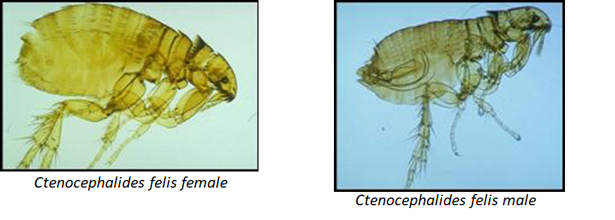
These insects are similar in size to sucking lice (sesame seed) but are rapidly moving. Under the microscope, the legs get larger posteriorly, with powerful posterior legs used for jumping from host to host. Fleas also have a pronotal and genial (it looks like a mustache!) comb that entomologists use to help speciate.
In the laboratory, you will not be asked to speciate fleas, but you will be required to distinguish a flea from other ectoparasites microscopically. The CAPC vet website is a great resource to learn more!
Table 10.10: Flea Species
Flea Species
Identifying characteristics
Transmission
Species commonly infected
Ctenocephalides felis
eggs: white, oval with rounded ends
larvae: found in the environment are maggot-like
pupae: white-colored
adults: eyes are present, comb located on the head with spines pointed horizontal, 6 legs
flea dirt: reddish-black pellets of dried blood excreted by adult fleas
Symptoms: Itching, redness, and in severe cases, flea allergy dermatitis.
Diagnosis: Visual examination and identification of fleas and flea dirt.
Ctenocephalides canis (Dog Flea)
Hosts: Dogs, cats, and occasionally humans
Symptoms: Similar to those caused by cat fleas.
Diagnosis: Visual examination and identification of fleas and flea dirt.
Pulex irritans (Human Flea)
Hosts: Humans and occasionally other animals
Symptoms: Itching and irritation.
Diagnosis: Visual examination and identification of fleas and flea dirt.
Diagnostics
Detection of flea dirt or frass
Flea dirt, also known as flea feces or flea droppings or frass, is a crucial indicator of flea infestation in pets and often the first diagnostic test that is done to determine if there is a flea infestation.
What is flea dirt or frass
Flea dirt is the fecal matter produced by fleas. It consists of digested blood that fleas consume from their host, typically cats, dogs, or other animals. Flea dirt appears as small, dark, granular specks on the pet’s skin and fur. It is often the first visible sign of a flea infestation.
Identifying flea dirt
Physical characteristics
Flea dirt can be identified by its distinct appearance and texture. Here are some key characteristics:
Color: Flea dirt is typically black or dark brown.
Size: The particles are very small, resembling ground pepper or tiny specks of dirt.
Shape: The granules are often irregular in shape, not perfectly round or uniform.
Common areas to check for flea dirt
Base of the Tail
Neck and Shoulders
Belly and Groin
Armpits and Between the Legs
Methods to detect flea dirt
Visual Inspection
A thorough visual inspection is the simplest and most direct method to detect flea dirt.
Part the Fur: Use your fingers or a fine-toothed comb to part the fur, exposing the skin.
Examine the Skin: Look for small, dark specks on the skin or in the fur.
Focus on Common Areas: Pay particular attention to the base of the tail, neck, and groin.
Use of a Flea Comb
A flea comb is a specially designed tool with closely spaced teeth that can capture flea dirt, fleas, and eggs.
Comb Through the Fur: Gently comb your pet’s fur, starting from the head and working your way down the body.
Check the Comb: After each stroke, inspect the comb for black or dark brown specks.
Use a White Surface: Shake the comb over a white surface (like a piece of paper or a towel) to better see the flea dirt.
The Wet Paper Towel Test
This test helps confirm whether the dark specks are flea dirt or just ordinary dirt.
Collect a Sample: Using a flea comb or your fingers, gather some of the black specks found on your pet.
Place on a Paper Towel: Put the specks on a wet paper towel or tissue.
Observe the Color Change: If the specks dissolve into a reddish-brown color, it is likely flea dirt, as it contains digested blood.
Confirming Flea Presence
While detecting flea dirt is a strong indication of a flea infestation, it is important to confirm the presence of fleas for effective treatment.
Look for Live Fleas
Comb or Part Fur: Use a flea comb or part of the pet’s fur and look for small, fast-moving insects.
Check Common Areas: Focus on areas where flea dirt is found, as fleas are likely to be nearby.
Check for Flea Bites
Red, Irritated Skin: Look for small, red bumps or areas of irritation, especially where flea dirt is present.
Excessive Scratching or Biting: Pets with fleas often scratch or bite at affected areas due to irritation.
Disease transmitted by fleas
Fleas have been transmitting terrible diseases to humans and animals for thousands of years (does anyone remember the Black Plague?) Below is a table of some diseases transmitted by fleas in general.
Table 10.11: Disease Transmitted by Ctenocephalides felis flea
Disease
Species affected
Type of disease transmitted
Bartonella henselae
cats, humans
bacteria
Dipylidium caninum
Dog, cat, humans
tapeworm (vector)
Summary
Fleas can cause significant discomfort and health issues for both animals and humans, including the diseases that they transmit. Early detection and effective treatment are essential to manage flea infestations effectively. An important part of management is environmental decontamination as fleas lay their eggs on the host and they will readily fall off contaminating the environment.
Knowledge check
Key Takeaways
Lice, mites, ticks, and fleas are frequently encountered ectoparasites that affect a variety of veterinary species, causing various health issues.
Mites possess 8 legs during their nymph and adult stages, but they have only 6 legs in their larval stage, which is an important detail for accurate identification.
Adult lice and fleas both have 6 legs, a characteristic feature that distinguishes them from arachnid ectoparasites like mites and ticks.
Lice and fleas are large enough to be observed with the naked eye, making them relatively easier to identify in a clinical setting.
Due to their small size, mites typically require microscopic examination for proper observation and identification.
You have now reached the end of Module 10. If you are enrolled in CVM 6925, please go to the Canvas page and take the quiz: “Module 10: Ectoparasites quiz.” There is an assignment that accompanies the in-person laboratory for this module.