Module 10: Veterinary Ectoparasites
Module 10.2: General anatomy of ticks
Introduction to Tick anatomy: Hard and Soft Ticks
Ticks are obligate hematophagous ectoparasites that belong to the order Ixodida and are divided into two main families: Ixodidae (hard ticks) and Argasidae (soft ticks). Both families are of significant medical and veterinary importance due to their role in transmitting a variety of pathogens to animals and humans.
Ticks belong to the arachnid class and are closely related to spiders, scorpions, and mites. They have four stages in their life cycle: egg, larva, nymph, and adult. Ticks are resilient and can survive in a variety of environments, though they are most commonly found in areas with high humidity and dense vegetation. Their survival and development are highly dependent on environmental conditions, including temperature and humidity.
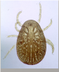
General anatomy of hard ticks (Family: Ixodidae)
Hard ticks, as their name suggests, possess a rigid, shield-like structure called a scutum that covers part of their dorsal surface. This distinguishing feature not only provides them with protection but also influences their feeding behavior and ecological niche. The life cycle of hard ticks typically involves three stages: larva, nymph, and adult, each requiring a blood meal from a host. Common examples include the deer tick (Ixodes scapularis), responsible for transmitting Lyme disease, and the dog tick (Dermacentor variabilis), known for spreading Rocky Mountain spotted fever.
- Scutum: The scutum is a hard, chitinous plate that provides protection and structural support. In males, it covers the entire dorsal surface, while in females, nymphs, and larvae, it covers only the anterior half, allowing for expansion as they engorge with blood. The scutum can be ornate or inornate.
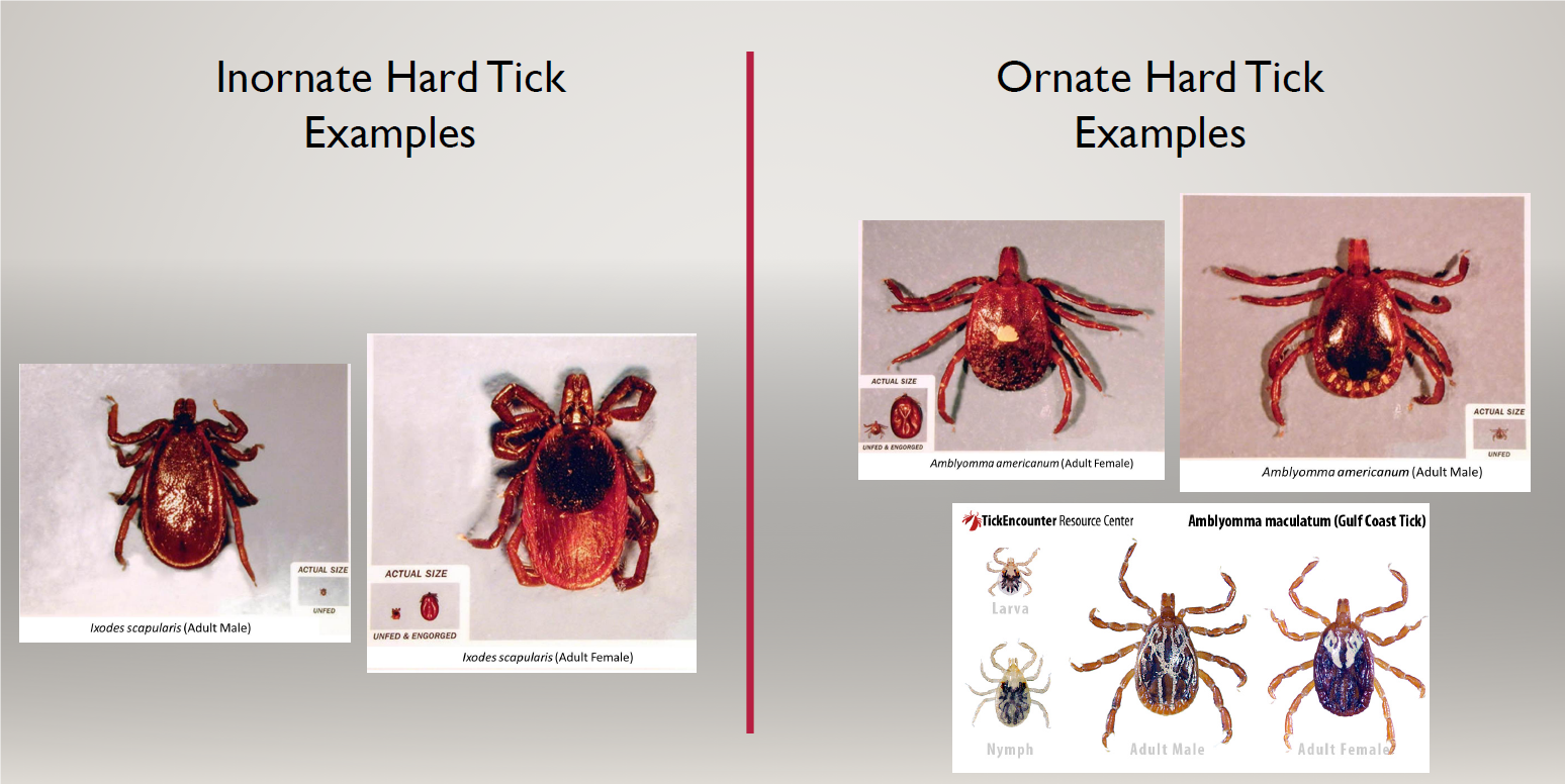
- Inornate: Lacks distinct patterns or markings.
- Ornate: Features distinctive white or iridescent patterns, aiding in species identification.
- Capitulum (Mouthparts): The capitulum consists of several structures used for attachment and feeding:
- Hypostome: A barbed structure that anchors the tick to the host’s skin.
- Chelicerae: Cutting appendages that pierce the host’s skin.
- Palps: Sensory organs that aid in locating feeding sites.
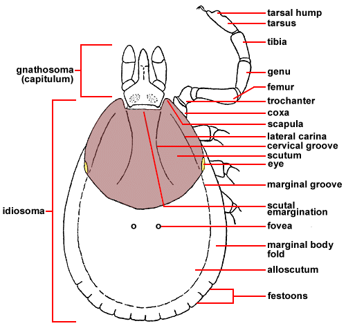
- Body: The body is broadly divided into the gnathosoma (capitulum) and idiosoma (main body). The idiosoma houses the digestive and reproductive organs and expands significantly during blood-feeding.
- Legs: Adult ticks have eight legs, each with claws and sensory structures at the distal end that help in host detection and attachment.
General anatomy of soft ticks (Family: Argasidae)
Soft ticks are less commonly encountered in veterinary practice compared to hard ticks. They lack a scutum, and their mouthparts are located ventrally, tucked underneath their bodies, which are adapted for a more cryptic lifestyle.
- Capitulum (Mouthparts): Unlike hard ticks, the capitulum of soft ticks is not visible from above and is located ventrally. This adaptation allows them to feed in sheltered environments.
- Hypostome: Less barbed compared to hard ticks.
- Chelicerae: Used for cutting the host’s skin.
- Palps: Similar sensory functions as in hard ticks.
- Body: The body of soft ticks is more flexible and leathery, facilitating their ability to hide in protected environments like crevices and burrows. Their body structure is more rounded and lacks the distinct segmentation seen in hard ticks.
- Legs: Like hard ticks, soft ticks have eight legs. However, their legs are adapted for crawling in confined spaces.
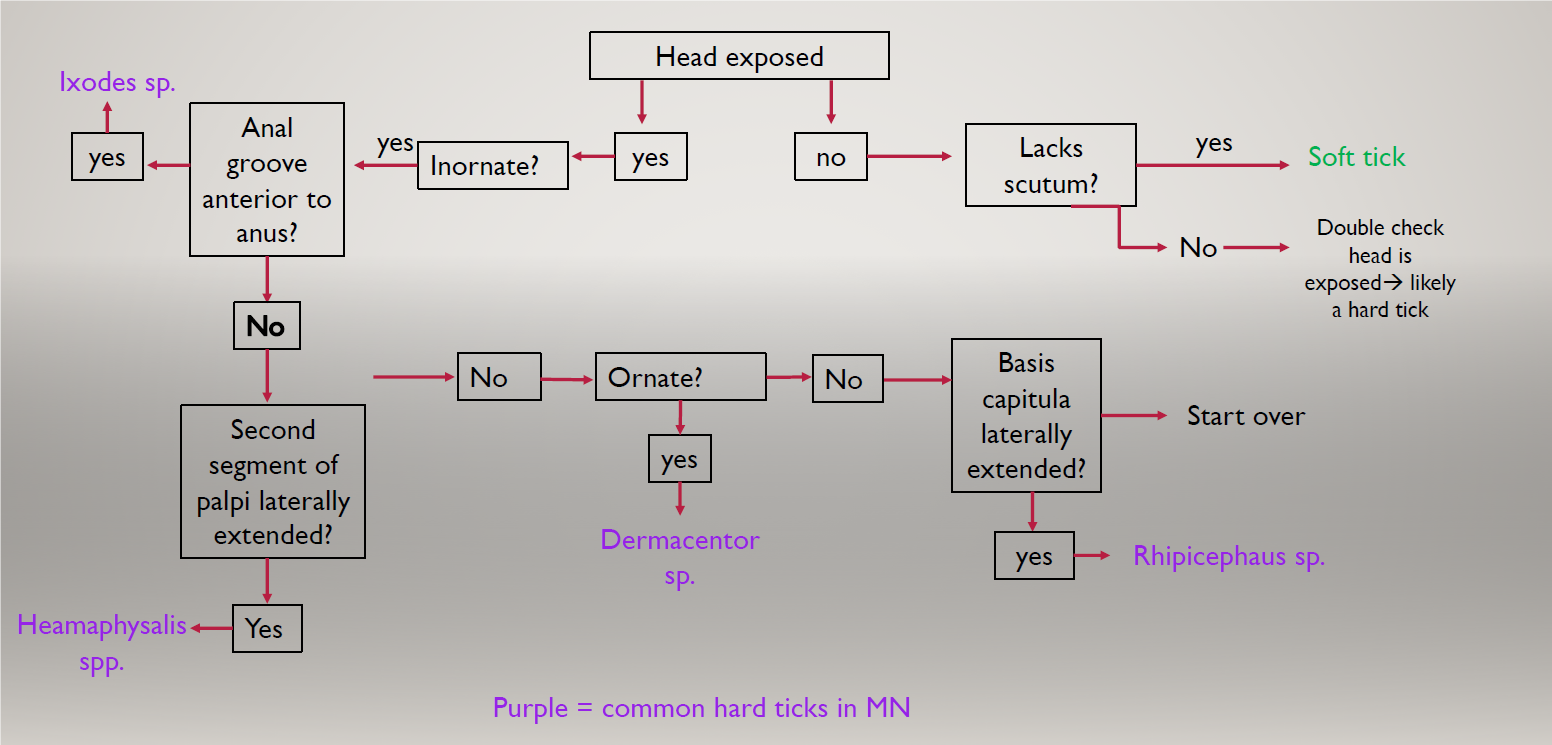
Summary table and charts
Tick Type |
Scutum |
Capitulum (mouthparts) location |
Habitat |
|---|---|---|---|
Hard Tick |
yes | Anterior of the body | Wide diversity |
Soft Tick |
no | Tucked underneath the body ventrally | Woods or rocks- need protection |