
Module 11: Rapid Point of Care (POC) Testing
Module 11.6: SNAP® Foal IgG Test
Equine failure of passive transfer testing
In your large animal medicine courses, you will learn about the failure of passive (FPT) transfer in more clinical detail than what will be covered in this course. Depending on the placental structure, gravid animals may or may not have the ability to transfer antibodies to their offspring in utero. For animals such as horses and cows, antibodies are not transferred to their offspring in utero during pregnancy making the ingestion of colostrum, which is rich in maternal immunoglobulins, immediately after birth essential to survival. This transfer of humoral immunity from the dam to the offspring is called passive transfer.
There are both foal and dam factors that may result in FPT. A few of those include:
Dam factors (failure to produce or deliver adequate colostrum to foal)
- Inadequate amount of immunoglobulins transferred into colostrum
- Loss of colostrum prior to parturition (g. premature lactation)
- Behavioral factors (unwillingness to accept foal or allow it to nurse)
- Dam death (orphan foal)
Foal factors (failure to suckle or absorb adequate colostrum)
- Congenital abnormalities that prevent the foal from standing or nursing
- Illnesses or injuries that prevent the foal from standing or nursing
- Inability to absorb immunoglobulins from the gut
To answer the questions in your laboratory activity. Please refer to the IDEXX website.
Knowledge check
SNAP® Foal IgG Test
There are several in-clinic diagnostic tests that can be used to screen for FPT in foals (and other species). These tests uses serum or whole blood is taken from the foal to detect IgG levels. Unlike the other SNAP tests that you will run in this laboratory, the SNAP Foal IgG test is semi-quantitative, as the intensity of color change compared to the calibration spot is used to semi-quantify IgG levels in the foal’s blood.
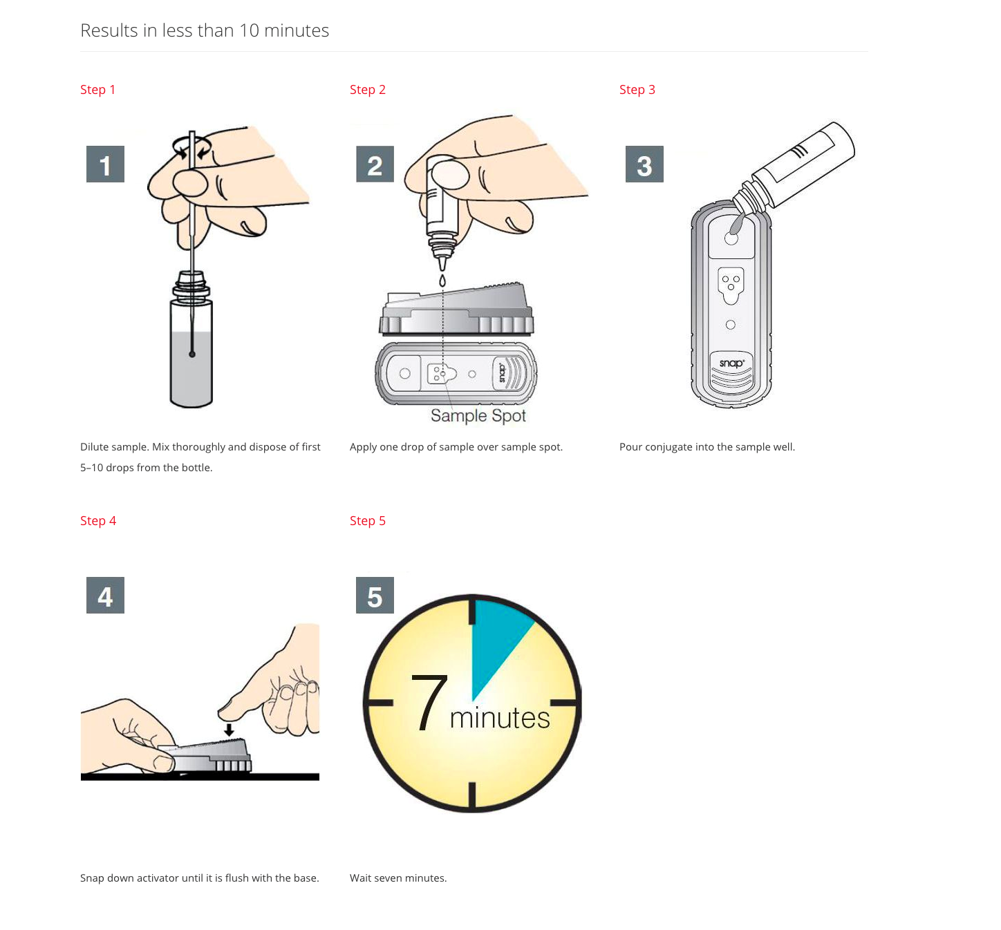
Procedure
- Remove the cap and dropper tip from the sample diluent bottle.
- Gently mix whole-blood samples by inverting in the EDTA tube
- Using the plastic sample loops provided, carefully immerse ONLY the loop tip into the blood or serum sample. Visually confirm that the loop is filled.
*For whole-blood samples, we recommend immersing the loop tip in the sample that remains in the cap of the sample collection container. Immerse the loop tip only. Do not immerse the loop handle in the sample.
*For serum and plasma, use one loop. For whole blood, use two separate loops.
- Transfer the filled loop by immersing and twirling the loop tip in the bottle of sample diluent.
- Firmly seat the dropper tip on the sample diluent bottle. Mix thoroughly by inverting five times. Hold the sample diluent bottle vertically and dispose of the first 5–10 drops from the bottle.
- Place the SNAP device on a flat surface. With the bottle tip ½ to 1 inch directly above the SNAP device, carefully apply one drop of the diluted sample directly onto the sample spot in the result window. Visually confirm that the drop of the diluted sample has wetted the sample spot completely. If the drop of the diluted sample has not wetted the sample spot completely, repeat the sample application using a new SNAP device.
- Remove the cap from the conjugate bottle and pour its contents into the sample well of the SNAP device. (Some of the contents will remain in the conjugate bottle.) The sample will flow across the result window, reaching the activation circle in 30 to 90 seconds. (Some conjugate will remain in the sample well at activation.) Watch the device carefully for color in the activation circle. When color FIRST appears in the activation circle, push the activator firmly until it is flush with the device’s body. Keep the device horizontal to ensure accurate results.
- Wait 7 minutes. Visually read the test result.
Knowledge check
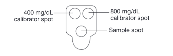
Interpretation of Foal IgG
The window of your SNAP test should have three BLUE dots following testing. These dots include the 400 mg/dL spot 800 mg/ dL spot, and our patient sample spot.
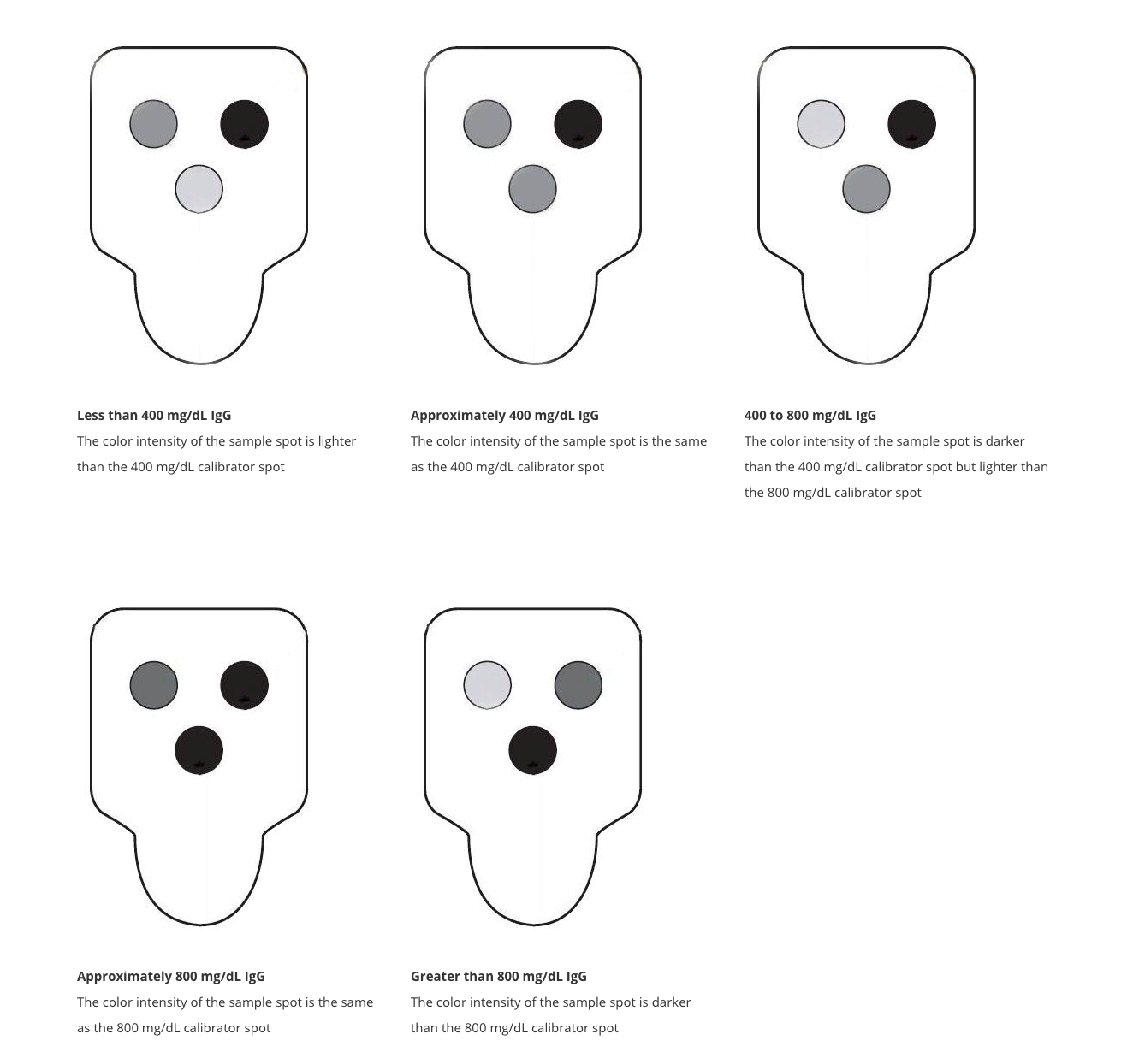
Depending on the intensity of your patient spot in comparison to the calibration spots is used to determine the blood levels of IgG in the foal. The sensitivity of this test is high but the specificity for FPT is lower meaning this test is only used on animals with a high index of clinical suspicion for FPT.
For example, if the patient sample spot is lighter blue than the 400 mg/dL and 800 mg/dL spots, then the foal is estimated to have <400 mg/dL of IgG in his/her bloodstream. This would be interpreted as the following: Since the sensitivity of this test is high, but the specificity is low, in a patient with a high clinical index of suspicious for FPT, following additional testing (CBC/ Chem), this patient is considered to likely be suffering from FPT.
On the reverse end of the testing, if the patient color spot is stronger blue than the 800 mg/dL spot, then the clinical signs observed in your patient are unlikely to be the result of FPT.
Quick and dirty interpretation
IgG > 800 mg/dL → adequate passive transfer (good/normal immune protection)
IgG between 400 – 800 mg/dL → partial failure of passive transfer (partial protection)
IgG < 400 mg/dL → complete failure of passive transfer (very susceptible to infection)
Knowledge check
Key Takeaways
- Rapid point of care tests commonly used ELISA techniques
- These tests can be used to detect a wide variety of things including antibodies, antigens, proteins, chemicals, etc.
- Most rapid point of care tests are used as screening tools
