
Module 8: Introduction to the Routine Urinalysis
Module 8.4: Common Structures Observed on the Sediment Exam
Epithelial cells
There are 2 types of epithelial cells that we can see in our urine sediment; transitional and squamous epithelial cells.
Squamous epithelial cells
- The external urinary tract (skin, vulva, prepuce, etc.) is lined by squamous epithelial cells.
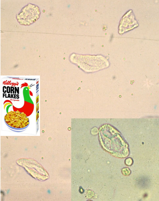
- These cells are anucleate, angular to polygonal, and vary greatly in size depending on how they exfoliated
- Some describe as resembling “corn flakes”
- Common to see in voided and catheterized samples, not cystocentesis
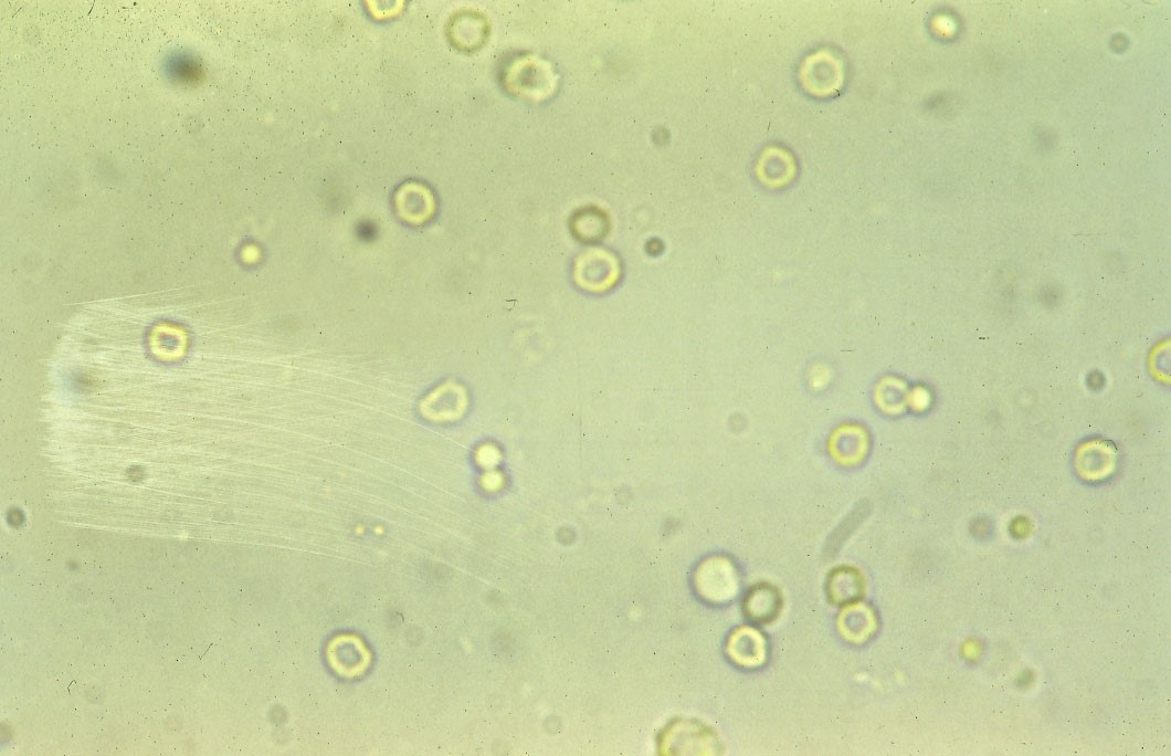
- In the image below, you can see several squamous epithelial cells at 10x and 40x (inset) magnification. These cells have not been stained.
- In the inset, you can also visualize 2 RBCs (smooth round structures directly adjacent to squamous epithelial cell) and 2 WBCs that are more irregular in shape and grainy appearance
Knowledge check
Round (transitional) cell epithelium
- The internal urogenital tract is lined by transitional epithelium (also referred to as urothelial cells and round epithelial cells in some texts, all these terms are interchangeable).
- In cytology textbooks, when describing unstained round in shape epithelial cells in urine samples, you will see these cells referred to as round or transitional epithelium. We assume based on their shape and location that these epithelial cells are of transitional cell origin, but this needs to be confirmed using Romanowsky-type stains.
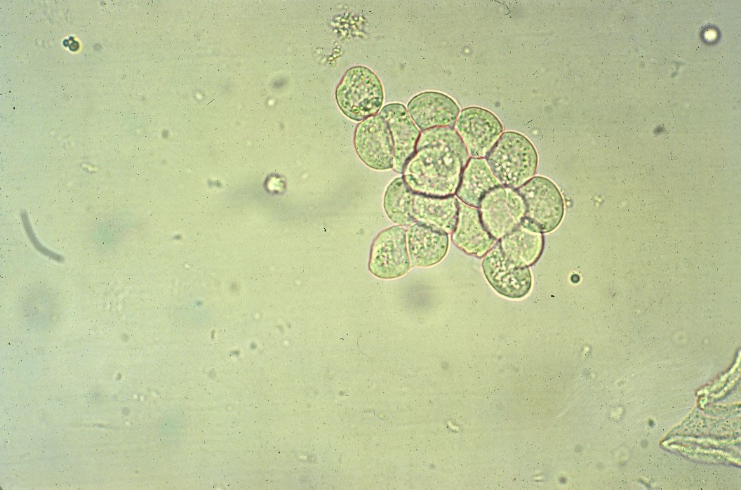
- These cells are normally shed into the bladder or along the urethra during normal cell turnover. Increased numbers in urine may indicate hyperplasia, inflammation, or neoplasia.
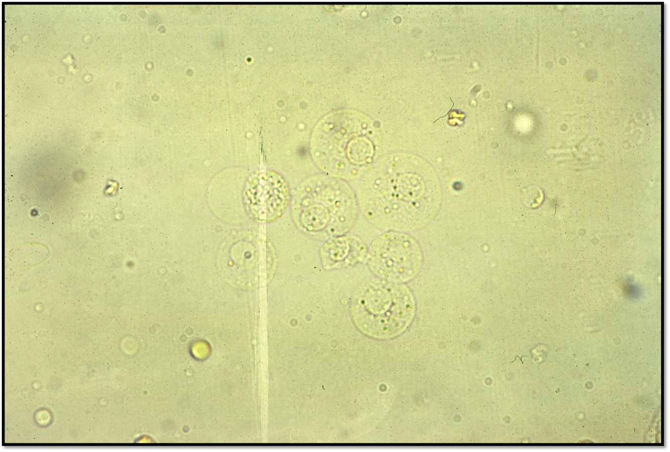
- These cells are typically 4-5x the diameter of a neutrophil with an eccentrically placed nucleus (see image). It is not unusual to see them individually or in small clusters.
Knowledge check
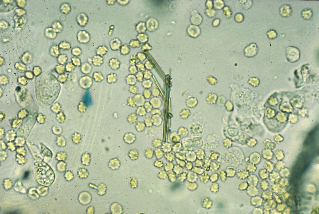
Crystalluria
- The medical term for crystal formation in the urine is crystalluria.
- Not all crystals are pathogenic
- Knowing the pH of urine is important to help determine the type of crystal you are seeing as certain crystal tend to only form in either acidic or basic urine
- Crystals form in vivo and vitro
Common causes for in vivo formation
- Diet
- UTI
- Abnormal urine pH
- High USG (very concentrated urine)
Common causes for in vitro formation
- Temperature
- Most commonly refrigeration
- Evaporation
- Urine pH
Please refer to Cornell University’s eClinPath website for images and guidance of what types of crystals are precipitated out in what types of scenarios.
Knowledge check
Urinary casts
Casts are cylindrical structures that are composed primarily of Tamm Horsfall Mucoprotein (THP) in the renal tubules. THP is secreted by renal epithelial cells. These casts are “molds” of the renal tubules. Cellular constituents can become entwined in THP during specific pathologies and characterize the type of cast found. The most common casts seen in the urine of domestic animals are “hyaline casts”. These casts can be found in the urine of healthy animals as they are comprised almost entirely of THP. More information and pictures of casts can be found at Cornell University’s eClinPath website. Casts can be seen at both 10x and 40x objectives.
Knowledge check
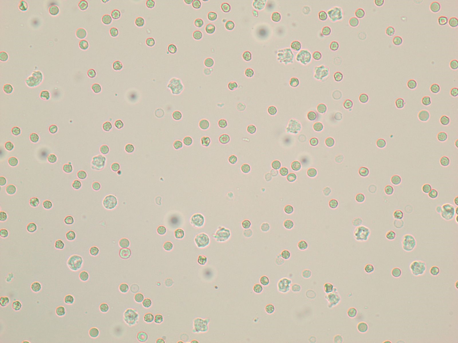
Red and white blood cells
Red blood cells
- Up to 5 RBC/hpf is generally considered acceptable for “normal” urine
- Cystocentesis may result in larger numbers of RBCs in the sample
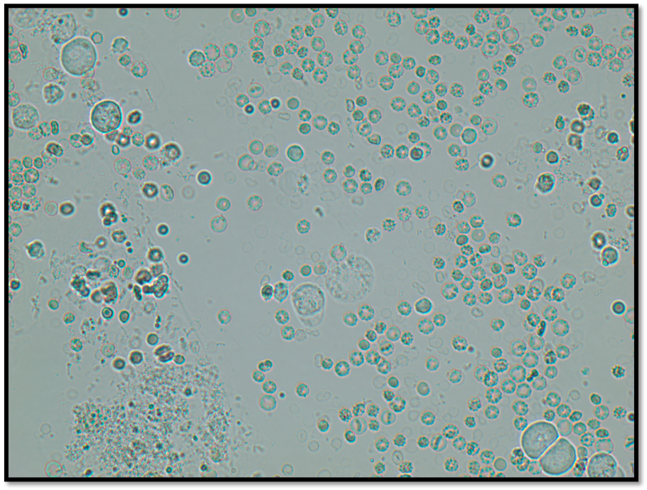
- Increased RBCs in the urine is called hematuria
- Can be secondary to hemorrhage, trauma, inflammation, necrosis, trauma, or neoplasia along the urinary tract
- The presence or absence of central pallor in the RBCs does not indicate the duration of time in the urine
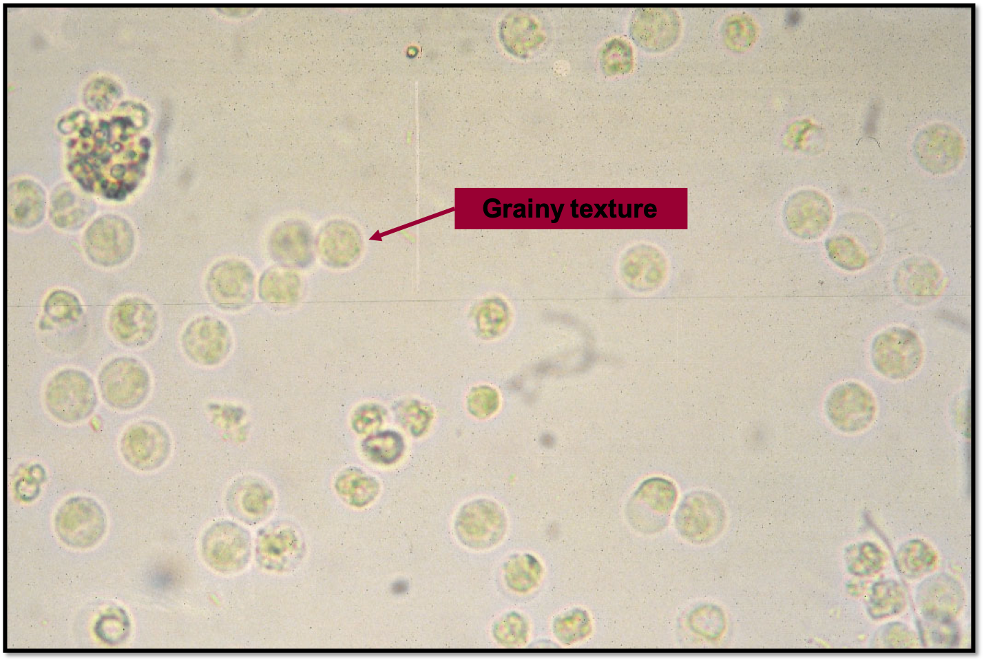
White blood cells
- Less than 5 WBC/hpf is generally accepted as “normal” in voided samples
- Large numbers of WBCs in urine is called pyuria
- Pyuria generally indicates inflammation along the urinary tract
Common causes of inflammation along the urinary tract include:
- Urinary tract infection (UTI)
- Neoplasia along the urinary tract
- Uroliths (bladder stones)
- Compared to RBCs, WBC’s (15um) appear grainy and are slightly larger than an RBC (8 um). It is important to remember both RBCs and WBC’s will flux in size depending on the osmotic concentration of the solution as the cell will take on or lose water.
Knowledge check
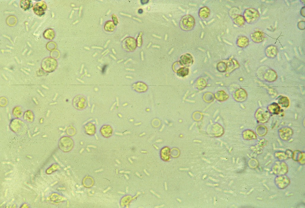
Bacteriuria
The urinary bladder is not sterile and has its own unique flora, just like the gastrointestinal tract. The overall biomass of bacteria in the bladder is minimal, thus it is unusual to see bacteria on routine urinalysis. When bacteria are observed in urine, it is important to determine if there is pyuria or hematuria present concurrently as that may indicate a urinary tract infection. The most common cause of urinary tract infections in dogs and cats is E. coli, a Gram-negative rod.
Lab procedure
In the laboratory, you will be working through several case-based dipstick cases and performing several sediment exams.
Knowledge check
Key Takeaways
- The two major parts of the routine urinalysis is the dipstick and sedimentation exam
- We use a refractometer, not the dipstic pad, to determine USG
- Not all the dipstick pad results are useful in veterinary species
- Low magification is used to detect large cellular features in urine (i.e. epithelial cells, cats, crystals)
- High magnification is used to detect hematuria, pyuria, and bacteriuria
Tamm-Horsfall protein (THP) is exclusively produced by renal tubular cells of the distal loop of Henle and is the most abundant urinary protein in mammals.
an abnormal increase in WBCs in the urine.
