
Module 8: Introduction to the Routine Urinalysis
Module 8.3: The Routine Urinalysis
The routine urinalysis
For standardization, in veterinary medicine, the urine sediment exam is prepared with 3-5mL of urine. This is important to help provide a semi-quantification of sedimentation results.
Routine urinalysis is comprised of these 4 steps:
- Assessment of visual urine attributes
- Assessment of concentrating ability
- Dipstick analysis
- Sediment examination
The following sections will guide you through the urinalysis process.
Knowledge check
Assessment of visual urine attributes
The first step of any urinalysis is an assessment of visual attributes such as color and turbidity. Observations of color and turbidity are determined using a well-mixed urine specimen.
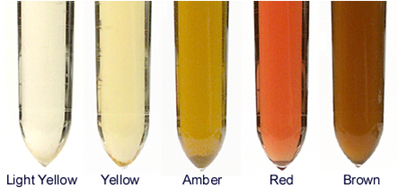
Color?
- Color change, in most cases, is a common reason that patients will present to you as this is something the owners will visually observe
- Color scale: Light yellow, yellow, amber, red, and brown
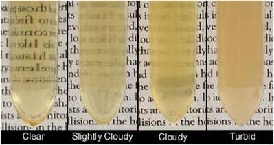
Turbidity?
- Evaluation of the clarity of the sample.
- For most species, we expect that the urine would be clear, but for some species (horse, rabbits) cloudy urine is normal. We use the ease with which an individual can read newspaper text behind the liquid to gauge clarity.
- Turbidity scale: Clear, slightly cloudy, cloudy, turbid
Knowledge check
Urine Specific Gravity (USG)
Urine Specific Gravity is a measure of the solute concentration in urine, and it is used to assess the ability of the renal tubules to concentrate or dilute the glomerular filtrate.
Two methods to determine USG:
- Refractometer
- Gold standard
- Uses the refractive index as a measurement of the density of urine compared to pure water
- Dipstick
-
- Unreliable in veterinary medicine as we expect animals to concentrate their urine well beyond the range the dipstick is able to detect (1.000-1.030). When USG’s get above 1.030 it is necessary to have a more quantitative value that cannot be achieved with the dipstick
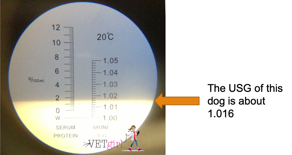
How do I read a refractometer?
Most refractometers are calibrated to measure both serum proteins and urine-specific gravity.
Steps:
- Place several drops of the sample liquid on the angled prism.
- Seal the clear plate on top of it.
- Look through the eyepiece while pointing the refractometer at a source of direct light.
- Follow the line of refraction across the scale to determine the USG in your patient. (See image below)
Knowledge check
Dipstick analysis
The dipstick examination is essentially the biochemical evaluation of urine using a urine dipstick pad. Dipsticks consist of various pads containing chemicals that provide a color change when a particular analyte is present in urine. This color change is converted to a semi-quantitative result for the analyte in question.
The common chemical constituents measured in veterinary medicine are:
- pH
- Glucose
- Ketones
- Bilirubin
- Protein
- Heme (Blood, Hemoglobin, and Myoglobin)
- The dipstick also detects leukocyte esterase, nitrites, urobilinogen; however, these 3 pads are unreliable in animals and we generally ignore their findings
- USG pad is also considered a fairly unreliable measurement of a patient’s concentrating ability. Additionally, it only measures to 1.030 and our patients should be concentrating well above that value.
Dipstick procedure
Please review the following video and handout on the dipstick procedure prior to the laboratory.
Urinalysis procedure- See Dipstick Assignment Canvas Page
Knowledge check
Sediment examination
The sediment examination is the microscopic evaluation of urine to identify cells, blood, crystals, bacteria, etc. of the urine that would not be detected using dipstick alone. The sediment exam is performed at low (10x obj.) and high (40x obj.) magnification. Subdued lighting is necessary to increase the refractivity of the unstained urine elements (lower the condenser and/or close down the substage iris diaphragm).
- Elements evaluated at 10x objective
- Squamous and round/transitional epithelial cells
- Crystals
- +/- casts (sometimes easier to see at high mag.)
- Parasite ova
- Elements evaluated at 40x objective
- Bacteria
- WBC
- RBC
- The purpose of this lab is not to interpret sediment findings, but rather to become comfortable with the identification of common sediments. In your clinical pathology and medicine courses, you will discuss their significance.
The following pages will guide you through common structures observed on the sediment exam, beginning with those that can be seen on the 10x objective and moving towards those seen with the 40x objective.
Knowledge check
(of a liquid) cloudy, opaque, or thick with suspended matter.
laboratory test that shows the concentration of all chemical particles in the urine.
