
Module 5: Introduction to Microbiology Stains
Module 5.2: Microbial Stains
Microbial stains
A few of the common microbial stains used in veterinary medicine include Gram stain and Acid-Fast stain. In our laboratory, we are going to focus on Gram’s stain and acid-fast.
Gram stain
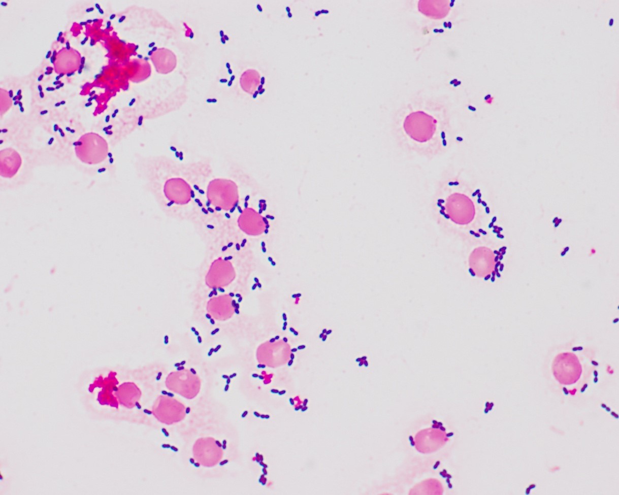
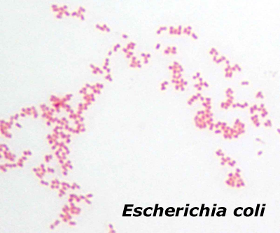
Gram stain is a common technique used to separate bacteria into two large categories; Gram-positive and Gram-negative based on cell wall characteristics. The thick peptidoglycan layer in the Gram-positive bacteria’s cell wall retains the crystal violet stain giving a deep purple color. Gram-negative bacteria have a thinner peptidoglycan layer that does not retain the crystal violet after a decolorization step and application of a red dye counterstain called Safranin, resulting in their red appearance. In veterinary medicine, we can use knowing if a bacterial population is Gram-negative or positive to help guide initial antimicrobial therapy while waiting for pending culture results.
It is important to remember that these color patterns are originally based on cellular properties following isolation from pure bacteria culture. While these features also remain in vivo, antimicrobial administration and phagocytosis of bacteria break down their cell wall resulting in inconsistent Gram staining features. I.E. Gram-positive bacteria may appear Gram-negative.
Procedure
In the laboratory, you will be given step-by-step instructions on how to perform a Gram stain. Here is a video that also shows the steps.
Knowledge Check
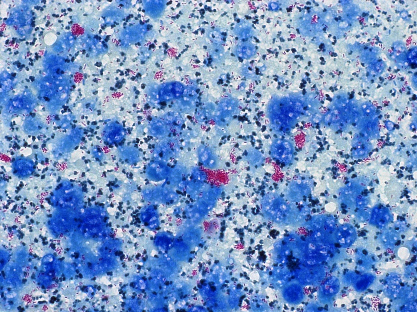
Certain bacteria, most notable Mycobacteria, have lipid components within their cell walls that make them resistant to taking up stains such as Romanowsky-type stains and Gram stains. Thus, acid-fast stains are used to differentiate acid-fast bacteria from non-acid fast bacteria.
The principle of these stains is the carbol fuchsin solubilizes the lipid present in the cell wall resulting in the bacteria appearing pink or red and retain that color after decolorization with alcohol. Non-acid fast organisms lack the lipid material in their cells wall following decolorization leaving them colorless. The sample is then stained with a counterstain, most commonly methylene blue or malachite green. Only decolorized cells absorb the counterstain and take its color and appear blue or green while acid-fast cells retain the pink or red color.
How the acid-fast technique is performed in a laboratory.
Summary of stains
Below is a summary of the staining characteristics of bacteria with each stain:
Gram-positive |
Gram-negative |
Acid-fast |
|
|---|---|---|---|
Romanowsky-type |
Purple | Purple | Colorless |
Gram Stain |
Purple | Red | Colorless |
Acid Fast |
Green/Blue | Green/Blue | Red |
Knowledge check
Key Takeaways
- Cytological stains, such as Romanoswsky stains, are designed to highlight cellular features (i.e. nucleus, cytoplasm, rod, cocci)
- Microbiology stains are often indicated when infectious agents, commonly bacteria, are seen using cytologic stains
- The most commonly used microbiology stains in veterinary medicine help differentiate bacteria based on cell wall characteristics
