
Module 11: Rapid Point of Care (POC) Testing
Module 11.1: Point of Care Diagnostic Testing
Introduction to point of care diagnostic testing
While there are many point-of-care diagnostic tests available on the market for both companion and production animals, we are going to focus on the screening tests that use plasma, serum, feces, and/or whole blood to generate a laboratory result. There are many examples of these tests available to veterinarians from several different pharmaceutical companies and new ones are always coming on the market. In the virtual laboratory, we are going to be using lateral flow assays (LFA), the most common point of care testing, on patient samples, and case studies. Other common names for these type of tests include the following:
-
- Lateral flow test (LFT)
- Lateral flow device (LFD)
- Lateral flow assay (LFA)
- Lateral flow immunoassay (LFIA)
- Lateral flow immunochromatographic assays
- Dipstick
- Pen-side test
- Quick test
- Rapid test
- Test strip
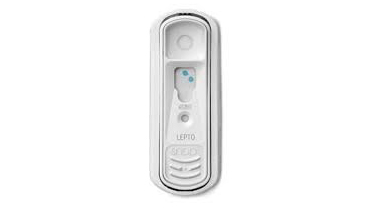
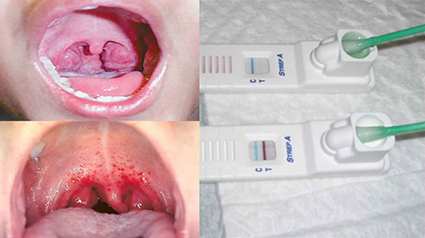
Below are images of common LFA used in everyday practice in the medical community. Examples in the images include a SNAP Test, a human pregnancy test, a rapid human Strep test, and a general heartworm test.

Methods of lateral flow testing
These tests use ELISA technology in which labeled antibodies react with a patient antigen or antibody that results in a visible color change at the test line or dot position.
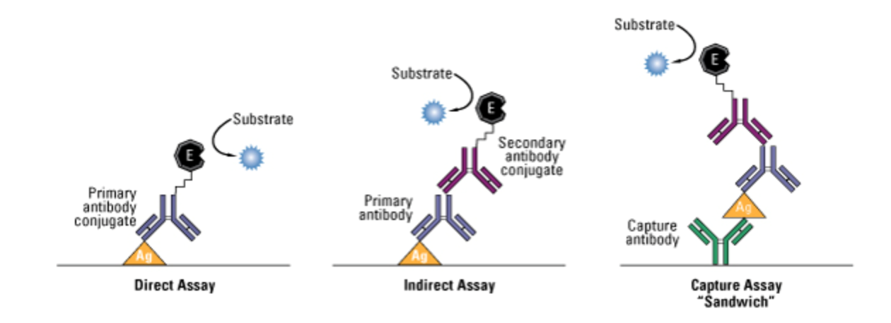
There are 3 main types of ELISA’s:
- Direct Assay
- Indirect Assay
- Sandwich Assay
The most common type of assay used in POC LFA is the Capture or Sandwich ELISA as the other two techniques often require a pure sample not easily achieved in a busy clinical setting.
How does this technology work?
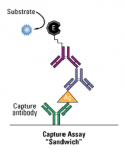
- The figure to the right demonstrates the reaction within the test
- This sandwich ELISA is detecting the patient’s blue antibody
- The capture antibody has an antigen attached to it that “captures” the patient’s blue antibody
- The magenta (purple) antibody is the secondary antibody conjugate that changes color following attachment to the patient’s antibodies.
- The description that follows is how the SNAP ELISA works
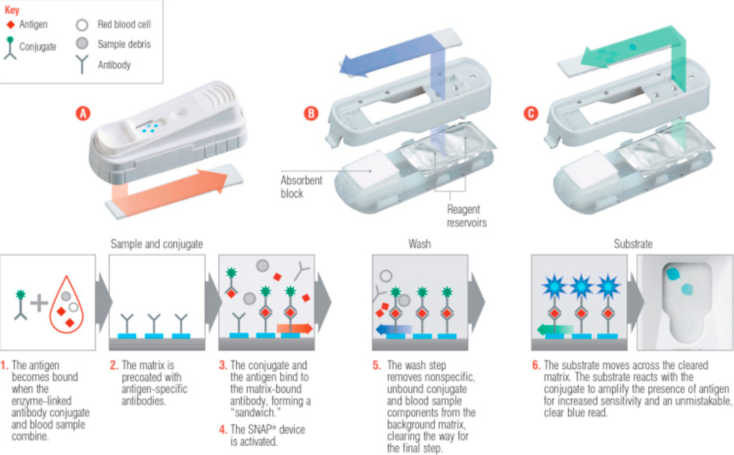
- The sample is dropped into the well of the test (below image “A”). The sample pad acts as the first stage of the absorption process, and in some cases contains a filter, to ensure the accurate and controlled flow of the sample.
Capture or Sandwich Assay - The conjugate pad, which stores the conjugated labels and antibodies, will receive the sample. If the target is present, the immobilized conjugated antibodies and labels will bind to the target and continue to migrate along with the test.
- As the sample moves along the device the binding reagents situated on the nitrocellulose membrane will bind to the capture antibodies at the test line or dot. A colored line or dot will form.
- The sample will continue passing through the nitrocellulose membrane into the absorbent pad. The absorbent pad will absorb the excess sample. The specification of the absorbent pad will have an impact on the volume of the sample the test can incorporate.
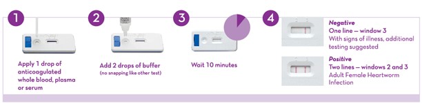
- If either a positive or negative sample does not result in activation of the positive control line or dot, then the test needs to be re-run
- The sample is dropped into the well of the test (below image “A”). The sample pad acts as the first stage of the absorption process, and in some cases contains a filter, to ensure the accurate and controlled flow of the sample.
How a lateral flow assay works
ELISA tests, in general, make great screening tests as they are often very sensitive, but not always very specific. This makes sense if we think about it from a strictly biological standpoint. In our bodies, antibodies are created by our B-cells in response to a myriad of causes for antigenic stimulation. In some cases, antibodies created to detect a certain protein or substrate secreted or on the surface of a pathogen or drug will have a similar molecular structure to that of a protein, peptide, receptor, etc. on one of our cells (aka molecular mimicry). This cross-reaction results in our cells being attacked and destroyed by immune cells. An example is this rheumatic fever in people with Strep A infections as the antibodies produced to attack the bacteria cross-react with proteins found in the synovium, myocytes, and heart valves resulting in immune-mediated polyathropathy (IMPA) and heart failure.
While the antibodies that are used in these tests have been produced in laboratory animals and then purified to increase specificity, they are still not perfect. Thus it is recommended that all point of care testing be confirmed with a secondary test. This confirmation test will vary depending on the positive result.
