Module 6: Blood Smear Technique and Reticulocyte Counting
Module 6.1: Preparing a Diagnostic Blood Smear
Making a blood smear or film is a skill that cannot be acquired in a single day. This skill must be practiced over and over again. However, hopefully, this exercise will give you the appreciation of how valuable hiring someone competent in making blood smears is and how challenging it can be to perform an accurate WBC and platelet count off of a sub-optimally prepared smear.
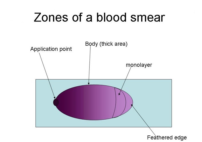
All blood smears have 3 essential components:
-
Feathered edge
The very distal region of the blood smear is formed at the end of your blood distribution. Under magnification, this area appears similar to the anatomy of a feather with the individual barbs. The feathered edge is useful for looking for platelet clumping, microfilariae, large neoplastic cells, and other large cells that are heavy and drifted to the edge.
-
Monolayer
The monolayer is the zone in which there is an equal distribution of erythrocytes (RBC) and leukocytes (WBC), and lies between the body and the feathered edge. Blood smears are shaped similar to a feather, so the monolayer itself is shaped in an arch. This region is where we perform 100 WBC count, platelet counts, and evaluation of erythrocyte morphology (i.e. schistocytes, spherocytes, acanthocytes, etc.). When performing a 100 nucleated cell count you will need to adjust to the monolayer as you go (i.e. you cannot just move straight across your slide)
-
Body
It can help you identify agglutination or rouleaux, but these findings should always be confirmed in the monolayer as abnormalities seen in the body will also be observed in the monolayer.